


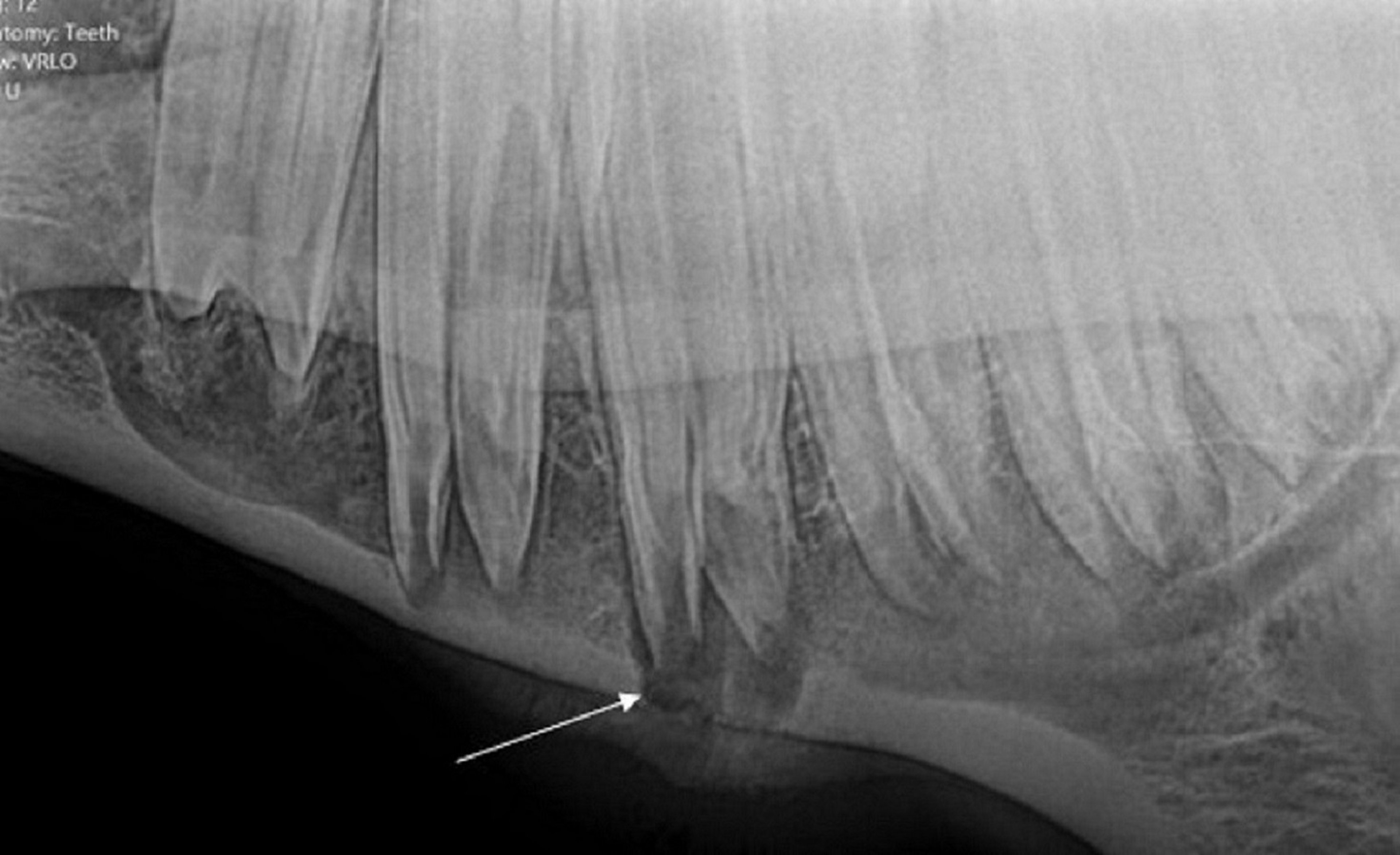
Courtesy of Dr. Jack Easley.
Periapical infection in horses most commonly affects the second and third cheek teeth (premolar 3 [Triadan 07s] and premolar 4 [Triadan 08s]) in both the upper and lower arcades. The pulp chamber of the teeth may become infected by various routes, including hematogenous (anachoretic pulpitis), periodontal, or from direct crown insult. In horses, hypoplasia of the cementum in the infundibula of the upper cheek teeth may predispose the animal to infundibular caries, leading to subsequent pulpitis and apical osteitis. The pathologic features of dental decay often affect tissues in the surrounding region; infection of the tooth may be accompanied by maxillary sinusitis, local cellulitis, periostitis, alveolar periodontitis, or fistula formation. Consequently, the specific tooth affected by apical infection in a horse or llama may be difficult to identify. Many animals are not examined until the infection is advanced. Tooth fractures discovered on examination may be secondary rather than primary. In some species (eg, the horse), apical osteitis and pulpitis may be initiated by abnormal eruption and dental impaction. The cause of apical osteitis in cattle and New World camelids may be similarly influenced.
When dental decay is advanced, extraction of the affected tooth is recommended. In horses, historically, decayed teeth were surgically repulsed from their roots into the mouth. Since the early 2000's, oral extraction under sedation has replaced repulsion in almost all cases, by use of specific equipment, careful techniques, sedation, and nerve blocks. These techniques limit the damage to the apical aspect of the alveolus (as occurs during repulsion) and enable a more controlled extraction of the tooth. Extraction techniques using intraoral forceps are the best way to remove a diseased cheek tooth with an intact clinical crown. Crown sectioning (coronectomy) may be required for oral delivery of some teeth that have crown or roots deformities.
After extraction, the socket should be cleaned carefully to remove all fragments of diseased bone and tooth. Dental acrylics, dental waxes, and wound packs should be used to protect the socket from food materials so that it can heal properly. After dental extractions, the adjacent teeth gradually move to close the gap in the dental arcade. However, this process is never complete, and the occluding arcade will not wear normally at the location opposite the missing teeth; the resulting irregularities should be corrected by grinding and realigning the arcades every 6–12 months.


