


Horses require regular dental care as part of an optimal preventive care program. As herbivores, efficient dental function is the key to food intake and to the maintenance of normal body condition. The variations in anatomical structure, dental formula, and eruption schedule for deciduous and permanent teeth are a fundamental part of veterinary knowledge and should be reviewed before dental care is performed.
The hypsodont (long-crowned) structure of herbivore teeth, coupled with their continuous eruption, provide a continually replenished grinding surface throughout the horse's life. The dental arcades consist of at least nine teeth (three incisors, three premolars, and three molars) in each quadrant of the mouth. These hypsodont teeth have regular serrations that expose sharp enamel edges for shredding and crushing cellulose. At the same time, the brittle nature of the tooth enamel is protected by the surrounding dentin and peripheral cementum. In the normal course of masticating forage, the rate of tooth eruption is matched by the rate of occlusal crown wear. Adult male horses have large permanent canine teeth situated in the interdental space. These are absent or very small in mares (reflecting sexual dimorphism).
Clinical Signs of Dental Disease
Dental disease (eg, broken teeth, periodontal disease, irregular dental arcade wear) is a common underlying cause of unthriftiness and loss of condition. Typical clinical signs of dental disease in horses include difficulty or slowness in feeding and a reluctance to drink cold water. While chewing, the horse may stop for a few moments and then start again. Sometimes the horse holds its head to one side as if in pain. Occasionally, the horse may quid; that is, it may pick up its food and form it into a bolus, but drop the bolus from the mouth after partial chewing. The semi-chewed mass of feed (called a "quid") may become packed between the teeth and the cheek or become lodged in the esophagus, leading to choke. Alternatively, to avoid using a painful tooth or a sore mouth, the horse may bolt its food and subsequently suffer indigestion, choke, or colic. Lack of desire to eat hard grain may be observed, and/or the presence of uncrushed (ie, not masticated) grain in the feces. Other clinical signs of dental disease in horses include excessive salivation and blood-tinged mucus in the mouth, accompanied by the fetid breath that accompanies dental decay. Extensive dental decay and accompanying periostitis and root abscessation may lead to sinusitis. There may be facial or mandibular swelling and development of mandibular fistulas from apical infections of the lower cheek teeth.
Horses may be reluctant to take the bit, may shake their head while being ridden, or may resist training techniques because of sharp edges on the maxillary cheek teeth and accompanying buccal mucosa laceration. The presence of small upper first premolar “wolf” teeth in horses may or may not be associated with resistance to the bit.
Dental Examination
In most cases, history, age, and clinical signs are correlated. A thorough physical examination should always be performed, followed by a detailed and thorough oral and dental examination. Most horses need to be sedated for the examination; certain patients may require general anesthesia. Rinsing the patient's mouth with warm water and illuminating the oral cavity with a bright headlamp while using an oral speculum may facilitate a thorough oral examination. A dental mirror or endoscopic camera greatly increases the quality of oral examination.
Detailed, specific record keeping is a requirement of the oral examination. Current convention is the use of the Triadan system of dental nomenclature, which refers to each tooth by a number: the arcades are numbered as 100 (right maxillary), 200 (left maxillary), 300 (left mandibular) and 400 (right mandibular). Each tooth within the arcade is then assigned an additional specific number, beginning with 1, from the midline: ie, the first incisor in the upper right quadrant is called "101", and in the upper left quadrant, "201". In this way, the horse with fully erupted dentition has 11 teeth or potential teeth (the canine and wolf teeth are still counted even if absent) in each arcade. When referring to a given tooth position in more than 1 arcade, the first digit of the Triadan number may be omitted (eg, "the upper 07s" instead of "teeth 107 and 207).
The oral dental examination is often aided by diagnostic imaging: radiography or other advanced imaging techniques, such as CT, scintigraphy, or MRI may be required.
Routine Dental Prophylaxis and Extractions
Routine dental prophylaxis consisting of a complete oral dental examination and odontoplasty of sharp enamel points is important in the health care of horses. Enamel edges should be removed twice yearly during establishment of the permanent dentition, and as frequently as needed thereafter, depending on how the horse is managed. Horses that graze on free range or grass usually require yearly dental prophylaxis; horses that are confined to stalls and essentially fed hay and grain may require twice-yearly oral examinations and dental prophylaxis.
The objective of dental prophylaxis is to remove sharp enamel edges of cheek teeth that might cause soft tissue irritation and any occlusal surface elongations. This procedure is often referred to as "floating" the teeth. Maintaining the normal occlusal surface inhibits the development of irregularities in wear on the dental arcades. Dental prophylaxis can usually be done with simple restraint and/or the use of sedatives. Power equipment is now used more frequently than handheld rasps to grind, balance, and realign the occlusal surfaces of the incisors and cheek teeth. Motorized dental instruments should be used carefully to avoid thermal and pressure trauma to dentin and pulp. This means using low-speed grinders (6,000–12,000 rpm) with short contact times, light pressure, and intermittent water irrigation, while removing no more than 3–5 mm of occlusal surface every 3–6 months.

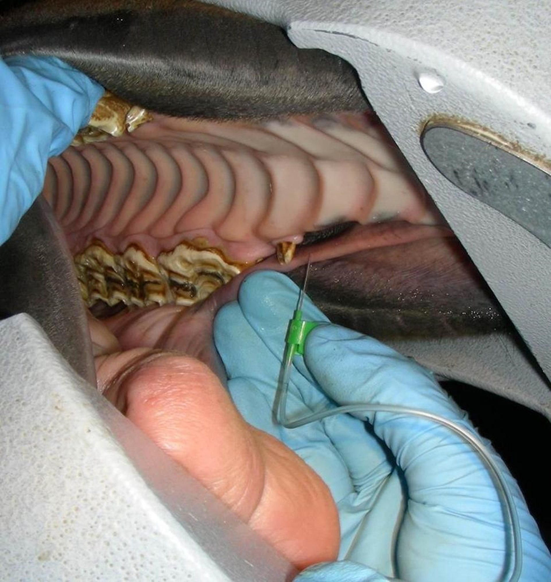
The upper first premolar (tooth 205), or "wolf tooth," of a horse is being anesthetized with a local anesthetic before extraction. Both left and right-sided wolf teeth are visible in this horse and they are fairly large; often wolf teeth are
Courtesy of Dr. Jack Easley.
Sharp edges on wolf teeth have been incriminated as a cause of bit resistance in horses. These small teeth, located just rostral to the upper cheek tooth row, are often extracted in performance horses. This procedure can be done in the standing, sedated horse with the aid of local infiltration anesthesia. The gingival attachment to the tooth is elevated, and a dental luxator or elevator is used to loosen the tooth. A small extraction forceps can be used to grasp the crown and pull the tooth from its socket. Socket healing requires minimal aftercare and minimal diet or work restrictions.
Most dental procedures can be performed on the standing, sedated horse with or without the use of regional anesthesia, but some major dental procedures (eg, extractions, repulsions and fracture repairs) require general anesthesia. Radiographic evaluation and protection of the airway from debris are necessary in most cases. Some decayed teeth can be extracted orally by the use of molar separators, extraction forceps, and elevators. In some cases, however, surgical exposure and tooth repulsion or sectioning and elevation are preferred over extraction. Tooth preservation by root-end resection and endodontic treatment has demonstrated that extraction is not required in all cases of dental decay in horses.


