


There are a variety of noninfectious disorders that can impair the urinary system. All of these diseases and conditions can be serious threats to the health of your dog.
Kidney Dysfunction
The kidneys’ most important function is to filter waste from the blood. When this does not happen, waste products can build to dangerous levels in the blood. This is called azotemia. Azotemia can be caused by many factors, including kidney disease, dehydration, shock, and congestive heart failure. Azotemia can also occur as a result of urine not being able to flow properly through the urinary tract.
Chronic Kidney Disease and Kidney Failure
Long-term, or chronic, disease can damage the kidney so severely that it is not able to function properly. This happens slowly. Chronic kidney disease often continues for months or years before a dog has any signs. There is rarely anything that a veterinarian can do to treat existing damage, and they can only slow the progression of (but not prevent) further damage once the process has started. Occasionally, chronic kidney disease results from a problem that is inherited or an abnormality present at birth. Most of the time, however, it is a problem in older animals. Starting at age 5 to 6, kidney disease becomes more common, affecting up to 10% of elderly dogs. Chronic kidney disease that is not inherited does not seem to be more common among certain dog breeds or among males or females.
Veterinarians classify chronic kidney disease into 4 stages based on laboratory tests, the results of physical examinations, and signs shown by the affected dog ( see Table: Chronic Kidney Disease Stages). In Stage I, the kidneys are damaged but azotemia (see above) has not yet developed and the dog has no signs. This is the stage at which treatment has the greatest chance of success, but because the dog has no signs, the disease is rarely diagnosed at this stage. In Stage II, the kidneys filter waste much more slowly than normal, and there is a buildup of waste chemicals in the blood, but many dogs still have no signs. Signs that may be present at this stage include an increase in the amount of water in the urine and an increased volume of urine. In Stage III, filtering slows even more, the waste chemicals are more concentrated in the blood, and the dog develops signs of disease. Stage IV, the final stage, reflects continued kidney damage and accumulation of waste products in the bloodstream. By this time the dog feels and acts very sick.
Veterinarians further classify dogs with kidney disease based on the presence of high blood pressure or protein in the urine. Approximately 20% of dogs with longterm kidney disease have elevated blood pressure (hypertension), which can cause further damage to the kidneys and also injure the eyes, brain, heart, and blood vessels. The risk of damage to the kidneys and other organs increases as the blood pressure increases. The presence of protein in your dog's urine can indicate that your dog has or may develop kidney disease, body-wide inflammation, metabolic disease, cancer, or infectious diseases. Your veterinarian will perform urine tests to learn how much protein is present, to help determine your dog's treatment and outlook.
Determining the cause of chronic kidney disease, especially in the early stages, will also help determine the appropriate treatment and provide an outlook for your dog. Some of the common causes include diseases of the circulatory system (such as high blood pressure, problems with blood clotting, and not having enough oxygen in the blood) or other diseases of the kidneys, such as pyelonephritis or tumors. Whatever the cause, chronic kidney disease usually results in scarring of the kidneys, which gets gradually worse.
Animals usually have no signs of kidney disease until they are at Stages III or IV, when their kidneys are working at less than 25% of their usual capacity. Exceptions to this include other illnesses that affect the entire body along with the kidneys, or kidneys that become unusually inflamed or sore and cause vomiting or pain. Veterinarians may be able to detect a problem in a blood test or on physical examination even before the dog starts to display signs of kidney failure. Usually, the earliest signs are excessive thirst and urination. However, these signs may signal other diseases as well, and they do not begin to appear until Stage II or III. After this, there are usually no new signs until Stage IV, when affected dogs vomit and are sluggish. As the disease progresses over months, other problems begin. These include loss of appetite, weight loss, dehydration, sores in the mouth, vomiting, and diarrhea.
To diagnose chronic kidney disease, veterinarians generally use a combination of x-rays, ultrasonography, urine and blood tests, blood pressure measurement, and physical examination. These tests are also used to check the response to treatment and monitor complications related to the kidney disease.
With proper treatment, even dogs with as little as 5% of normal kidney function can survive for a long time. The recommended treatment depends on the stage of disease. Identifying and treating complications, such as high blood pressure or urinary tract infections, needs to be done as well. All dogs with kidney disease should see their veterinarian every 3 to 6 months, or more frequently if there are problems. During these visits, the veterinarian will do tests on the dog’s blood and urine and may measure blood pressure.
Although there is no way to prevent chronic kidney disease from getting progressively worse, there are some things you can do to slow the process. These include making sure the dog’s diet does not contain too much phosphorus, supplementing your dog’s food with fish oil, and giving all medications as directed. Your veterinarian may suggest special food which has been designed for animals with kidney disease. If there are problems with the acidity of your pet’s blood, or if the levels of phosphorus in your pet’s blood are unhealthy, your veterinarian may prescribe a supplement or vitamin.
In the later stages of kidney disease (III and IV), the dog should be taken to the veterinarian every 1 to 2 months. At this stage, treatments will focus on easing some of the signs of the disease. Some approaches include limiting the amount or type of protein in your dog’s diet (your veterinarian can suggest special food formulated for pets with kidney disease) and medications. Sometimes veterinarians will recommend intravenous fluids or feeding tubes. At this point, there are very few options. Dialysis machines, which do the job of the kidneys by filtering the blood, can prolong life, but dialysis is not feasible for most dogs. Kidney transplants are rarely done and require immune-suppressing drugs to prevent the body from rejecting the new kidney, which can cause other problems.
Acute Kidney Injury
Acute (short-term, or sudden) kidney injury is the result of sudden, major damage to the kidneys. This damage is usually caused by toxic chemicals either consumed by your pet or built up by an abnormal condition in your pet’s body. Kidney function can also be affected when the kidneys do not receive sufficient oxygen, such as when a blood clot blocks the flow of blood to the kidneys. Some infections (such as leptospirosis or Lyme disease) can also cause an acute kidney injury.
Some dogs consume toxic chemicals, such as antifreeze, or poisonous plants that can damage the kidneys. Certain medications, such as nonsteroidal anti-inflammatory drugs or certain antibiotics, can also cause kidney damage. There are many substances and foods in the average home that may be safe for humans but dangerous for dogs and other pets ( see Introduction to Poisoning). For example, grapes and raisins can potentially cause significant kidney damage. Alternatively, some toxic chemicals come from inside the dog’s own body. For example, there could be a buildup of calcium or other substances due to a disease in another part of the body. In these cases, the effects on kidney function can last from 1 to 8 weeks, depending on the chemical(s) that caused the injury.
Mild kidney disease often goes unnoticed. However, repeated occurrences can lead to chronic kidney disease. The stages of acute and chronic kidney disease are the same (see above). Usually, acute kidney disease becomes obvious only in Stage IV, when the signs include loss of appetite, depression, dehydration, sores in the mouth, vomiting, diarrhea, and a smaller than normal volume of urine.
It is important to determine whether the kidney disease is acute or chronic, as well as the cause of the disease. This information will help your veterinarian determine the most appropriate treatment. Usually, veterinarians can identify acute kidney disease by taking a urine sample and asking thorough questions about exactly what your pet has eaten, what medications your pet may have taken, and how your pet has been acting in the months and weeks prior to becoming ill.
An injured kidney can often regain some or most of its function. The uninjured part of the kidney (or remaining uninjured kidney) helps compensate for the injured organ. To determine how much potential your dog’s kidneys have to regenerate, your veterinarian may need to do a kidney biopsy.
If the cause of the kidney injury can be determined, treatment will be aimed at this cause. Dogs that are dehydrated or not eating may require intravenous fluids or a feeding tube. Your veterinarian may suggest treatment to promote urination in dogs that are not urinating enough or not urinating at all. This treatment involves intravenous fluids, inserting a catheter into your dog’s bladder, and, occasionally, medication. If none of the available treatments work, and your dog is simply not producing urine, the only remaining options are kidney dialysis or euthanasia. About 50% of dogs with a severe acute kidney injury will survive.
Glomerular Disease
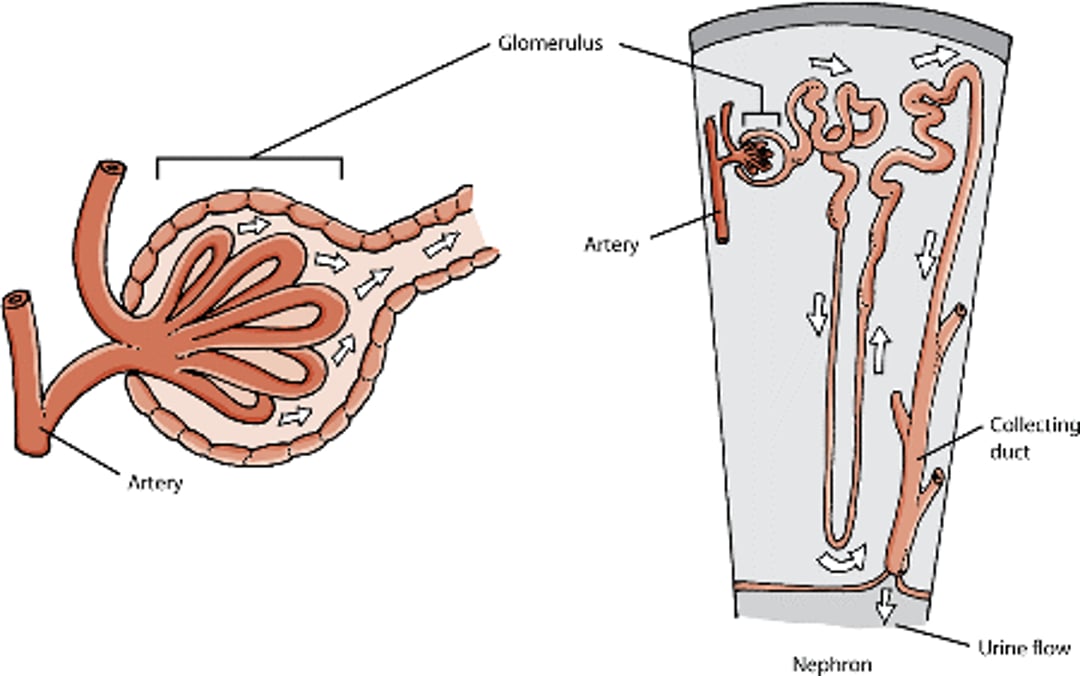
The glomerulus is one of the structures that are essential to kidney function. It is made up of special blood vessels that help filter blood. Each kidney contains thousands of these structures. Glomerular disease is a common cause of longterm kidney disease in dogs. It can also cause short-term kidney injury. Damage to parts of the glomerulus can cause protein in the urine and low levels of a protein called albumin in the blood. This can lead to other problems, such as swelling of the legs, high cholesterol, and blood clots.
Glomeruli

Glomerular disease can occur due to the long-term effects of high blood pressure. However, glomerular disease can also occur along with other kidney disorders. Some glomerular disease is immune-mediated, that is, caused by the dog’s immune system attacking parts of its own body. Tumors, rickettsial infections, lupus, heartworm disease, and other infections have been identified in connection with immune-mediated glomerular disease.
Glomerular disease may also be caused by hyperadrenocorticism, which is an excess of a hormone called cortisol. Cortisol is produced by the adrenal gland, and is one of the hormones that helps convert food into energy and regulate blood sugar. Animals with glomerular disease caused by hyperadrenocorticism frequently also have diabetes.
Some types of glomerular disease are inherited. Dog breeds that are more prone to inherit this illness include Bernese Mountain Dogs, English Cocker Spaniels, English Springer Spaniels, Doberman Pinschers, Greyhounds, Lhasa Apsos, Poodles, Rottweilers, Samoyeds, Shih Tzus, and Soft-coated Wheaten Terriers.
Some glomerular disease is caused by amyloidosis, or deposits in the kidneys of a misfolded protein called amyloid. Glomerular amyloidosis usually causes protein in the urine. Amyloidosis is sometimes inherited in Chinese Shar-Peis. The noninherited form of amyloidosis usually affects dogs that are middle-aged or older. Certain breeds, such as Beagles, Collies, and Walker Hounds, are at increased risk. Dogs with the inherited form of the disease are usually diagnosed at a young age.
Signs and Diagnosis
Disease in the glomerulus often leads to protein in the urine, low levels of protein in the blood, a buildup of fluid in the abdomen (which can cause visible swelling), shortness of breath, and swelling of the legs. Taken together, these signs are called the nephrotic syndrome. (Nephrotic means relating to the kidneys.) The loss of protein through the urine can cause loss of muscle tissues. Severe or longterm glomerular disease can cause chronic kidney disease, usually Stage 3 or 4. High blood pressure is more likely in dogs that lose protein into their urine.
Protein in the urine can lead to a loss of an important protein that helps the blood to clot properly. This can cause strokes, heart attacks, and other problems that occur when the blood clots too easily. For example, if blood clots form in the lungs, dogs may have severe difficulty breathing.
Your veterinarian will look for abnormal levels of protein and other chemicals in your dog’s urine and blood. Physical examination usually reveals that something is wrong; however the signs are often nonspecific and could point to any of a wide variety of problems. A biopsy of the kidneys is often required to determine the cause of glomerular disease. Additional tests may be required in some cases, including x-rays, ultrasonography, blood pressure measurements, and special blood tests.
Treatment and Outlook
Treatment for glomerular disease varies according to the cause. If the glomerular disease is immune-mediated, then the cause of the problem should be treated. If a cause cannot be determined, drugs that suppress the immune system may be used in an attempt to limit damage. If the dog has signs of nephrotic syndrome (see above), your veterinarian will probably recommend a special food and possibly a diuretic. For a dog with a low level of protein in the blood, your veterinarian might recommend a medication that thins the blood. Because protein in the urine can cause scar tissue to build up in the kidneys, your veterinarian will probably attempt to limit how much protein is shed from the body into the urine. Options for this include limiting the amount of protein in the dog’s diet and prescribing certain medications. Complications of chronic kidney disease also need to be monitored and treated.
Dogs with glomerular disease live an average of about 3 months once the illness is diagnosed. However, if the problem can be diagnosed and treated early, dogs can live much longer. Survival times for animals with amyloidosis vary widely, with reported times ranging from 49 days to 20 months.
Renal Tubular Problems
Renal tubules are structures in the kidneys that help filter blood.
Renal Tubular Acidosis
Healthy kidneys help rid the body of acid by producing urine that is acidic. Diseased kidneys cannot get rid of acid properly, and instead of being eliminated in the urine, this acid builds up in the blood, leading to a condition called uremic acidosis. This condition can also occur when there are defects in the renal tubules, in which case it is called renal tubular acidosis. These defects are rare in dogs.
Renal tubular acidosis can cause kidney stones and can cause the bones to become soft and easily breakable. To treat this condition, your veterinarian will probably prescribe medication to help rebalance the amount of acid in the blood. However, this treatment is not effective for all cases.
Fanconi Syndrome
Fanconi syndrome is a condition in which the kidneys cannot properly absorb certain chemicals. The chemicals are glucose, sodium, potassium, phosphorus, uric acid, bicarbonate, albumin (a type of protein), and amino acids. All of these are required to meet the body’s needs, but animals with Fanconi syndrome cannot reabsorb them through the kidneys. Instead, they are lost in the urine. Dogs can develop Fanconi syndrome in one of several ways. They can have a bad reaction to gentamicin (a type of antibiotic) or certain foods (such as chicken jerky treats). The syndrome can appear suddenly and for no apparent reason, or it can be inherited. Basenjis are most likely to inherit the condition; in this case, the condition develops gradually in adults.
Signs of Fanconi syndrome include excessive thirst, excessive urination, and weight loss. There may be a buildup of toxic chemicals in the blood, called uremia. Your veterinarian can diagnose Fanconi syndrome by doing tests on your dog’s blood and urine.
There is no way to correct the tubular defect that causes Fanconi syndrome. Veterinarians can try to balance the amount of acid and electrolytes in the blood by prescribing supplements. Treating the signs of disease can make your dog more comfortable. However, ultimately there is little a veterinarian can do beyond keeping the dog as comfortable as possible. Most dogs with the inherited form of Fanconi syndrome die from uremia.
Obstructions of the Urinary Tract
Even when the kidneys are functioning normally, a blockage in the urinary system at any point below the kidneys can lead to a backup of toxic wastes that can damage the kidneys and cause illness. In dogs, the most common cause is a kidney stone that blocks the urethra. Other possible causes include tumors, stones, or blood clots in the ureters or urethra.
If the flow of urine is blocked, the kidney becomes abnormally enlarged. When this happens suddenly to both kidneys, especially when the urine is completely blocked, the dog does not live long. When the blockage is only partial, or only occurs on one side, the dog often survives, but the kidneys often sustain permanent damage. The affected kidneys eventually become giant, useless sacs filled with urine and may become infected. The ureter may also become enlarged due to a backup of urine. This often occurs when the blockage is located far down the urinary tract and away from the kidneys.
Signs and Diagnosis
Dogs with a blockage in their urethra urinate small amounts quite frequently, urinate slowly and painfully (some dogs may whine or cry), and typically have blood in their urine. They may also have a painful abdomen. Uremia, the buildup of toxic waste in the blood, happens quickly. The signs of uremia include vomiting, dehydration, a drop in body temperature, and severe depression. The bladder is bloated and painful. The heart can be affected by the buildup of potassium in the blood and start to beat abnormally. In dogs with only one blocked kidney, the other kidney can compensate for the blockage. The blockage in these dogs often goes unnoticed unless there is another kidney disease or your veterinarian notices it during a physical examination or on an x-ray or ultrasound examination.
Your veterinarian may be able to diagnose obstructions based on the signs and physical examination. Sometimes, however, ultrasonography or other special tests are required. Blood tests can show an elevated level of potassium in the blood. In cases where the heart is affected, a heart test called an electrocardiogram (EKG) may be required.
Treatment
To restore normal urine flow, the blockage must be removed. In most cases, intravenous fluids will be used to restore the balance of various chemicals in the blood. Because of increased production of urine after the blockage is removed, it is normal for a dog to urinate more than usual for 1 to 5 days after treatment. During this period, it is important that your pet not get dehydrated. Your veterinarian may want to monitor your dog daily during this time, and adjust the amount and type of fluids your pet is receiving.
Surgery is often required to remove the blockage. If the blockage is caused by stones, the stones may naturally pass and eliminate the need for surgery. However, the damage caused by the stone may require part of the ureter or urethra to be removed. In some cases, a kidney is so damaged that it needs to be removed. This is possible only if the other kidney is healthy.
If a dog has signs of a blocked urethra, it is critical to seek veterinary care immediately. Dogs with a complete blockage may die without treatment.
Tumors
Tumors of the kidneys and urinary tract are not common in dogs. Tumors can be benign (harmless) or malignant (cancerous).
Kidney Tumors
Kidney tumors are uncommon; only about 1 to 2% of all tumors in dogs involve the kidneys. It is unusual to find benign kidney growths because they rarely affect the health of an animal. They are usually discovered only by accident and do not require treatment.
Malignant tumors that begin in the kidneys (as opposed to those that begin in other organs and spread to the kidneys) are most common in middle-aged to older dogs. In general, no breed is more prone to kidney tumors than others; however, German Shepherds can inherit a tendency to develop a very specific kind of cancer known as a cystoadenocarcinoma. This cancer involves many small tumors on both kidneys and usually appears when the dog is between 5 and 11 years old.
The most common malignant kidney tumor is a carcinoma that starts in the lining of the renal tubules. Usually, the tumor appears on only one kidney. Cancerous tumors that begin in the kidneys spread quickly to other organs, especially the opposite kidney, the lungs, adrenal glands, and the liver.
Blastomas are tumors composed of previously healthy young cells that never mature normally. Instead, these cells mutate into cancer. Those that originate in the kidney are known as nephroblastomas. (Other names for this type of tumor are embryonal nephroma and Wilms’ tumor). Dogs with this type of cancer are usually diagnosed when they are less than 1 year old. Males are affected twice as often as females. Nephroblastomas usually occur in only one kidney, but occasionally they affect both kidneys. They can become quite large; it is not uncommon for a single nephroblastoma to take up all of the space inside the affected dog’s abdomen. Nephroblastomas typically spread to nearby lymph nodes, the liver, and the lungs.
Transitional cell carcinomas are cancers that appear in the lining of certain parts of the urinary tract including the ureter, bladder, urethra, or the center of the kidney (referred to as the renal pelvis). The lining in these parts of the urinary tract is called transitional epithelium and is different from the lining of other organs because it is very stretchy.
Other malignant tumors rarely originate in the kidneys, but can include hemangiosarcomas (tumors from the lining of the blood vessels), fibrosarcomas (tumors of connective tissue), leiomyosarcomas (tumors of the smooth muscle), and squamous cell carcinomas (tumors of the outer layer of the kidney surface).
When cancer spreads from one organ to another, it is said to metastasize and the cancer itself is described as metastatic. The kidneys are a common second site for metastasis of cancers that begin in other organs, such as the lymph nodes. Up to half of dogs with cancer of the lymph nodes (called lymphosarcoma) also develop cancer in their kidneys. In some cases, the cancer remains only in the lymph nodes and the kidneys. In others it also affects the brain. When cancer spreads to the kidneys, it usually takes the form of many small tumors. It can affect both kidneys and may result in the kidneys being unusually large and irregularly shaped.
Signs of kidney tumors are usually general and can point to many different illnesses. Common signs include weight loss, loss of appetite, depression, and fever. Your veterinarian will need to eliminate other causes of these signs before confirming cancer. Occasionally, tumors that appear in both kidneys can cause enough damage that the dog will develop signs of late-stage chronic kidney disease and failure (see above). Pet owners who pay close attention may notice “lumps” in the dog’s belly or an enlarged belly. There may be blood in the urine, but it is usually too tiny an amount to see with the naked eye.
Your veterinarian may suspect a tumor in the kidneys based on physical examination and careful consideration of your dog’s signs in the weeks and months prior to becoming ill. This suspicion can be confirmed with ultrasonography, x-rays, or contrast x-ray tests of the urinary tract. Cancer cells can also occasionally be found in the urine. A biopsy of the tumor is usually necessary to determine its type.
Most types of kidney tumors must be surgically removed. Usually it is necessary to remove the entire affected kidney. Tumors in the lymph nodes around the kidney are usually treated with chemotherapy instead of surgery. If your dog develops a urinary cancer, the veterinarian will assess the severity of your pet’s condition, the outlook for your pet, and other factors when recommending a treatment program.
Lower Urinary Tract Tumors
Tumors in the ureters, bladder, and urethra are not common in dogs. The average age of affected dogs is 9 years old. Tumors that begin in (as opposed to those that spread to) the lower urinary tract are more likely to be malignant than benign. Benign tumors that can be found in the lower urinary tract include papillomas (warts; tumors of the lining of organs), leiomyomas (smooth muscle tumors, also called fibroids), neurofibromas (tumors of the protective sheath that surrounds nerves), hemangiomas (blood vessel tumors), rhabdomyomas (another type of smooth muscle tumor), and myxomas (tumors of primitive connective tissue).
The most common type of malignant tumor that begins in the lower urinary tract is transitional cell carcinoma. Transitional cell carcinomas are cancers that appear in the lining of certain parts of the urinary tract including the ureter, bladder, urethra, prostate, and renal pelvis. Transitional cell carcinomas may appear as a single tumor, or as multiple wart-like growths that are visible on the membranes lining the urinary tract. Alternatively, these carcinomas may develop all over the ureter, bladder, prostate gland, or urethra. Once they appear, they have the tendency to grow and spread quickly, most often to the nearby lymph nodes and lungs.
Tumors in the ureter and bladder can cause blockage of urine, which can back up into the kidneys and cause damage ( see Obstructions of the Urinary Tract, above). Tumors in the urethra are more likely than tumors in the ureter and bladder to suddenly cut off the passage of urine. Tumors in the bladder and urethra are usually accompanied by urinary tract infections that will not go away despite treatment with antibiotics.
Other types of malignant tumors that can originate in the urinary tract include squamous cell carcinomas (cancer of the outer layer of an organ), adenocarcinomas (cancer of the mucus-secreting glands of an organ), fibrosarcomas (cancer of the connective tissue), leiomyosarcomas (cancer of the smooth muscles), rhabdomyosarcomas (cancer of smooth muscle), hemangiosarcomas (cancer of the blood vessels), and osteosarcomas (bone cancer).
The most common signs of cancer of the lower urinary tract include blood in the urine; painful, slow, or difficult urination; and excessive urination. Dogs with one blocked ureter may have a painful abdomen, and a veterinarian may be able to feel an enlarged kidney. Dogs with blocked ureters on both sides or a blocked urethra may show signs of uremia (a buildup of the toxic chemicals usually eliminated in the urine). While doing an examination, a veterinarian may be able to detect a thickened bladder wall, an irregular urethra, or masses on the urethra.
Laboratory tests on the dog’s urine usually reveal blood in the urine, and sometimes reveal a bacterial or other infection that has developed in addition to the tumor. It is occasionally possible to mistake tumors in the lower urinary tract with simple urinary tract infections, especially those that do not go away or keep coming back. Thus, your veterinarian may order additional tests to confirm the diagnosis of cancer. Sometimes cancer cells can be found in the urine, especially when the cancer is in the transitional cells. Your veterinarian may use ultrasonography or specialized x-rays to locate and assess the severity of the tumor. A biopsy of the tumor is required to identify its type.
Surgical removal of the tumor, if possible, is the best treatment. Transitional cell carcinomas are frequently located in critical parts of the bladder or urethra, and removing them requires reconstruction of the lower urinary tract. Survival time tends to be short for these dogs, even with surgery, because the tumors spread quickly and often reappear. Chemotherapy and/or radiation therapy may give the dog more time but can have serious side effects. You and your veterinarian will want to discuss the treatment options and the quality of life for your pet both during and after treatment.
Problems with Urination
Urination problems can be grouped into problems with storing urine and problems with eliminating urine. Urinary incontinence is the inability to prevent or control urination. Incontinent animals leak urine constantly or on and off without realizing it. An incontinent dog may leave a pool of urine where it has been lying or dribble urine while walking. The fur around the vulva or penis may be wet, and the constant dribbling of urine can cause inflammation and urine scalding of the skin in these areas.
Problems with Urine Storage
Problems with urine storage are identified by inappropriate leakage of urine, and can be caused by several different disorders. These include failure of the muscles in the bladder to relax appropriately, urethral muscles that do function properly, birth defects, injury or damage to the urethra or other parts of the urinary system, and overflowing of the bladder.
Urge incontinence occurs when urine leaks during the times when an animal feels the urge to urinate as opposed to urine which leaks when an animal is unaware of it. Urge incontinence is usually caused by irritation of the bladder muscle that forcibly expels the urine. This is usually due to inflammation of the bladder. Unusually low levels of sex hormones in neutered dogs are another common cause. This type of incontinence, called hormonal--responsive urethral incompetence, is particularly common in female dogs. Problems with the urethral sphincter (the muscle that allows urine to pass through the urethra) can also cause urine-storage incontinence.
Incontinence that results from birth defects or malformation of the urinary system usually becomes obvious while the dog is still young. For example, a dog that was born with an ectopic ureter on one side might urinate normally but dribble urine on and off, whereas dogs with ectopic ureters on both sides are less likely to be able to urinate normally at all. Although it seems contradictory, a dog can also become incontinent if its urethra is partially blocked; the blocked urethra can cause urine to back up and the bladder to overflow.
Problems with Urine Elimination
Problems with urine elimination can have many causes, including a physical blockage of the urethra by stones, growths, or scar tissue; problems related to the nervous system; or a lack of muscle tone in the muscle that controls the bladder. Dogs that cannot urinate normally will usually try to urinate often, but the urination will be slow and painful, and only small amounts of urine will come out. Dogs with urine elimination problems may also develop incontinence over time, if the bladder does not empty properly. The bladder can become stretched out and begin to overflow and leak.
Neurologic Problems
Neurologic problems with urination can be caused by damage to the lower half of the spine, damage to the major nerve in the pelvis, or a lack of muscle tone in the muscle that controls the bladder. Dogs with one of these injuries may have an enlarged bladder that empties easily when squeezed by your veterinarian. Other neurologic problems with urination are caused by damage to the upper half of the spine or disease in the brain. Dogs with these disorders have an enlarged bladder that does not empty easily when squeezed by your veterinarian.
Another neurologic cause of urination problems is poor muscle coordination. The various muscles involved in the different steps of urination do not work together normally. Dogs with this condition usually urinate extraordinarily often, with the stream of urine being cut short. Some dogs with neurologic problems may leak urine. Animals with any neurologic urination problem may develop incontinence, especially if the bladder becomes too full and begins to overflow and leak.
Diagnosis and Treatment of Urination Problems
A thorough physical examination and a history of your dog’s behavior can help your veterinarian determine whether your dog has problems related to urination. Your veterinarian will probably also want to watch your pet urinate. Specialized tests, such as ultrasonography, x-rays, cystoscopy (visualizing the inside of the urethra and bladder with a camera), or neurologic tests, may be helpful in some cases.
Dogs with incontinence that is caused by imbalances in sex hormones may be prescribed hormones in order to re-establish the proper balance. Urethral incontinence can be treated with medication that targets the membrane inside the urethra (called alpha-adrenergic agonist drugs). Urge incontinence can be treated with medication that targets certain nerves (called anticholinergic drugs). Weakened bladder muscles can be treated with medications that target slack muscles (called cholinergic drugs). Medications may be useful in cases where muscle coordination issues are identified.
Complete physical blockage of the urethra is a medical emergency. The treatment varies, depending on the circumstances. A catheter may be used to push the blockage backwards out of the urethra and into the bladder. The blockage may have to be removed during surgery. Dogs with bladder muscles that have been weakened by overfill and stretching may require a special catheter that remains in place, or is placed at regular intervals every few hours for 3 to 7 days. This allows the bladder to empty properly and regain some muscle tone.
In dogs in which the bladder has lost its muscle tone due to neurologic problems, there are few medical options to restore muscle tone. For these dogs, it is usually necessary to empty the bladder several times a day for the rest of its life. In these cases, you will need to be trained to properly insert and remove a catheter or to express the bladder with your hands.
Urinary Stones (Uroliths, Calculi)
Minerals that naturally occur in urine clump together to form tiny crystals. When crystals clump together. they form uroliths (also known as stones or calculi). These stones can develop anywhere in the urinary system, including in the kidneys, ureters, bladder, or urethra.
Veterinary researchers do not completely understand what causes stones to form. There are many different types of stones, each formed from a complex mixture of various minerals. The most common types are made of struvite, calcium oxalate, or urate. Each type of stone develops only under certain conditions. Uroliths only form when the components of the stones are present in adequate amounts and when crystals remain in the urine for an adequate length of time. For some stones, the correct environment must also be present , such as the proper level of acidity. These conditions can be affected by urinary tract infections, diet, digestion, the amount of urine that a dog produces, how frequently a dog urinates, medications, and genetics.
Dogs with crystals or very small stones in the urinary system do not usually have any signs. However, larger stones in the lower urinary tract may interfere with urination or irritate the lining of the bladder or urethra. In turn, these problems can cause painful urination, blood in the urine, and slow or painful urination. Kidney stones usually cause no signs unless the kidney becomes inflamed or the stones pass into the ureter. If a ureter becomes blocked by a stone, it can cause vomiting, slowness or tiredness, and pain in the abdomen in the area around the kidneys. This sign is particularly common when a ureter is suddenly and completely blocked and a backup of fluids causes the kidney to become enlarged. Pain may be the only sign of stones in the ureter on only one side; however, pain can be difficult to detect in dogs. If the blocked ureter is not diagnosed right away, kidney damage occurs. Ultimately, the blocked kidney is destroyed.
Veterinarians can sometimes detect stones in the bladder by pressing on the dog’s abdomen. Stones in the urethra may also be detected during a rectal examination or when attempting to insert a catheter. There may be many stones present at once, so if one stone is located, it is important to examine the entire urinary tract to look for others. X-rays can reveal stones as small as 3 millimeters in size. A veterinarian will also need to do tests on the dog’s urine and may need to do ultrasonography or other specialized tests.
The treatment of stones, and preventing their return, depends on their type and location. Treatment and prevention may include surgery, lithotripsy (a procedure that uses sound waves to break apart stones), a special diet, and medication. When stones are removed, the veterinarian will probably send them to a laboratory to be analyzed. Knowing what types of minerals are in the stone can provide the information needed to prescribe medication to help prevent the formation of more stones. Dogs undergoing treatment will need to be monitored closely and return at regular intervals for additional testing.
Urethral Obstruction
Uroliths can become lodged in the urethra and block the flow of urine out of the bladder. This is called a urethral obstruction and is common in male dogs. It may occur suddenly or may develop slowly across several days or weeks. At first, the dog may frequently try to urinate and produce only a fine stream, a few drops, or nothing. Dogs may also have extreme pain and cry out when trying to urinate. Complete obstruction causes toxins to build up within the body in 1–2 days, which leads to depression, lack of appetite, vomiting, diarrhea, dehydration, coma, and death within about 3 days. Urethral obstruction is an emergency condition, and your dog needs to be treated by a veterinarian immediately.
Treatment involves relieving the obstruction, either by pushing the urolith back into the bladder with a catheter or by removing the stone surgically. If the stone is flushed back into the bladder, surgery to remove the stone is usually necessary so that it does not pass into the urethra again.
For More Information
Also see professional content regarding noninfectious diseases of the urinary system.


