


Common Parasites of the Abomasum in Small Ruminants
Nematodes of the abomasum in small ruminants cause erosion and ulceration of the gastric mucosa leading to gastritis. Damage to the gastric mucosa decreases the amount of pepsin and hydrochloric acid produced, resulting in a potentially less acidic abomasum. Some abomasal nematodes cause profound anemia and ill thrift. All the abomasal nematodes described below have the typical life cycle and egg appearance of a trichostrongyle, as described in Etiology.
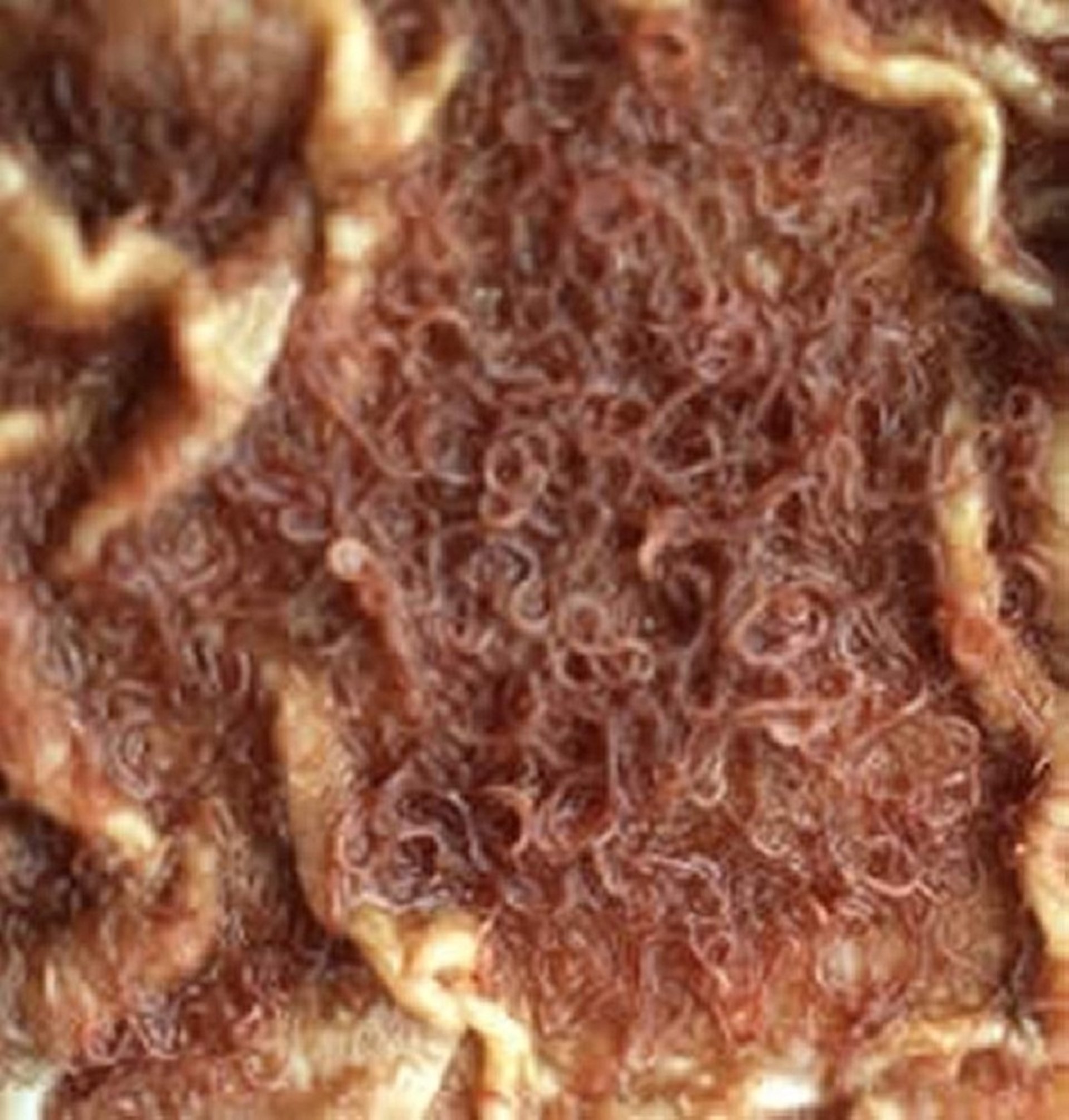
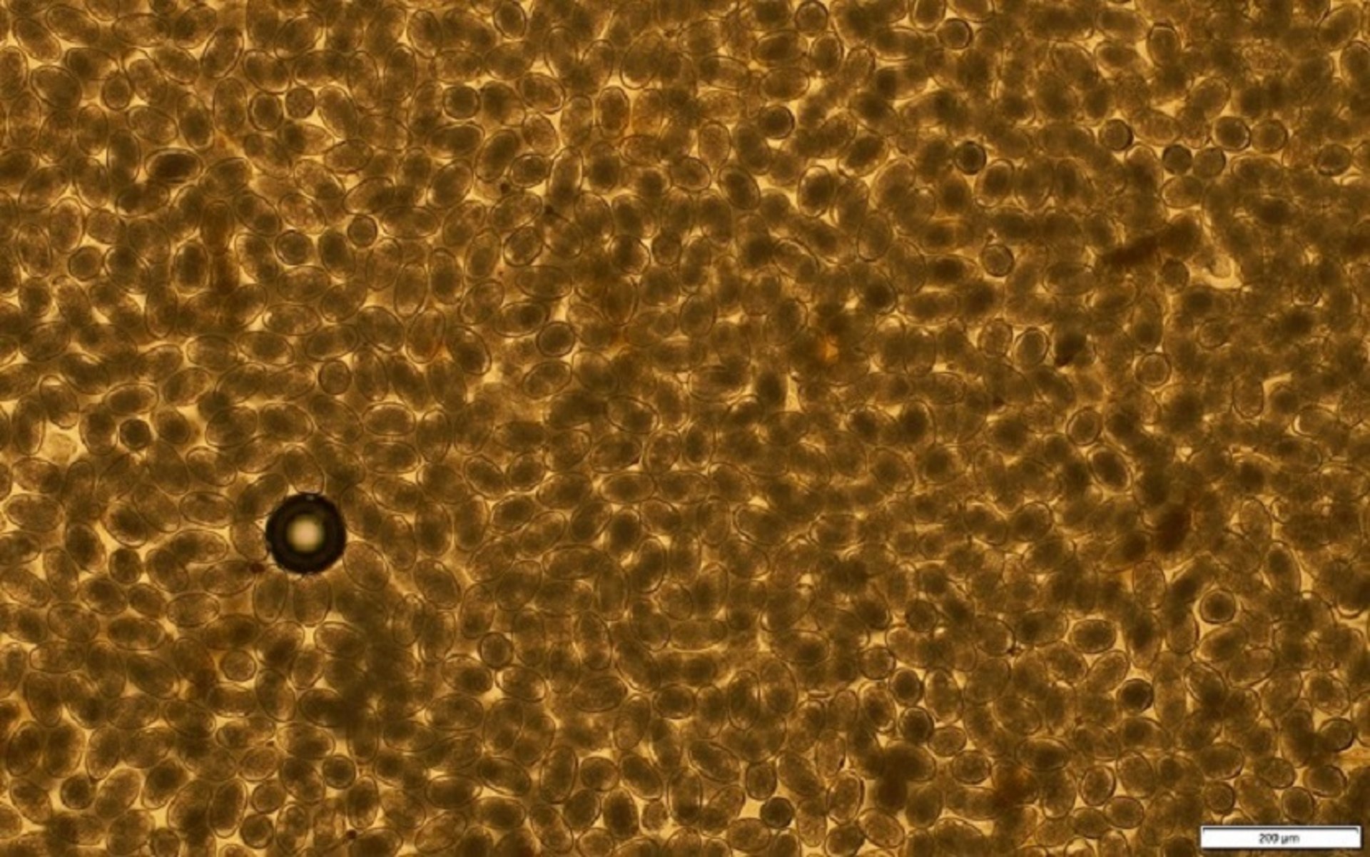
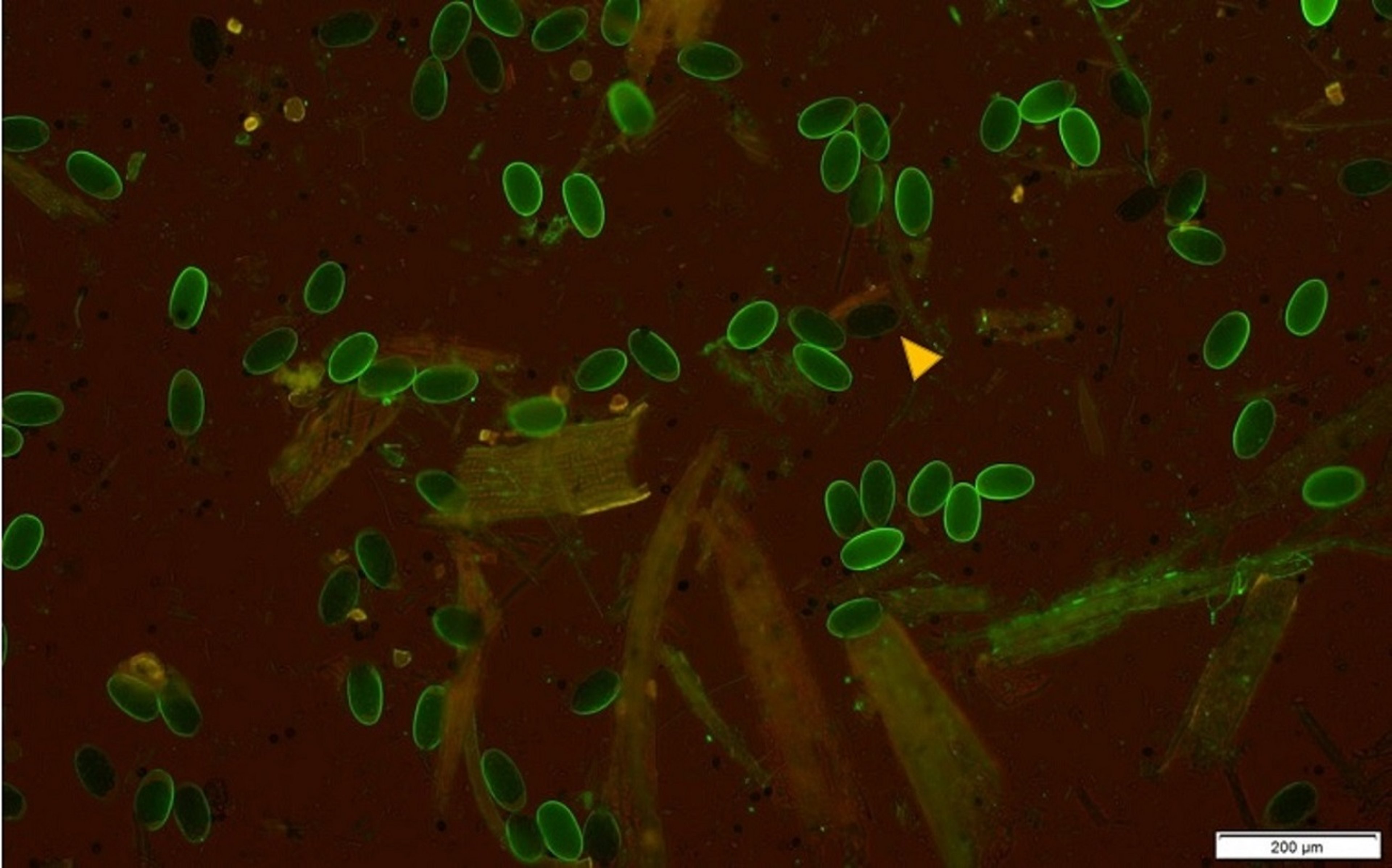
Haemonchus spp Abomasum Parasites of Small Ruminants

Courtesy of Dr. Raffaele Roncalli.

Courtesy of Dr. Grace VanHoy.

Courtesy of Dr. Antoinette Marsh.
Haemonchus is prevalent in tropical or subtropical regions and in regions with summer rainfall, and H contortus is the most common pathogenic GI parasite in small ruminants. For descriptions and life cycles, see Common Parasites of the Abomasum in Cattle.
Haemonchosis in sheep may be classified as hyperacute, acute, or chronic. In hyperacute disease, death may occur within 1 week of heavy infection without notable clinical signs. Acute disease is characterized by severe anemia accompanied by generalized edema. Anemia is also characteristic of chronic infection with low worm burdens, and is accompanied by progressive weight loss.
Diarrhea is not a characteristic clinical sign of pure Haemonchus infection; when diarrhea is present, a mixed infection with other worm genera should be suspected. The abomasum is edematous, and in the chronic phase, gastric pH increases, leading to abomasal dysfunction. Periparturient rise occurs commonly with Haemonchus infections, and mature sheep and goats may develop fatal infections in late pregnancy and early lactation.
FAMACHA scoring (see Monitoring) is an important tool in the diagnosis, monitoring, and control of haemonchosis in herds of goat and sheep.
Teladorsagia (Ostertagia) circumcincta and Ostertagia trifurcata Abomasum Parasites
The brown stomach wormsTeladorsagia (formerly Ostertagia) circumcincta and Ostertagia trifurcata are more common in cooler areas with wet winters and temperate zones than are Haemonchus species. The lesions, pathogenesis, and clinical signs of infection by these parasites are similar to those of ostertagiasis in cattle (see Common Parasites of the Abomasum in Cattle). Teladorsagia adults are 14 mm long and brown, and the female’s tail is annulated.
Even subclinical infection depresses appetite, impairs gastric digestion, and decreases nutrient utilization. Teladorsagia is also involved in the periparturient rise in fecal egg counts in sheep and goats, and heavy infections may lead to diarrhea and depress milk production in dams. The output of eggs is the main source of contamination in lambs and kids. The same type of inhibited larval development that occurs in cattle has been observed with both Ostertagia and Haemonchus in small ruminants.
Trichostrongylus axei Abomasum Parasite of Small Ruminants
Trichostrongylus axei is also more common in temperate zones than are Haemonchus species. T axei infections in small ruminants show the same lesions, pathogenesis, and clinical signs as infections in cattle (see Common Parasites of the Abomasum in Cattle).
Mecistocirrus digitatus Abomasum Parasite of Small Ruminants
Mecistocirrus digitatus is a hematophagous trichostrongyle with a pathology similar to that of Haemonchus; however, M digitatus adults are much larger, ~40 mm. This parasite is typically present only in tropical climates.
Common Parasites of the Small Intestine in Small Ruminants
Small intestine nematodes cause enteritis and varying amounts of protein-losing enteropathy in small ruminants, depending on their virulence and the worm burden. Small intestine cestodes (tapeworms) rarely cause disease in small ruminants; however, they can cause ill thrift and weight loss in very heavy infections of juveniles.
Trichostrongylus spp Small Intestine Parasites of Small Ruminants
Trichostrongylus colubriformis, Trichostrongylus vitrinus, and Trichostrongylus rugatus are found in the small intestine of small ruminants. The life cycle and appearance of eggs on fecal flotation are typical of a trichostrongyle, and the prepatent period is 18–21 days.
Anorexia, persistent diarrhea, and weight loss are the main clinical signs of Trichostrongylus infection. Villous atrophy (or stunting of villi) impairs digestion and malabsorption; protein loss occurs across the damaged mucosa.
Strongyloides papillosus Small Intestine Parasite of Small Ruminants
Strongyloides papillosus is not a trichostrongyle, and it has a complex life cycle (seeCommon Parasites of the Small Intestine in Cattle). Heavy infections by adult worms cause a disease resembling trichostrongylosis. The means of infection is usually skin penetration; however, S papillosus can also infect the animal via milk.
Damage to the skin between the claws, produced by skin-penetrating larvae, resembles the early stages of footrot and may aid penetration by the causative agents of foot rot. Most infections are transitory and inconsequential.
Bunostomum spp and Gaigeria spp Small Intestine Parasites of Small Ruminants
Bunostomum trigonocephalum (hookworm) adults are found in the jejunum. The life cycle and clinical findings are essentially the same as for the cattle hookworm, with as few as 100 worms causing clinical signs.
Gaigeria pachyscelis is found in Africa and Asia and resembles Bunostomum in size (2–3 cm) and form. Larvae of G pachyscelis infect the host only by skin penetration. It is a voracious bloodsucker and highly pathogenic.
Nematodirus spp Small Intestine Parasites of Small Ruminants
The species of Nematodirus found in the small intestine of sheep and goats are similar in morphology and life cycle to the Nematodirus spp found in cattle (see Common Parasites of the Small Intestine in Cattle). Clinical infections of Nematodirus are of considerable importance in the UK, New Zealand, and Australia, where lamb mortality may reach 20% in affected flocks if animals are untreated. The parasites are also endemic in some parts of the US, where they occasionally cause disease in young animals. The life cycle can be broken if lambing or kidding takes place in different areas each year. Most clinical infections occur in animals 6–12 weeks old.
Nematodirus battus is an important parasite of lambs in the UK and other parts of Europe and North America. Eggs hatch after a period of chill and then a rise in ambient temperature to a day/night mean of 10°C (50°F), which in temperate areas occurs in late spring. These hatching requirements mean that there is generally one generation of N battus per year; in the UK, however, occasional outbreaks in the fall have been reported.
The parasite can be highly pathogenic because large numbers of larvae hatch over a short period at a time when young lambs are beginning to consume sizable quantities of grass. Disease may be associated with developing larval stages and may occur within 2 weeks of challenge (the prepatent period is 15 days). Other Nematodirus spp often are found in low-rainfall regions (eg, the Karoo in South Africa and inland Australia) where other parasites are rare.
Nematodirosis is characterized by sudden-onset unthriftiness, profuse diarrhea, and marked dehydration, with death as early as 2–3 days after an outbreak begins. Nematodirosis is commonly confined to lambs or weaner sheep, but in low-rainfall country where outbreaks are sporadic, older sheep may have heavy infections.
Nematodirosis lesions usually consist of dehydration and a mild catarrhal enteritis; however, acute inflammation of the entire small intestine may develop. Counts of ≥ 10,000 worms, together with characteristic clinical signs and history, are indicative of clinical infections. Affected lambs may pass large numbers of eggs, which can be identified easily. If the onset of disease precedes maturation of the female worms, eggs will not be identified on fecal examination.
Aonchotheca (Capillaria) longipes Small Intestine Parasite of Small Ruminants
Aonchotheca (formerly Capillaria) longipes is closely related to Trichuris, but its eggs are slightly smaller and lighter brown, and the bipolar plugs are less prominent. A longipes has been detected on routine fecal examination; however, no specific pathogenicity has been reported in small ruminants. For more information regarding egg identification, seeCommon Parasites of the Small Intestine in Cattle.
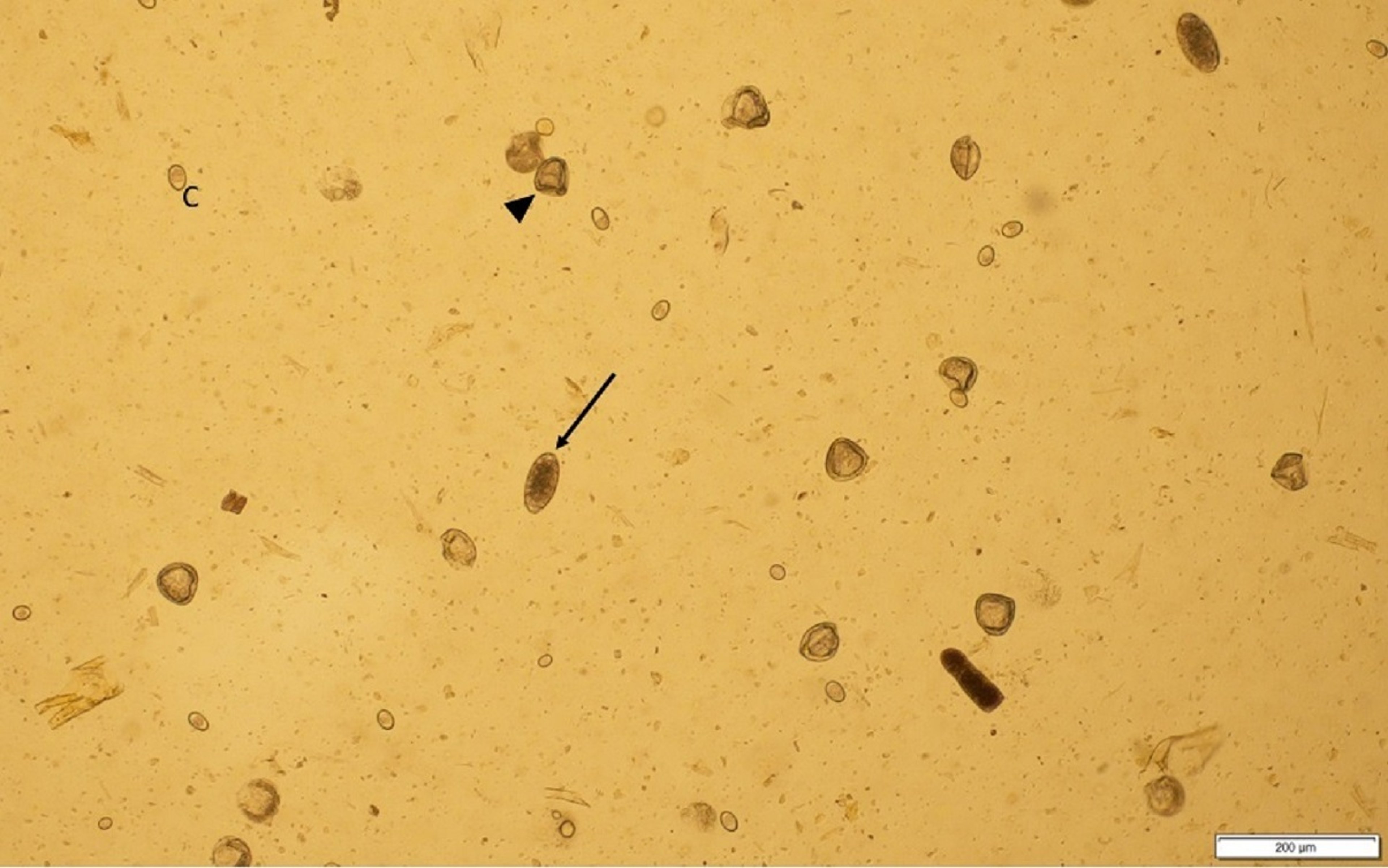
Moniezia spp Small Intestine Parasites of Small Ruminants

Courtesy of Megan Lagatta, RVT.

Courtesy of Dr. Grace VanHoy and Dr. Andrew Muir.
Moniezia expansa, Moniezia benedeni, and Moniezia caprae can all be found in small ruminants; however, the pathogenicity of Moniezia has long been debated. Many earlier observations, which associated Moniezia infection with diarrhea, emaciation, and weight loss, did not accurately differentiate between tapeworm infections and infection with certain small nematodes (eg, Trichostrongylus colubriformis).
Tapeworms are relatively nonpathogenic, but heavy infections can result in mild unthriftiness and GI disturbances. Diagnosis may be made if individual segments (which are much wider than they are long) are found in the feces, if lengths of adult tapeworms are observed protruding from the anus, or if the characteristic square to rectangular eggs are evident on fecal examination.
The life cycle of Moniezia spp is indirect and involves an oribatid mite that lives on the pasture. The prepatent period is 6–7 weeks. Infections are seasonal, aligned with mite activity, and unusual in animals older than 5–6 months.
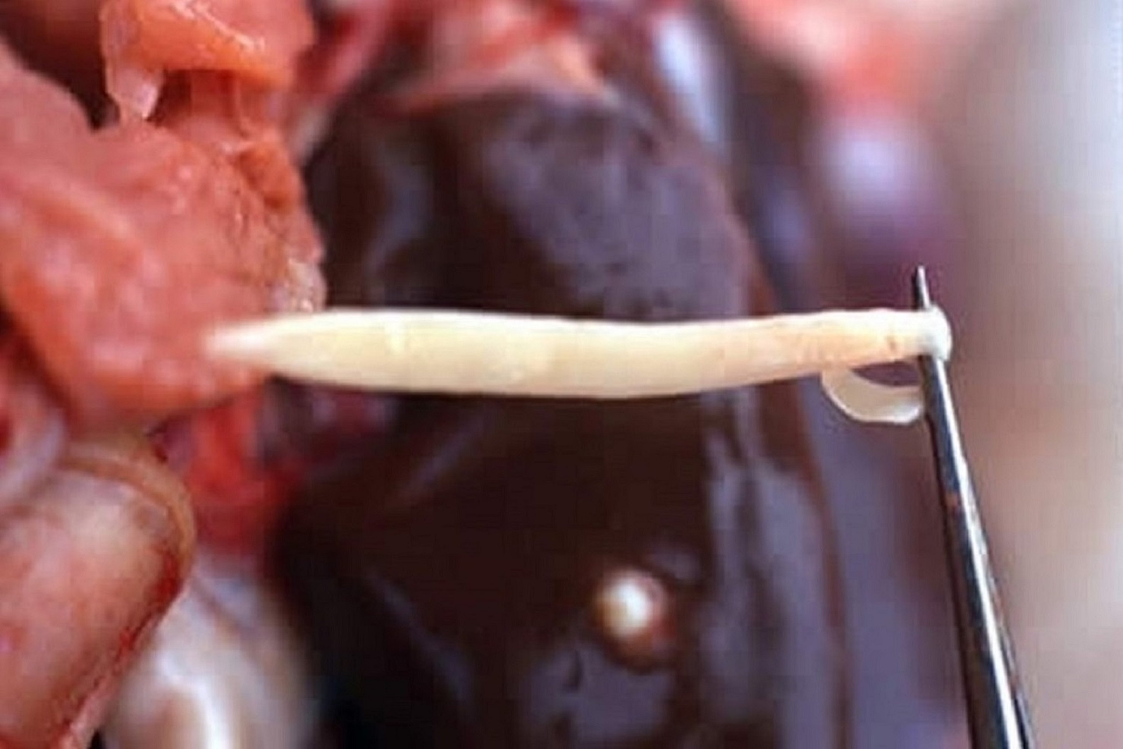
Thysanosoma actinioides Small Intestine Parasite of Small Ruminants

Courtesy of Dr. Raffaele Roncalli.
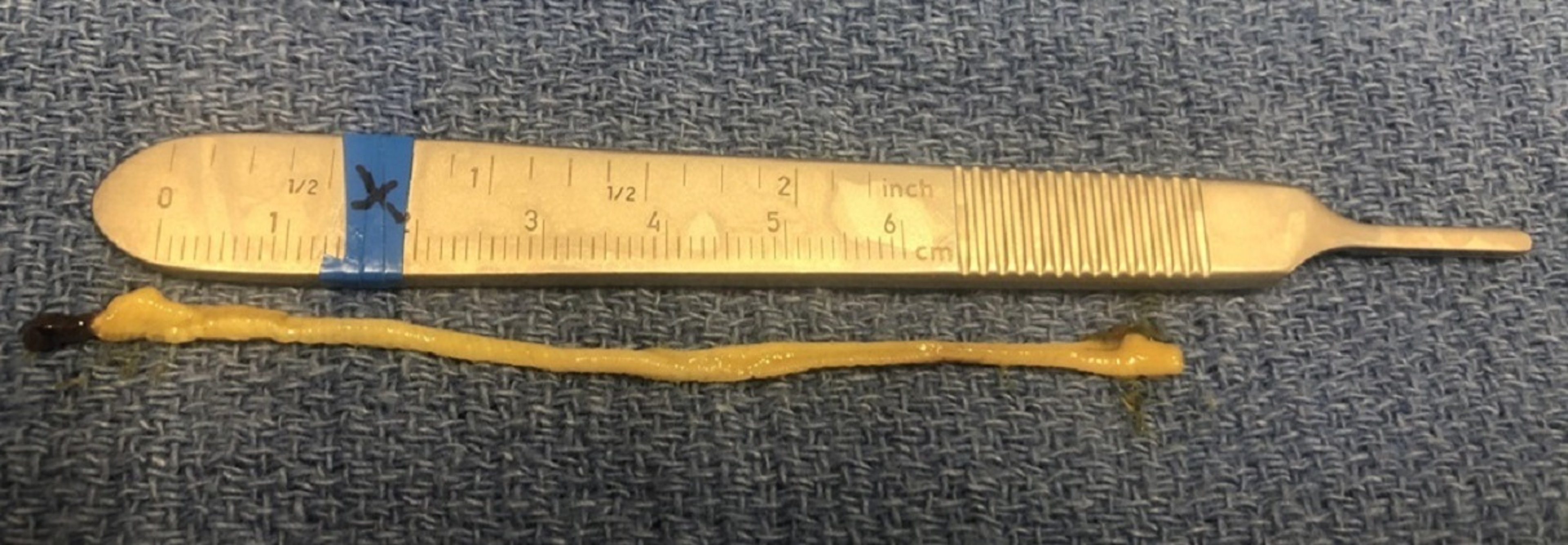
Thysanosoma actinioides inhabits the duodenum, the bile ducts, and the pancreatic ducts of small ruminants. It is commonly found in sheep from the southern and western parts of the US and South America. The adult tapeworm can be up to 50 cm long, the head (scolex) is 1.5 mm, and the segments (proglottids) are much wider than they are long and have fringed posterior borders.
Although it has not been associated with disease, T actinioides is of economic importance because livers are condemned when tapeworms are found in the bile duct.
Common Parasites of the Large Intestine in Small Ruminants
Oesophagostomum columbianum Large Intestine Parasite of Small Ruminants
The nodular worm of small ruminants, Oesophagostomum columbianum, has a morphology and life cycle similar to those of the nodular worm of cattle (see Common Parasites of the Large Intestine in Cattle).
Diarrhea usually develops during the second week of infection by O columbianum. The feces may contain excess mucus, as well as streaks of blood. As the diarrhea progresses, animals become emaciated and weak. These clinical signs often subside near the end of the prepatent period; however, the continuing presence of numerous adult worms may result in a chronic infection in which clinical signs may not develop for several months. Animals with chronic infection become weak, lose weight despite a good appetite, and show intermittent diarrhea and constipation.
As immunity develops, nodules form around the larvae; they may become caseated and calcified, and during active infections they may contain a single larva. Nodule formation usually is more pronounced in sheep and goats than in cattle. Stenosis and intussusception of the small intestine may develop in severe cases. Diagnosis is difficult during the prepatent period, at which time it must be based largely on clinical signs.
Chabertia ovina Large Intestine Parasite of Small Ruminants
Heavy infection with Chabertia ovina adults can severely damage the mucosa of the colon, with resulting congestion, ulceration, and small hemorrhages. Infected small ruminants are unthrifty, and their feces are soft, contain mucus, and may be streaked with blood. Immunity develops quickly, and outbreaks occur only in young animals or under conditions of severe stress. For more information on the life cycle of C ovina, see Common Parasites of the Large Intestine in Cattle.
Trichuris spp Large Intestine Parasites of Small Ruminants
Heavy infections with Trichuris spp (whipworms) are not common; however, they may occur in young lambs or kids or during drought conditions when animals are fed supplemental feed on the ground. The eggs are very resistant to environmental conditions and can remain persistent on farms with contamination. Congestion and edema of the cecal mucosa develop, accompanied by diarrhea and unthriftiness. Hypoproteinemia is a prominent feature of heavy Trichuris infection. For more information on the life cycle, see Common Parasites of the Large Intestine in Cattle.
Skrjabinema spp Large Intestine Parasites of Small Ruminants
Skrjabinema ovis (parasites of sheep) and Skrjabinema caprae (parasites of goats) are nonpathogenic pinworms. Adults are 8–10 mm long and live in the rectum and anus. Eggs are not commonly observed on fecal flotation, but clear tape applied to the perianal region and then to a glass slide will demonstrate ovoid eggs that are slightly flattened on one side and ~55 mcm long. Clinical signs include possible anal pruritus.
Diagnosis, Treatment, and Control of Gastrointestinal Parasites in Small Ruminants
For the diagnosis, treatment, and control of GI parasites in small ruminants, see Overview of Gastrointestinal Parasites of Ruminants.


