


Until recently, geriatric medicine had been a neglected area of avian medicine. Infectious diseases, inadequate diets, and poor husbandry meant that most pet birds did not live long enough to develop geriatric conditions. As the knowledge base of avian medicine, nutrition, and proper husbandry has grown, so has the life span of pet birds increased. Most pet birds have the potential to live 20−80 years, depending on their size (with smaller birds having a shorter life span and larger birds a longer life span). With pet birds living longer, the incidence of geriatric-onset diseases, including cataracts, neoplasia, arthritis, and cardiovascular disease, has increased.
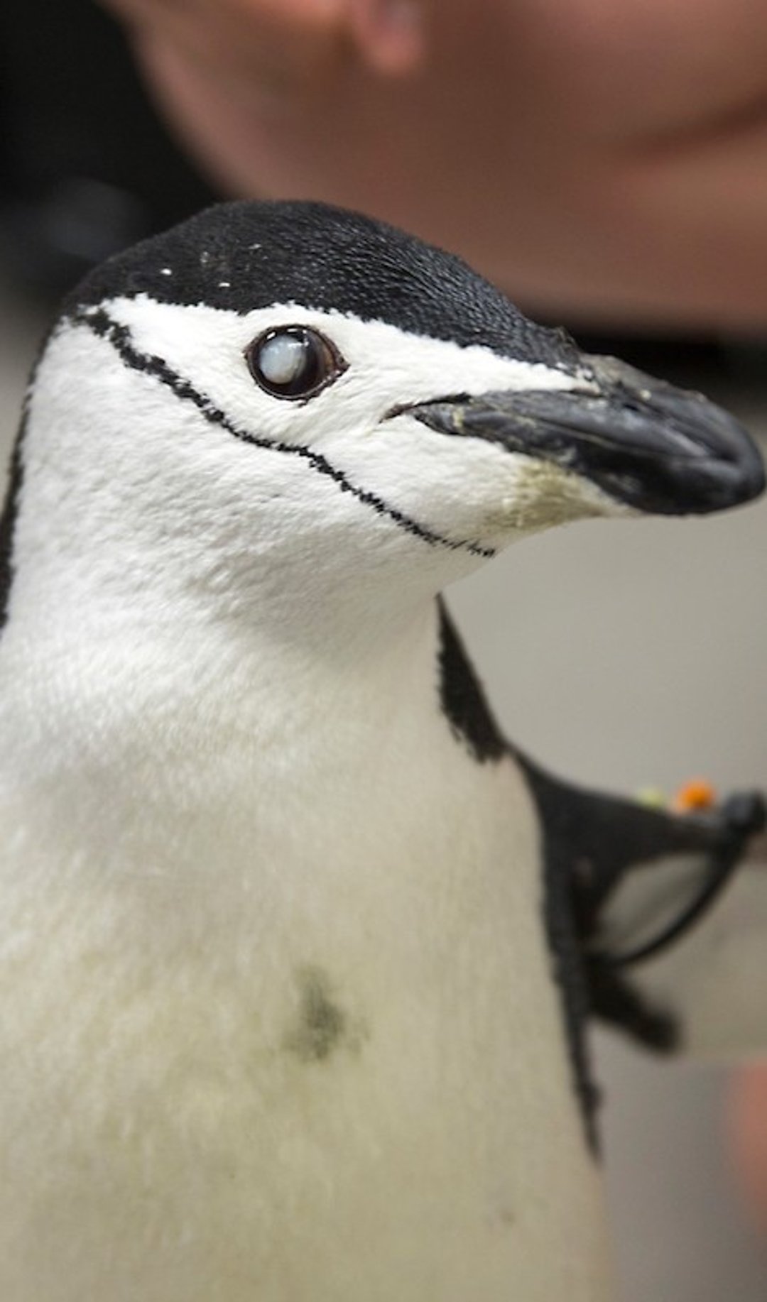
Cataracts

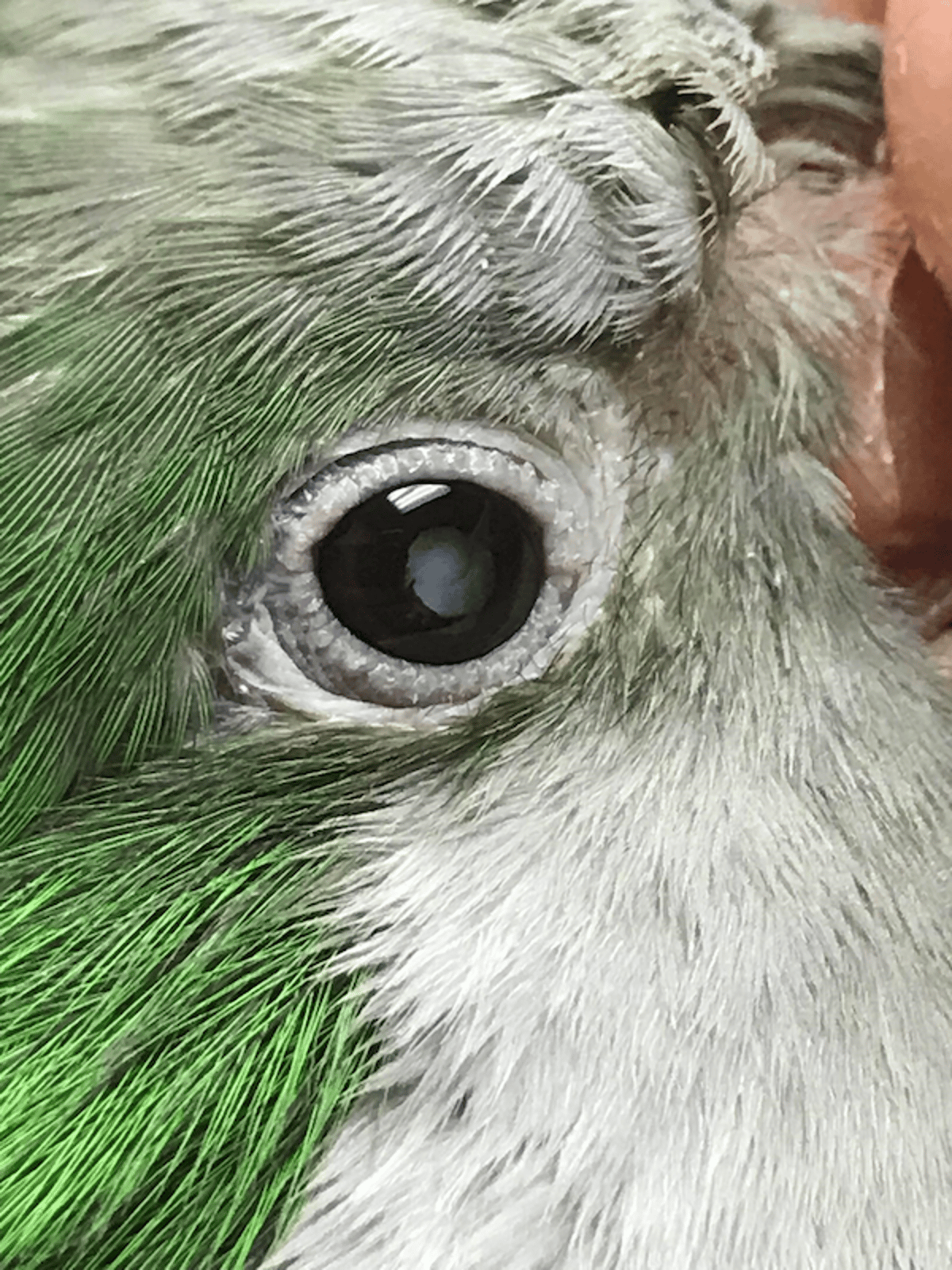
Courtesy of Dr. Bob Monaco.

Courtesy of Dr. Sharman Hoppes.
Cataracts develop in many species of psittacine birds as they age, notably macaws, Amazon parrots, and cockatiels. These species may be prone to cataracts or may be overrepresented in the older pet bird population. If the onset of cataracts is gradual, adaptation to decreased vision usually occurs; if not, clinical signs can be depression, inactivity, and reluctance to come out of or move around in the cage.
The eyes of older birds should be examined annually to detect early changes in lens opacity. Because of the small size of the exposed cornea and pupil in psittacines, and the numerous acquired diseases that can occur, screening by an ophthalmologist is recommended. Cataracts often develop secondary to infection or trauma or may be age related. Uveitis may also be present.
Additional ophthalmic conditions that may be encountered in geriatric birds are keratoconjunctivitis sicca, corneal ulcerations, third eyelid abnormalities, hypopyon, anterior uveitis, conjunctival granulomas, infection of the conjunctiva (eg, Chlamydia, Mycoplasma, poxvirus), Harderian gland adenoma, and lymphoma.
In large psittacine birds, surgical removal of cataracts is successful in many cases. The bird’s general health and the degree to which the cataracts affect its quality of life should be evaluated before surgery. Commonly used mydriatics are not useful in birds because of the skeletal (as opposed to smooth) muscle found in the iris. In any bird with decreased vision, minimal alteration of the home environment is critical. Early cataracts, especially if uveitis is present, may be painful. NSAIDs, either ocular drops (flurbiprofen) or systemic (meloxicam, celecoxib), or both, can be used to reduce inflammation and pain.
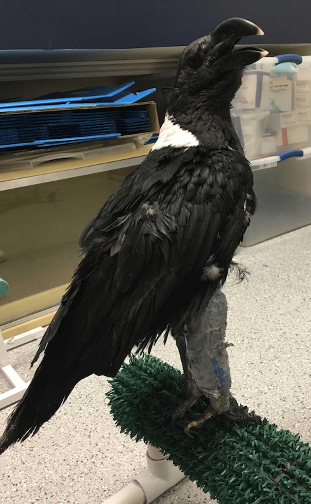
Arthritis

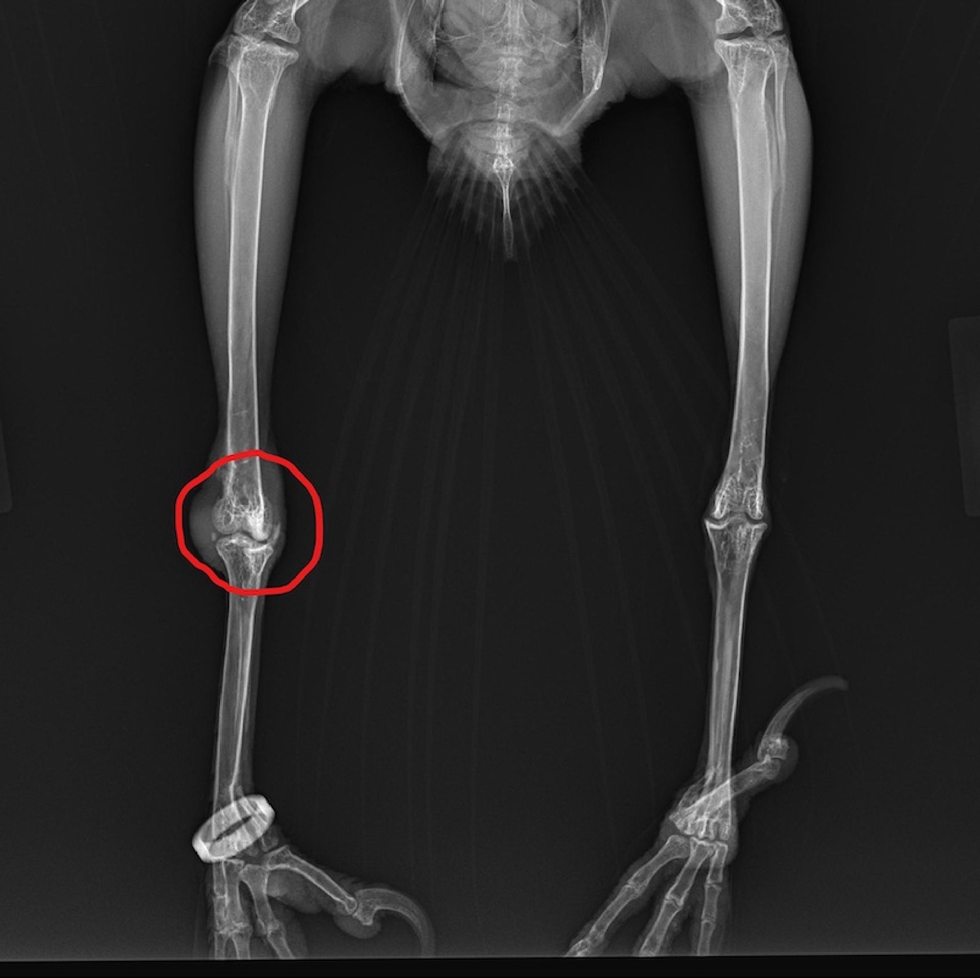
Courtesy of Dr. Sharman Hoppes.

Courtesy of Dr. Sharman Hoppes.
Septic and traumatic arthritis may occur at any age. Septic arthritis is most common in the digits. Osteoarthritis is also common in geriatric birds and can lead to other issues such as pododermatitis if not caught early and treated. The weight of the bird, its general physical condition, previous injuries, and any concurrent medical conditions can all contribute to the onset and severity of arthritis. Concurrent pododermatitis is often present and may be both a cause and result of decreased activity. Malnutrition, which decreases the integrity of the plantar epithelium, and concurrent obesity are often present in affected birds. The cage environment, especially the variety, diameter, and texture of perches, can be important in providing comfort and stability for arthritic birds while preventing or minimizing pododermatitis. If possible, the nails should be left with sharp points to add strength and stability to the grip. Wings should be minimally clipped, to help with balance.
Clinical signs vary, depending on the location of the arthritis and the severity of disease. Birds may exhibit lameness or be less active. A flighted bird may not want to fly or may not fly as well. The bird may not be perching normally or may fall off perches. Other signs of arthritis are swollen or warm joints, decreased range of motion, feather picking or mutilation, or excessive vocalization.
Diagnosis is based on clinical signs, physical examination findings, and imaging (radiographs or CT scan). Radiographic lesions include narrowing of the joint space, sclerosis of the subchondral bone, misalignment of the joint, and osteophyte formation. CT scans help determine the severity of the bony changes. Commonly affected joints are the tarsus, stifle, and phalangeal joints. The joints of the thoracic limb appear to be less commonly affected.
A multimodal treatment plan is recommended, incorporating both medical and nonmedical modalities. Medical treatment includes the use of NSAIDs, chondroprotectants, and possibly opioids. The most common NSAID used in avian medicine is meloxicam, a COX-2 inhibitor. Potential adverse effects of NSAIDs are renal ischemia, so these drugs should be used with caution longterm and at the lowest therapeutic dose possible. Anecdotally, glucosamine or polysulfated glycosaminoglycan has been used successfully. The latter should be used carefully, because some birds have had fatal coagulopathies from the injections. Gabapentin in conjunction with NSAIDs has been effective in relieving arthritic pain.
Opioids may be necessary for acute exacerbations of a chronic arthritic condition or for conditions not responding initially to NSAIDs. Tramadol or butorphanol may be used until the NSAIDs take effect.
Additional management includes husbandry changes, a weight loss and exercise plan, a healthier diet (rich in omega-3 fatty acids), and physical therapy. Encouraging flighted birds to fly in a safe environment is the best form of exercise. If a safe environment is not possible, encouraging climbing, walking, or even stepping up multiple times can be exercise for parrots. Foraging for food, by putting multiple foraging boxes on opposite sides of the cage or enclosure, promotes exercise. If the bird is overweight, then weight loss is essential, because studies have shown that obesity is a risk factor for osteoarthritis in many species. This may involve converting the bird slowly to a pelleted diet with added essential fatty acids. Fatty acids may have an anti-inflammatory effect and be renal protective. Flax seed oil or an omega supplement is recommended as the best source of fatty acid supplementation for birds. Other husbandry changes, such as changes in perch texture or diameter or padding perches, can be helpful in birds with weak or painful legs or feet.
Articular gout is also common in older birds. Differentiation between arthritis and articular gout is critical because of the vast differences in progression, quality of life, and prognosis.
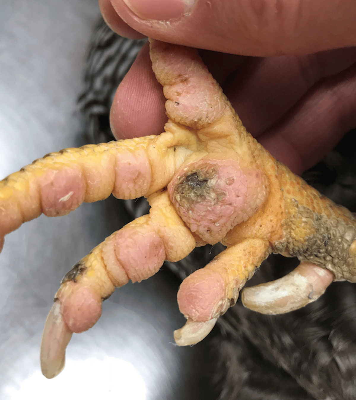
Pododermatitis
Pododermatitis, or bumblefoot, is a relatively common condition of older pet birds. It is a general term for any inflammatory or degenerative condition of the avian foot and can range from mild redness to bony changes.
Pododermatitis develops most commonly when birds are either housed with inappropriate perching or secondarily to an injury in one leg, which causes the bird to shift its weight to the other (good) leg, creating increased pressure and potential ulceration on the plantar surface of that foot. Birds most at risk are birds with leg fractures; arthritis of a hip, stifle, or tarsal joint; obese birds; or birds on a poor diet (eg, vitamin A deficiency). These are the same conditions that can predispose a bird to arthritis.
Pododermatitis is often a sequela of osteoarthritis and a progressive disease. A localized hyperemic lesion develops, followed by ulceration and, if untreated, can lead to abscess formation and osteomyelitis. Initially, the skin on the metatarsal and digital pads becomes flattened and smooth. The skin may become proliferative and then ulcerate, allowing bacterial access, which leads to inflammation and infection. As the infection progresses, tendon sheaths become affected, and osteomyelitis and septic arthritis develop.
Birds may present with lameness, depression, and anorexia due to the inflammation, pain, and infection. Diagnosis is based on clinical signs, physical examination findings, radiographs, and culture results. Affected birds should be examined thoroughly for predisposing injuries or illness.

Courtesy of Dr. Sharman Hoppes.
Treatment includes correcting inappropriate husbandry (adding padded perches or perches covered with artificial grass) and conversion to a healthier, preferably formulated diet. Weight loss and exercise should be encouraged in obese birds (flying, climbing, or walking). In early cases, this may be all that is necessary.
As the disease progresses, a bandage may be necessary to relieve pressure on the lesion. The lesions should be kept clean. Strict sanitation of the perches and feet is important to prevent bacterial infections. If a scab is present, it should be softened and removed or surgically debrided. Antimicrobial use should be based on results of culture and sensitivity testing. Staphylococcus spp is most commonly identified; other reported bacteria include Escherichia coli and Proteus spp.
Effective antimicrobials are amoxicillin/clavulanate (125 mg/kg, PO, 3 times a day), enrofloxacin (10–15 mg/kg, PO, 2 times a day), and marbofloxacin (5 mg/kg/day) for 10–14 days. Pain management is important and includes a combination of NSAIDs and/or opioids, depending on the severity of disease and after any surgical debridement. One regimen is meloxicam (0.5–1 mg/kg, 1 to 2 times daily) along with tramadol (15–30 mg/kg, PO, 2 to 3 times a day), and, in severe cases or after surgery, butorphanol (0.5–3 mg/kg, IM or intranasal, every 4 hours, depending on species). Local anesthetics may be helpful after surgery.
Cardiac Disease
As birds live longer and diagnostic techniques improve, cardiac disease is being diagnosed more frequently. It can be difficult to detect and may mimic other problems, such as respiratory disease. Cardiac disease has been associated with atherosclerosis in pet birds, and potential risk factors are a sedentary lifestyle, a high-fat diet, and hypercholesterolemia. Most affected birds are >15 years old, and incidence is higher in females.
Clinical Findings of Cardiac Disease in Pet Birds
Clinical signs of cardiac disease in pet birds are weakness, depression or lethargy, increased respiratory rate and effort, and /or tachycardia. With right-side heart disease, hepatomegaly and ascites are common. Signs are often subtle and insidious until the disease process is advanced. Birds with subclinical disease may arrest when diagnostic tests or treatments are attempted. In birds, right-side cardiac disease is more prevalent than left-side.
Diagnosis of Cardiac Disease in Pet Birds
Cardiac disease is underdiagnosed in the avian patient, and consultation with a cardiologist for any avian patient with suspected cardiac disease is advised. Diagnosis of the cardiovascular abnormality and forming a therapeutic plan requires knowledge of avian anatomy and physiology and a cardiologist’s diagnostic skills and pharmacologic recommendations.
Diagnosis of cardiac disease begins with a thorough history and physical examination. Initial diagnostic evaluation should include a CBC, a biochemistry profile, imaging, and echocardiogram. Hematology and biochemistry values are not always helpful in diagnosing cardiovascular disease, although myocardial disease may cause increases in concentrations of AST, CK, and LDH. Lipemia may be noted, and marked increases in cholesterol and triglyceride concentrations may be seen with atherosclerosis.
Radiographs will aid in general assessment of the cardiac size, contour, and radiodensity of the cardiac silhouette and great vessels (brachiocephalic trunk, aorta, pulmonary arteries, and veins). Other radiographic abnormalities that may be present with cardiac disease are an enlarged hepatic silhouette, increased radiodensity of the pulmonary parenchyma, coelomic air sac compression, or loss of coelomic detail. Increased radiodensity, tortuosity of the great vessels, and focal or linear mineralization of vessels is suggestive of atherosclerosis, although assessment of increased radiodensity and opacity is somewhat subjective and varies with radiographic technique. Mineralization of vessels in psittacines appear most often along the aorta and brachiocephalic trunks but may be seen along smaller arteries.
CT is useful in determining cardiomegaly, ventricular dilatation, pericardial effusion, pulmonary edema, ascites, and venous congestion. CT angiography is useful to image the vascular system and can more readily help diagnose vascular disease, including arterial calcification and luminal stenosis related to atherosclerosis, aneurysms, and congenital vascular abnormalities.
Although radiographs and CT scans have value in assessing cardiomegaly and hepatomegaly, coelomic ultrasound can aid in confirmation and assist in determination of the etiology of the disease. Coelomic ultrasound can allow for assessment of chamber size, wall thickness, contractility valvular morphology, and function. Chamber dilatation, myocardial hypertrophy, valvular insufficiency and abnormalities, septal defects, cardiac masses, pericardial effusion, ascites, and aneurysms can be identified with ultrasound assisted by color Doppler. Ascitic or pericardial fluid may be sampled by fine-needle aspirate.
An ECG is used to characterize arrhythmias and can provide information on cardiac abnormalities and chamber enlargement. It is used in conjunction with imaging and ultrasound to aid in diagnosis of specific cardiac disease. Severe disease can exist in the absence of ECG changes. Studies have consistently shown that blood pressure values obtained with indirect methods do not agree with direct systolic measurements and may be of little value as a diagnostic tool in the clinical setting. But, in general, hypotension is defined when a systolic blood pressure is < 90 mmHg with a mean < 60 mmHg. Systolic values >200 mmHg are considered hypertension.
Treatment of Cardiac Disease of Pet Birds
Treatment of cardiac disease of birds includes supportive care and oxygen therapy for congestive heart failure. The mainstays of treatment in both small animal and avian patients include diuretics, ACE inhibitors, and positive inotropes. Decreasing hypervolemia, edema, and effusion is an immediate treatment priority in a bird presenting with heart failure. This is accomplished by administration of diuretics, most commonly furosemide at 1–5 mg/kg, IM. Initially, this may need to be repeated every 2 hours until the bird is stable; the dose can then be reduced to every 6–12 hours. Once the bird is stable, furosemide may be administered orally every 8–12 hours. The goal is to administer the lowest dose that controls clinical signs of congestion.
ACE inhibitors are an essential component of longterm medical management of congestive heart failure. Enalopril 1.25–5 mg/kg, PO, every 12 hours has been the most commonly administered ACE inhibitor, and empirical evidence suggests it is both safe and effective.
Positive inotropes are used to increase myocardial contractility and are used for heart failure due to systolic dysfunction. They are contraindicated in cases of hypertrophic cardiomyopathy and outflow obstruction, and they may not be appropriate in birds with heart failure secondary to atherosclerosis. Both digoxin 0.01–0.02 mg/kg, PO, every 12 hours, and pimobendan 0.1–0.25 mg/kg, PO, every 12 hours, have been administered for treatment of heart failure in avian species, but data are lacking as to their efficacy and safety. Negative inotropes (beta-blockers) are a central component of heart failure treatment in humans but have not been commonly used in avian patients. The beneficial effects of beta-blockers have been documented in furazolidone-induced dilated cardiomyopathy in turkey poults, but no data are available for psittacines.
Long-term care may include exercise restriction, cage rest with appropriate housing, and conversion to a healthier diet, along with weight loss if obese. Because many of these birds will need to be on long-term medications, positive reinforcement training for targeting and syringe training may aid in administration of medications. Medications should be formulated or compounded to be as palatable and concentrated as possible to provide for better acceptance and reduced volume dosing. Essential fatty acids (flaxseed oil at 0.1 mL/kg, PO, once a day and omega supplement at 0.22–0.44 mL/kg, PO, once a day) have also been advocated and used to reduce cholesterol and inflammation.
Atherosclerosis of Pet Birds
Atherosclerosis is a proliferative lesion of the tunica media and tunica intima of elastic and muscular arteries. Atherosclerotic plaques cause abnormal vascular flow and loss of endothelial integrity. These changes in vessel walls can initiate thrombosis. In birds, lesions are primarily in the aorta and brachiocephalic arteries. Atherosclerosis is common in psittacine birds. It is generally a geriatric condition, except in African grey parrots, in which this disease has been observed in very young birds. Amazon parrots, Quaker parrots, macaws, and African grey parrots seem to be particularly susceptible. Atherosclerosis is an underlying factor in many of the noninfectious cardiovascular diseases seen in pet birds.
Predisposing factors include age (most affected birds are 10–15 years old), sex (females have a higher incidence), sedentary lifestyle, and a high-fat diet. Clinical signs are rarely reported in birds, and the condition is often associated with sudden death. Clinical signs that may be seen are exercise intolerance, dyspnea, episodic weakness, and neurologic signs (eg, seizures, tremors, paresis). At necropsy, grossly thickened arterial walls are seen.
Diagnosis can be difficult. Radiographically, the right aortic arch may be enlarged, with increased density. Lipemia is often present, and marked increases in cholesterol and triglyceride concentrations may be seen. Unfortunately, definitive antemortem tests are lacking. A CT scan may reveal narrowing of some of the major vessels affected.
Medical treatment is anecdotal. A variety of treatments have been advocated to lower cholesterol levels, but none appears to be consistent in its efficacy. Converting the bird to a low-fat diet and increasing exercise may aid in reducing cholesterol and triglyceride levels. Isoxsuprine (10 mg/kg/day, PO), a peripheral vasodilator that causes vascular smooth muscle relaxation, has been used anecdotally with some success in birds. Statins (lipid-lowering drugs used in humans with atherosclerosis) have been used in birds, but there is little information to support their use. Essential fatty acids (flax seed oil at 0.1–0.2 mL/kg/day, PO, or a veterinary supplement) have been advocated and used to reduce cholesterol and inflammation.
Kidney Disease
Kidney disease may be seen at any age, but older birds are more likely to develop renal insufficiency. Causes are multiple and include glomerulonephropathies, renal tubular gout, and chronic bacterial nephritis.
On physical examination, certain abnormalities may indicate kidney issues. Most birds with articular gout have some form of kidney disease. Also, unilateral lameness or paresis may indicate compression of the lumbar/sacral plexus from an inflamed or enlarged kidney. Clinical signs include weight loss, depression, polyuria, polydipsia, and dehydration. Diagnosis is made based on a persistent hyperuricemia before and after fluid therapy. Other laboratory findings may include anemia or increased CPK and urinary gamma glutamyl transferase concentrations. Imaging (radiographs or CT scan) may demonstrate small or large kidneys with or without mineralization. Occasionally, ureteroliths may be seen. Kidney biopsy is necessary for definitive diagnosis.
Treatment includes supportive care (fluid therapy) and antimicrobials as needed based on diagnostics. Colchicine (0.04 mg/kg, PO, 2 times a day) and allopurinol (10–30 mg/kg, PO, 2 times a day) have successfully reduced uric acid concentrations in certain disease processes. After the bird has been stabilized, conversion to an appropriate diet and supplementation of vitamin A, if warranted, should be initiated. Essential fatty acids at 0.22–0.44 mL/kg/day, PO, with low-dose aspirin (0.5–1.0 mg/kg, PO, every 12 hours) have been used anecdotally to manage kidney disease in birds.
Liver Disease
Liver disease is common in pet birds and can be related to viral disease (eg, polyoma and avian herpesvirus), bacterial disease (eg, chlamydiosis), and/or hepatic lipidosis. Hepatic lipidosis is most commonly seen in the older pet bird that has been on a poor diet (seed mix or unhealthy table foods) and has a history of obesity. Chronic liver disease can result in fibrosis of the liver and decreased liver function and failure.
Clinical Findings and Diagnosis of Liver Disease of Pet Birds
Clinical signs of liver disease in birds are anorexia, lethargy, overgrown beak and nails with bruising, biliverdinuria, discolored feathers, and, in later stages, ascites. Birds often are overweight or have a history of being obese. Clinical pathology may reveal high liver enzyme levels (AST concentration) and bile acids, a nonregenerative anemia, and high triglyceride and cholesterol concentrations. Imaging (radiographs or CT scan and/or ultrasound) may reveal hepatomegaly or microhepatica. Liver biopsy may be necessary to confirm the diagnosis and determine etiology and prognosis.
Treatment of Liver Disease of Pet Birds
Treatment involves supportive care (fluid therapy), antimicrobials as needed, and medications to support hepatic function such as silymarin (milk thistle) (100–150 mg/kg, PO, 2 times a day). Other drugs used to treat liver disease in birds include ursodeoxycholic acid (10–15 mg/kg/day, PO), colchicine 0.01–0.2 mg/kg, PO, every 12–24 hours, and lactulose 150–650 mg/kg, PO, every 8–12 hours. Longterm modifications of the diet may include converting to a formulated diet.
For More Information
Also see pet health content regarding disorders and diseases of pet birds.


