


Sugar sliders are small, nocturnal marsupials native to Australia, Indonesia, and New Guinea that have become popular pets. Their name derives from the thin membrane of tissue (the patagium) that extends from the wrists on their forelimbs to their ankles on their hindlimbs that allows them to glide from tree to tree in the wild. Wild sugar gliders live in large social groups and fare better in captivity when housed in pairs. They are omnivorous hindgut fermenters that rely on bacterial cecal fermentation to digest carbohydrates. In the wild they eat a variety of plants, sap, and invertebrates. This diet is hard to mimic in captivity, predisposing captive gliders to nutrient deficiencies and disease.

Courtesy of Dr. Laurie Hess.
Courtesy of Dr. Laurie Hess.
Sugar gliders (Petaurus breviceps) are small, nocturnal marsupials native to Australia, Indonesia, and New Guinea that live in eucalyptus and acacia forests. They belong to the family Petauridae, which includes the wrist-winged gliders. Gliders in this family possess a gliding membrane (patagium) that runs from the wrist of the forelimb to the ankle of the hindlimb that allows them to glide as far as 50 m and forage for food using less energy. They use their tails as stabilizing rudders that enable them to change direction easily. The second and third toes on their hindfeet are fused to form a "grooming comb" that helps them clean their fur.
Females are seasonally polyestrous and have two lateral vaginas, a central vaginal canal, two uteri, and a pouch containing four teats; they often have twin births. After 16 days of gestation, the young (joeys), each weighing only 0.2 g, migrate to the pouch to develop further and finally leave the pouch after 70–74 days. They remain in the nest until 110–120 days of age, when they are weaned. They stay with the colony until 7–10 months old.
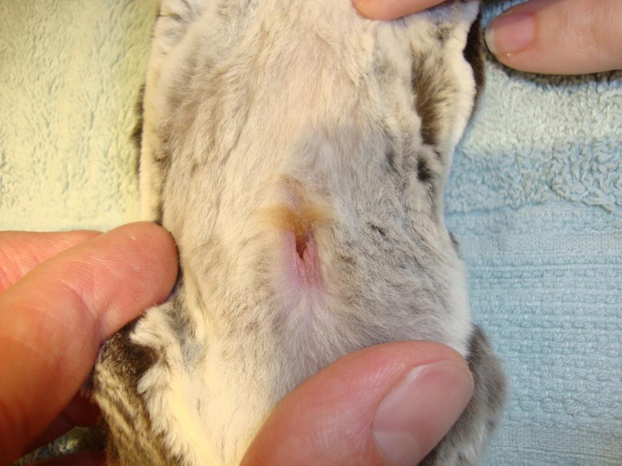
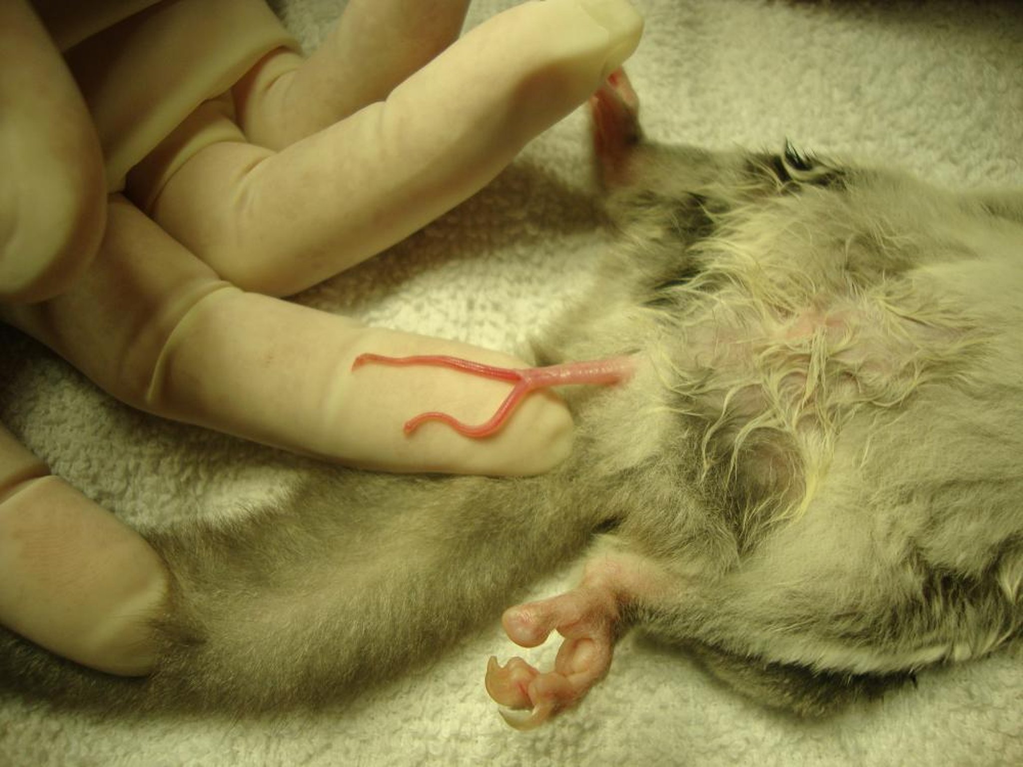
Males have a forked penis (to match the female's double vagina) and a pendulous scrotum containing two testicles. Males do not urinate from the forked end of the penis but from the proximal end. Both males and females have paracloacal scent glands adjacent to the vent (the cloacal opening or common opening of urinary, reproductive, and GI tracts) with which they mark territory and each other, and males also have frontal scent glands on their foreheads and glands on their throats and chests. Sparse fur and an oily discharge are normal on the frontal and sternal glands of postpubescent males. These glands give both sexes a musky odor.
Sugar gliders have large, protruding, widely spaced eyes, giving them a wide field of vision, especially at night. Their ears move independently and are highly sensitive to sound. They also have a great sense of smell to locate food, sense predators, and recognize both their territory and their colony-mates.
Wild gliders have gray fur and a central black stripe dorsally on their heads; domesticated gliders may look similar to the wild type but also come in several color variations. Sugar gliders are polygamous, territorial, and live in colonies of 5 to 12 individuals with a dominant male. They sleep in tree hollows by day and between foraging trips at night. They tolerate a wide range of environmental temperatures and go into torpor to conserve energy in very cold conditions.
Sugar gliders are omnivorous and feed on sugar-rich plant and insect exudates (sap, gum, nectar, manna, pollen) and invertebrates as a source of protein. They are hindgut fermenters and possess a well-developed cecum that utilizes bacterial fermentation to break down complex polysaccharides contained in gum. Also see Table: Selected Physiologic Data for Sugar Gliders, which summarizes important biologic and physiologic data for sugar gliders.
Physical Examination of Sugar Gliders
For a full clinical examination of sugar gliders, anesthesia with isoflurane via face mask may be required if gliders are very stressed or biting. More docile animals may be examined while wrapped in a small towel and cupped in the palm of the hand. Gliders inside fabric bags or pouches may be partially exposed, one body part at a time, for examination. Because gliders are nocturnal, it may be best to schedule clinical examinations earlier in the day when they are less active.
If possible, the sugar glider should be first observed moving in its cage to assess posture, coordination, and demeanor. If the glider is anesthetized, cloacal temperature, heart rate, and respiration rate can be recorded, and the heart and lungs assessed with a pediatric stethoscope. The fur and skin should be examined for ectoparasites, traumatic injury, fur loss, and degree of hydration; the oral cavity for broken teeth, dental abscesses, or tartar build up; and the eyes and ears for any abnormalities. The cloacal area should be examined, and the penis of males extruded. The abdomen should be palpated, and the pouch in females examined. Major joints should also be palpated, and digits and toenails checked for evidence of trauma.
Diagnostic and Treatment Techniques in Sugar Gliders
If a sugar glider is dehydrated, isotonic fluids up to 10% of body weight can be administered SC over the shoulder region. Care should be taken not to induce edema to the patagium. Fluids also may be administered intraosseously in the proximal femur or tibia. SC injections may be given in the same area that SC fluids are administered. IM injections may be given in the epaxial muscles of the neck and dorsal thorax. IV injections are very difficult to perform but may be accomplished in cephalic or lateral saphenous veins in an anesthetized glider.
Radiographs generally require the glider be anesthetized for proper positioning. Pulmonary diseases are easiest to detect with radiographs. Feces may be easily sampled and checked annually with a fecal flotation and direct smear for parasites; animals with diarrhea should have their feces cultured and tested for appropriate antibiotic sensitivity.
Blood Collection, Hematology, and Biochemistry of Sugar Gliders
Reference ranges for sugar gliders are presented in Selected Hematologic and Serum Biochemical Values for Sugar Gliders. Chemical restraint, essential to allow blood collection, is most safely achieved with isoflurane/oxygen administered via mask and T-piece.
To assist in making clinical diagnoses, blood samples may be obtained from the cranial vena cava, jugular vein, medial tibial artery, or lateral tail vein. Blood volumes of up to 1% of body weight can be collected; typically 0.5–1 mL is obtained.
To access the cranial vena cava, a 27- or 25-gauge needle on an insulin syringe may be inserted in the thoracic inlet just lateral to the manubrium, with the needle directed at a 30° angle from midline toward the opposite hindleg. The vena cava is not visualized directly during venipuncture but is accessed blindly using the manubrium as a palpable landmark.
The jugular vein can be visualized if the hair is clipped and the vein is forced to fill by applying gentle digital pressure at the thoracic inlet; it sits midway between the point of the shoulder and the mandibular ramus. The needle can be bent at its base to facilitate venipuncture at this site.
The medial tibial artery, which runs very superficially just distal and medial to the stifle joint, is easier to access and can be sampled with a 27- or 25-gauge needle and 0.5–1 mL syringe. Pressure after sampling is required to prevent hematoma formation. The size of the lateral tail vein is most suited to skin prick and droplet collection into capillary tubes. Small blood samples (0.25 mL) may also be obtained from the cephalic, lateral saphenous, femoral, and ventral coccygeal veins with a 27-gauge needle on an insulin syringe.
Anesthesia and Surgery in Sugar Gliders
Anesthesia in sugar gliders should be approached the same way it is in other small mammals. Gliders ideally should be fasted for at least 4 hours preoperatively. Preoperative analgesics, sedatives to reduce preoperative stress, and local and gas anesthetics all may be used. Care must be taken to keep gliders warm during surgery to ensure rapid recovery. Gliders undergoing surgeries that last >1 hour or that require intra-abdominal access should be administered fluids intraosseously (in the femur or tibia) throughout the procedure if IV access is impossible because of small size. Shorter, simple procedures may necessitate only SC fluids,
Commonly used pre-anesthetics include atropine or glycopyrrolate, midazolam, and either butorphanol or buprenorphine. Local anesthetic injections of a 50:50 mixture of 2% lidocaine with 5% bupivacaine may be used to infiltrate the incision site to help reduce the chance of postoperative self-mutilation. To facilitate castration, this mixture may be diluted with sterile water and infused into the base of the scrotal stalk nearest to the abdomen while a low concentration of gas anesthetic is administered. Both isoflurane and sevoflurane may be administered via small face mask or via a large face mask used as an induction chamber. For surgery, gliders may be maintained on gas anesthesia with a face mask or intubated with a 1-mm Cook endotracheal tube threaded with a stylet.
During surgery, blood loss should be monitored carefully; use of radiosurgery may help minimize bleeding. Because sugar gliders tend to chew incisions after surgery, ideally subcuticular sutures and skin glue should be used to close skin incisions. Analgesics should be administered before surgery to minimize pain immediately after surgery. Gliders should recover from surgery in temperature-controlled incubators.
Orchiectomy and scrotal ablation are commonly performed on male sugar gliders to prevent breeding and to decrease sexual frustration. Hair is clipped around the base of the scrotal sac and stalk, and the skin is cleaned without alcohol so as not to lower body temperature. A local anesthetic is administered at the base of the stalk, and with the glider under gas anesthesia, the incision is made over the scrotal stalk ~2–3 mm from the body wall. The spermatic cords are exposed by blunt dissection and are clamped and ligated with 5-0 polydioxanone suture or cut and cauterized with radiosurgery. The scrotal sac containing the testicles and distal spermatic cord is then removed, and the ligated stalk can be sutured to the abdominal wall fascia to prevent herniation. The skin is then closed with tissue glue.
Standard ovariohysterectomy is not routinely performed because of the internal position of the female reproductive organs, which are difficult to access beneath the pouch. With ovariohysterectomy, the area around the pouch is clipped and scrubbed in a routine manner, and a 1–2 mm incision is made paramedian to the pouch. The linea alba is bluntly dissected and incised, and the bladder is exteriorized from the incision to reveal the ovaries beneath. The ovarian arteries are ligated, as is the uterus proximally to the lateral vaginal canals. After resection of both ovaries and uteri, the linea is closed, and the skin is sutured subcuticularly and closed with skin glue.
Distal penile amputation is another common surgery. Sexually frustrated males may self-mutilate the distal end of their penises or develop paraphimosis. Because males urinate only from the proximal end, the distal, forked segment may be safely amputated.
Other surgeries performed in sugar gliders include cystotomy to remove uroliths, urethrostomy to alleviate urinary tract obstruction from uroliths in males, and surgical removal of impacted paracloacal glands. The skin over impacted glands is infiltrated with a small volume of local anesthetic and incised; the gland is bluntly dissected and removed without rupturing it. The blood vessel supplying the gland may be ligated with 5-0 or 6-0 polydioxanone or cut and cauterized with radiosurgery. The skin is closed with tissue glue.
Drug Dosages for Sugar Gliders
Very few pharmacologic studies have been performed in sugar gliders, and most published drug dosages have been extrapolated from those determined for cats, ferrets, and hedgehogs. For drugs and dosages commonly used in sugar gliders, see Table: Commonly Used Drugs in Sugar Gliders.
Nutrition and Housing of Sugar Gliders
Ideally, sugar gliders should be maintained as a group with one male and a number of females. If breeding occurs, the young should be removed soon after weaning, or violent attempts to disperse them may occur.
Because sugar gliders are arboreal (tree-dwelling), a large cage (ideally of PVC-coated stainless steel) is best for nocturnal climbing. As large a cage as possible should be provided, with a minimum size of 36 × 24 × 36 in. Bar spacing should not be more than 1 × 0.5 in. wide, or feet and heads may become entrapped. Caging containing vertical bars should be avoided, because vertical bars do not facilitate climbing. A wooden nest box (made for birds) or a fabric pouch positioned high up in the cage should be provided for hiding and sleeping. Cages should contain numerous branches (commercially available for bird cages) and horizontal shelves to promote climbing. Swings and chew toys made for birds are ideal for gliders to play with. Exercise wheels containing smooth interiors (so as not to entrap toes) should be provided for physical and mental stimulation. The cage bottom may be covered with newspaper or other recycled paper product that is nontoxic if ingested. Several food and water bowls should be provided throughout the cage, with designated areas for eating, drinking, exercising, and hiding.
The recommended diet for captive sugar gliders includes calcium-loaded insects (crickets, mealworms, waxworms, cockroaches, moths) to promote dental health, as well as a daily nectar/sap substitute (eg, fructose/sucrose/glucose or honey diluted to 10% with water). Nectar should account for ~50% of the diet. Several nectar substitutes are commercially available, including Gliderade nectar supplement (Exotic Pet Nutrition Company, Newport News, VA), acacia gum powder, and nectar diets meant for lory birds. Other protein sources in addition to insects include eggs, lean meat, newborn mice, and commercial pelleted diets meant for sugar gliders. Commercial diets or homemade insectivore or omnivore mixes should be provided in addition to live food, with insects and pelleted food accounting for nearly 50% of the total diet. Fruits, nuts, and vegetables should be offered only in moderation (<10% of total diet), because many fruits and vegetables lack essential vitamins, minerals, and protein and contain mostly water.
Several commercial diets and recipes for homemade diets are available; no single diet studied has yet proved to be ideal for captive sugar gliders. Traditionally, before the availability of commercial pelleted diets, a homemade formula called Leadbeater's mixture was fed successfully in addition to insects, fruits, and vegetables. The mixture contains 150 mL of warm water mixed with 150 mL of honey, one hard-boiled shelled egg, one teaspoon of multivitamin/mineral supplement, and 25 g of high-protein dry human infant cereal. An alternative to the homemade Leadbeater's diet is commercially available High Protein Wombaroo Diet (Wombaroo Food Products, Glen Osmond, South Australia), a powdered protein supplement that is mixed with water before feeding. In addition, if not provided in the diet, a multivitamin and mineral supplement with calcium should be sprinkled on food daily. Food should be offered in the evening, when sugar gliders are active, on an elevated platform, because gliders feel more secure eating up high, as they would in trees in the wild.
Sugar gliders are generally robust in captivity when proper husbandry practices are followed; however, nutritional deficiencies, especially of calcium and protein, are commonly seen in captive gliders fed inappropriately. Obesity also is common in gliders fed excessive amounts of treats high in fat and protein and not provided with the opportunity to exercise. Iron storage disease, in which excessive dietary iron accumulates in the liver (hemochromatosis), spleen, and other tissues, has been reported in captive gliders and may be associated with wild gliders' evolutionary adaptation to extract limited iron available in natural diets. Hepatocellular toxic changes and cirrhosis from iron deposition may lead to death, unless the disease is caught and treated early with chelation and supportive care.
Key Points
Sugar gliders are marsupials whose juveniles complete development in the maternal pouch.
They have unique anatomic structures, such as double vaginas and uteruses in females, a forked penis in males, and numerous scent glands, including paracloacal glands.
They are omnivorous hindgut fermenters that consume a variety of plants, insects, and sap/gum.
For More Information
Also see pet health content regarding sugar gliders.


