


In general, diagnosis and treatment of common disorders in zoo species is based on translation from closely related commonly managed species or medicine in humans. Commonly encountered medical problems include:
acute or chronic gastroenteritis
enteric parasites
traumatic injuries, including bite or gore wounds, lacerations, or bone fractures or joint luxations
bacterial abscesses
obstetric problems, like dystocia
degenerative joint disease or osteoarthritis
GI foreign bodies
bacterial pneumonia, including aspiration pneumonia
Avian Aspergillus infection generally results in chronic respiratory tract disease. Affected birds may exhibit weight loss, markedly increased WBC counts, and in later stages, dyspnea. Death can also occur peracutely if there is a localized aspergilloma that occludes the trachea or invades a major blood vessel. Antemortem serology and sensitivity and specificity show wide species variation; however, radiographic or CT imaging is often diagnostic, even if treatment can prove frustrating. Nebulization with antifungal agents has been well described, as have oral regimens, which may include itraconazole, voriconazole, and/or terbinafine. Caution must be exercised when using voriconazole, because dose-dependent toxicity has been noted in penguin species. Aspergillosis is often associated with microclimates of reduced ventilation, increased crowding, and other stressors, emphasizing an immunosuppressive effect. Postmortem examination generally demonstrates extensive fungal granulomas in the air sacs and lungs that seem to outpace clinical signs. Zoo species that are more sensitive to Aspergillus infections include penguins (Spheniscoformes), birds of prey, pheasants (Phasianinae), certain passerines, such as birds of paradise (Paradisaeidae), and some waterfowl. Some zoos use prophylactic management with itraconazole during time of stressors, like shipment or other moves.
Pododermatitis (bumblefoot) is a common disorder of birds. It can be either unilateral or bilateral and is characterized by lameness, inflammation, and swelling of the footpad due to localized bacterial infection. Sequelae of infection can include chronic pododermatitis, septicemia, or amyloidosis. It can occur due to injury, infection, inappropriate substrate, obesity, or unilateral limb problems (trauma, arthritis) that result in excess and abnormal weight bearing on the contralateral foot. Research into risk factors for specific orders (Spheniscoformes, Phoenicopteriformes) can help identify and mitigate causes in these birds; the most important part of treatment is management of the underlying medical or husbandry issue. Treatment of the lesion itself may involve local or systemic antimicrobials—including more recently, success with regional limb perfusion, anti-inflammatories and/or analgesic medications, or surgical correction for more advanced cases.

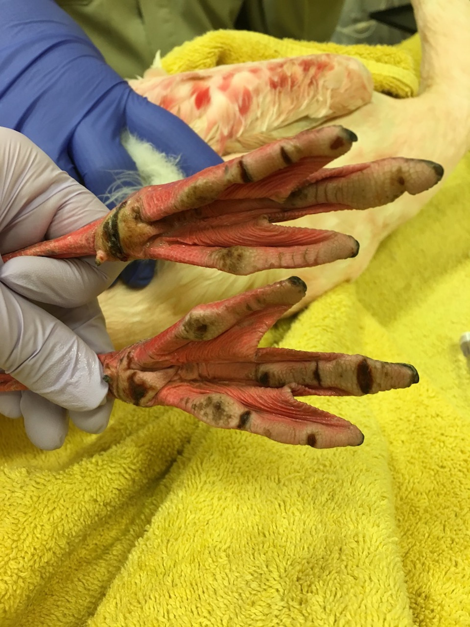
Courtesy of Dr. Meredith M. Clancy.
Atypical mycobacteriosis is a chronic problem in many bird collections. Extensive research indicates this is an environmental issue rather than a classically infectious disease, and so enclosure mates are often exposed from the same source of infection in the environment rather than from an infected individual bird. Crowding and stressors can increase risk of infection from the environment; however, control measures to try to identify chronically infected birds are historically unreliable, and attempts to sanitize the environs may prove frustratingly inadequate because of the ubiquity of atypical mycobacterial pathogens. Avian species with noted sensitivity include the Guam kingfisher (Todiramphus cinnamominus), the white-winged duck (Cairina scutulata), and the whooping and sandhill cranes (Grus americana, G canadensis), with mycobacteria responsible for major challenges in conservation of these species.
Marsupials, especially the tree kangaroo (Dendrolagus spp), are exquisitely sensitive to infection, resulting in spinal arthropathy, and infections have been reported in other mammals, including primates and hoofstock. For this reason, sensitive species are often not exhibited in mixed-species exhibits or where avian fecal matter or contaminated water sources may be present. Atypical mycobacteriosis often presents as bone lesions or caseated abscesses in the abdominal viscera, although it may present with lung lesions. Atypical mycobacteriosis may confound tuberculin skin testing in hoofstock or primates, so comparative testing should be considered, especially in orangutan (Pongidae), because most captive specimens are reported to have a nonspecific tuberculin test response.
Atypical mycobacteriosis is also commonly seen in aquatic settings, because of the bacteria's presence in biofilm and water. These agents can cause signs in fish and frogs ranging from localized inflammation and nonhealing wounds to systemic abscesses and death. Acid-fast staining may provide a more rapid presumptive diagnosis; however, as in other species, treatment of the clinical case may be fruitless and management of the environment may not result in elimination of points of infection.
Mammalian tuberculosis remains a disease of major concern in zoos because of the zoonotic nature of the pathogen. It is most often found on routine screening rather than as clinical cases in primates, hoofstock, and elephants. Tuberculin testing of hoofstock in the US is regulated by the USDA, with Category II licensure required to perform testing. In elephants, the USDA has established testing and treatment protocols based on zoo veterinary stakeholder input. Primates, however, are often routinely tested, and unless they are part of a CDC-regulated import, no regulatory requirements exist.
Intradermal tuberculin testing is the basis of testing for hoofstock of the family Bovidae (and Giraffidae, if they are tested) and primates. Serology and other immunologic tests exist and are especially important in deer (Cervidae). Multiple and frequent changes in the recommended testing protocols for cervids have occurred in recent years, so consultation with a regulatory veterinarian is advised before testing cervids, especially those intended for shipment. Any potential positive intradermal tuberculin test should be followed up with subsequent testing, whether via a comparative cervical test in hoofstock performed by a regulatory veterinarian or additional diagnostics, such as imaging or gastric and bronchial lavage for cytology and culture in primates.
Tuberculosis testing in elephants relies most heavily on routine trunk washes (as mentioned in Regulatory Issues), although serologic testing is advocated by some as more sensitive in detecting early cases. Most cases of tuberculosis in elephants are caused by Mycobacterium tuberculosis; however, other species, including M bovis, have also caused clinical disease. Asian elephants are more frequently infected than African elephants. Clinical signs are nonspecific and usually present only in advanced cases; they include chronic weight loss, anorexia, weakness, exercise intolerance, discharge from the trunk, cough, and dyspnea. Extensive personal protective equipment is required to manage a tuberculosis-positive elephant. Therapeutic regimens have been established, and many antituberculin drugs have documented pharmacokinetics in both healthy and infected animals to guide therapy.
Prevention of flight in birds is accomplished by amputating one wing just distal to the radiocarpal joint (pinion) or, less commonly, by performing a tenectomy and fusing the radiocarpal joint. Pinioning of young birds soon after hatching is easier and more successful and widely used in the US, although new regulations on deflighting techniques in the EU may reduce its use outside the US. Newer technologies of ablating the feather follicle using laser or other methods are gaining acceptance, although techniques and success rates vary.
Hoof and nail trims are part of routine care of many zoo species. The hooves of exotic hoofstock are designed for movement and substrate that may not always be perfectly mimicked in a captive setting; therefore, hoof overgrowth may occur. The skills of an equine farrier may prove useful in management of hoof-related issues, and establishment of routine trimming schedules are recommended to avoid excessive overgrowth. Elephant foot care is especially important to prevent chronic musculoskeletal problems and can usually be accomplished in an awake elephant via training. Many other species require chemical immobilization for foot care, although, increasingly, institutions are working to perform routine hoof care on animals under a combination of physical restraint, training, and behavioral conditioning.
Dentistry in zoo animals presents unique problems. Understanding the anatomy and dental formulae of the differing zoo species is essential before embarking on any dental procedure. The roots of canine teeth in primates and carnivores are more extensive than the exposed crown. Simple traction and rotation cannot remove such teeth intact; dislodging with a dental elevator is essential. A small electric drill or bone chisel is used to remove a section of alveolar bone around the root. Root canals are indicated when a large canine tooth is fractured and viable pulp exposure occurs. Specialized long dental instruments are required to remove the nerve tissue from these elongated canals. Tusks, elongated canines, or incisors, present in many zoo species, require additional specialized instrumentation if extraction is required, as does management of molar abnormalities in elephants, who experience molar progression, as do macropods. The incisor teeth of rodents, such as beavers, porcupines, and capybaras, grow continually; unless these animals are supplied with coarse feed or logs to gnaw on, their incisors grow excessively and interfere with their ability to feed. Periodontal disease in zoo animals is treated by routine cleaning (under general anesthesia) and by providing adequate chewing substances to supplement the soft, prepared diets fed to many zoo animals.
Mandibular osteomyelitis (lumpy jaw) is a common problem of small ruminants and macropods (wallabies and kangaroos). It can occur secondarily to coarse feed, oral trauma, or dental disease. Animals generally present with localized facial swelling and a foul oral odor or discharge. Treatment consists of lancing the abscess, debriding infected bone, removing affected teeth if indicated by imaging (radiography or CT scans), and treating with systemic antimicrobials.
Elephant endotheliotropic herpesvirus infection (EEHV) is one of the most important disease threats to captive elephants. Documented as the number one cause of death in juvenile Asian elephants (Elephas maximus, 2–8 years of age), mortality even with aggressive intervention occurs in up to 85% of cases. EEHV primarily causes an acute hemorrhagic disease with initial nonspecific clinical signs that occur during early viremia. Routine blood sampling for CBC and viral qPCR assay has improved early detection and allowed for early intervention with fluids and antivirals (famciclovir or ganciclovir), plasma transfusions, and analgesia, because once severe clinical signs (including edema of the head and body, bruising, cyanosis of the tongue, hemorrhage, and oral ulcers) have developed, treatment is rarely successful. Recently, EEHV has also been documented to cause disease and death in juvenile and adult African elephants (Loxodontal africana). This emergence of EEHV as a cause of severe morbidity and mortality in African elephants is evolving, with ongoing research and development of best practices. See The EEHV Advisory Group website for the most up-to-date recommendations.
Since the emergence of West Nile virus in the US in 1999, it has been detected in multiple orders of birds, mammals, and even reptiles. West Nile virus vaccination is a hallmark of preventive medicine programs in multiple captive bird populations, focusing on montane and non-African species with known susceptibilities. Mammals with reported clinical infections in zoos include alpacas, sheep, reindeer, harbor seals, Indian rhinoceroses, a polar bear, a wolf and several domestic canids, a Barbary macaque, white-tailed deer, and a killer whale. Clinical signs have also been seen in alligators.
Highly pathogenic avian influenza and virulent Newcastle Disease are threats to zoo bird collections. Zoos should develop response plans and establish a dialogue with state and national agricultural regulatory agencies to mitigate the impact of these diseases on zoo collections. A great resource for zoo veterinarians is the Zoo and Aquarium All Hazards Preparedness, Response, and Recovery Fusion Center (ZAHP Fusion Center Resources )
Iron storage disease (hemachromatosis)is an important disease of captivity in certain zoo species, including the black rhinoceros (Diceros bicornis), bottlenose dolphin (Tursiops truncatus), Egyptian fruit bat (Rousettus aegyptiacus), and multiple Asian hornbill species (Bucerotidae) and toucan species (Rhamphastidae). Associated with insulin resistance in dolphins, hemachromatosis in other captive species is thought to originate from constitutively high levels of iron absorption from the diet, which in the wild would have far less iron or far more chelation from other components. Phlebotomies have been successfully used to manage cases in dolphins and black rhinos, although consideration for the dietary iron content is the most common way to mitigate or prevent hemachromatosis in all affected species.
Because of excellent management, husbandry, nutrition, and veterinary care, many zoo animals live to advanced ages. Care of geriatric specimens is becoming increasingly common, with major attention paid to the optimization and routine evaluation of quality of life and opportunities to thrive in these individuals. Age-related or degenerative disorders, including joint disease, neoplasia, heart disease, and endocrine disorders, require specialized management in zoo species, with newer modalities of therapy coming from both the domestic animal and human medicine realm that may be successfully applied to the care of zoo animals. Quality of life assessments, whether qualitative or quantitative, should be performed on geriatric patients or those with chronic diseases or morbidities to ensure that zoo animals are thriving under human care. Once the decision is made that the quality of life is compromised, euthanasia should be performed in an AVMA-approved method for the taxa (see AVMA Guidelines for the Euthanasia of Animals: 2020 Edition).


