


Erysipelas in swine is caused primarily by Erysipelothrix rhusiopathiae, a bacteria carried by up to 50% of pigs. Possible clinical manifestations are cutaneous erythema, including characteristic diamond-shaped lesions, septicemia, arthritis, and endocarditis. Erysipelas is a common cause of carcass condemnation at abattoirs. Diagnosis is by bacterial culture from fresh tissues, fluid, or blood or by molecular testing (ie, demonstration and identification of E rhusiopathiae). E rhusiopathiae is susceptible to beta-lactam antibiotics, and penicillin is the most commonly recommended treatment. Vaccines are generally effective in preventing acute disease.
Etiology and Pathogenesis
Infectious disease caused by E rhusiopathiae in pigs is known as erysipelas and is one of the oldest recognized diseases that affect growing and adult swine. Up to 50% of pigs in intensive swine production areas are considered to be colonized with E rhusiopathiae. The organism commonly resides in the tonsillar tissue. These typical healthy carriers can shed the organism in their feces or oronasal secretions and are an important source of infection for other pigs. Infection is by ingestion of contaminated feed, water, or feces and through skin abrasions. When ingested, the organism can survive passage through the hostile environment of the stomach and intestines and may remain viable in the feces for several months.
On farms where the organism is endemic, pigs are exposed naturally to E rhusiopathiae when they are young. Maternal-derived antibodies provide passive immunity and suppress clinical disease. Older pigs tend to develop protective active immunity as a result of exposure to the organism, which does not necessarily lead to clinical disease. Recovered pigs and chronically infected pigs may become carriers of E rhusiopathiae. Healthy swine also may be asymptomatic carriers.
Clinical Findings of Swine Erysipelas
Disease outbreaks may be acute or chronic, and clinically inapparent infections also occur. Acute outbreaks are characterized by sudden and unexpected deaths, febrile episodes, inappetence, painful joints, and skin lesions that vary from generalized cyanosis to the often-described diamond skin (rhomboid urticaria) lesions. Chronic erysipelas tends to follow acute outbreaks and is characterized by enlarged joints and lameness. A second form of chronic erysipelas is vegetative valvular endocarditis. Pigs with valvular lesions may exhibit few clinical signs; however, when exerted physically they may show signs of respiratory distress, lethargy, and cyanosis, and possibly suddenly succumb to the infection.
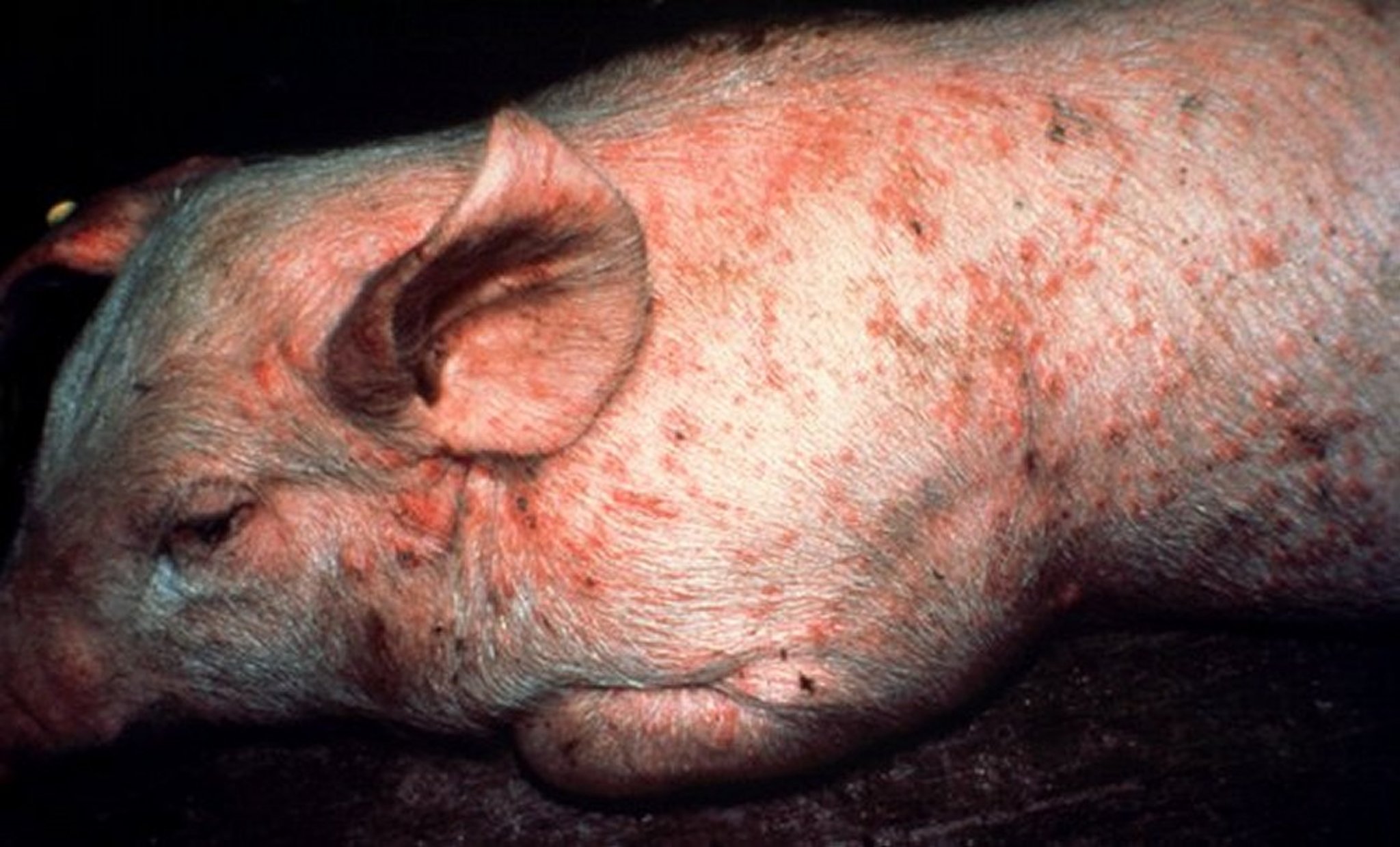
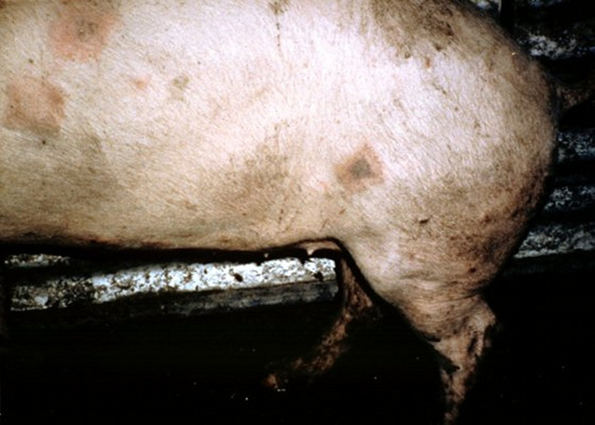
The acute and chronic forms of swine erysipelas may occur in sequence or separately. Pigs that succumb to the acute septicemic form may die suddenly without previous clinical signs. This form occurs most frequently in growing and finishing pigs. Outbreaks may be related to stressful conditions, such as extreme heat during transportation. Acutely infected pigs are depressed, febrile (104°–108°F [40°–42°C]), and reluctant to stand and move. Affected pigs squeal excessively when handled, require assistance to stand, and prefer to lie down soon after being forced to stand. Affected pigs may also walk stiffly on their toes and shift weight from limb to limb when standing. Anorexia and thirst are common, and febrile pigs will often seek wet, cool areas to lie down. Skin discoloration may vary from widespread erythema and purplish discoloration of the ears, snout, and abdomen, to diamond-shaped skin lesions almost anywhere on the body, but particularly on the lateral and dorsal regions. The lesions may occur as discrete, pink or purple areas of varying size that become raised and firm to the touch within 2–3 days of illness. They may disappear over the course of a week or progress to a more chronic type of lesion, commonly referred to as diamond skin disease. If untreated, necrosis and separation of large areas of skin can occur, and the tips of the ears and tail may become necrotic and slough.
Clinical disease is usually sporadic and affects individuals or small groups, but sometimes larger outbreaks occur. Mortality is variable (0–100%), and death may occur up to 6 days after the first signs of illness. Acutely affected pregnant sows may abort, probably due to the fever, and lactating sows may show agalactia. Untreated pigs may develop the chronic form of the disease, usually characterized by chronic arthritis, vegetative valvular endocarditis, or both. Such lesions may also be seen in pigs with no previous signs of septicemia. Valvular endocarditis is most common in mature or young adult pigs and is frequently manifest by death, usually from embolism or cardiac insufficiency. Chronic arthritis, the most common form of chronic infection, produces mild to severe lameness. Affected joints may be difficult to detect initially but eventually become hot and painful to the touch and later visibly enlarged. Dark purple, necrotic skin lesions that commonly slough may be seen. Mortality in chronic cases is low, but growth rate is retarded.
Lesions
At necropsy, acutely infected pigs may exhibit skin lesions, enlarged and congested lymph nodes, edematous and congested lungs, splenomegaly, and hepatomegaly. Petechial hemorrhages may be seen on the kidneys and heart.

Courtesy of Dr. Ranald D. A. Cameron.

Courtesy of Dr. Ranald D. A. Cameron.

Courtesy of Dr. John Prescott.
In chronic erysipelas, valvular endocarditis is seen as proliferative, granular growths on the heart valves, and embolisms and infarctions may develop. Arthritis may involve joints of one or more legs, and the intervertebral articulations may be involved. Affected joints may be enlarged, with proliferative, villous synovitis and increased viscosity of synovial fluid, inflammatory exudates, and thickening of the joint capsule. Proliferation and erosion of articular cartilage may result in fibrosis and ankylosis of the joint.
Diagnosis of Swine Erysipelas
Clinical signs and/or gross lesions
Response to antimicrobial therapy
Demonstration of the bacterium or DNA in tissues (bacterial culture and/or molecular tests)
Diagnosis of erysipelas is based on clinical signs, gross lesions, response to antimicrobial therapy, and demonstration of the bacterium or DNA in tissues from affected animals. Acute erysipelas can be difficult to diagnose in individual pigs showing only fever, poor appetite, and listlessness. However, in outbreaks involving several animals, the presence of skin lesions and lameness is likely to be seen in at least some cases and would support a clinical diagnosis. Rhomboid urticaria or diamond skin lesions are almost diagnostic when present; however, similar lesions can also be seen with classical swine fever virus infection, Actinobacillus suis septicemia, or the porcine dermatitis and nephropathy syndrome.
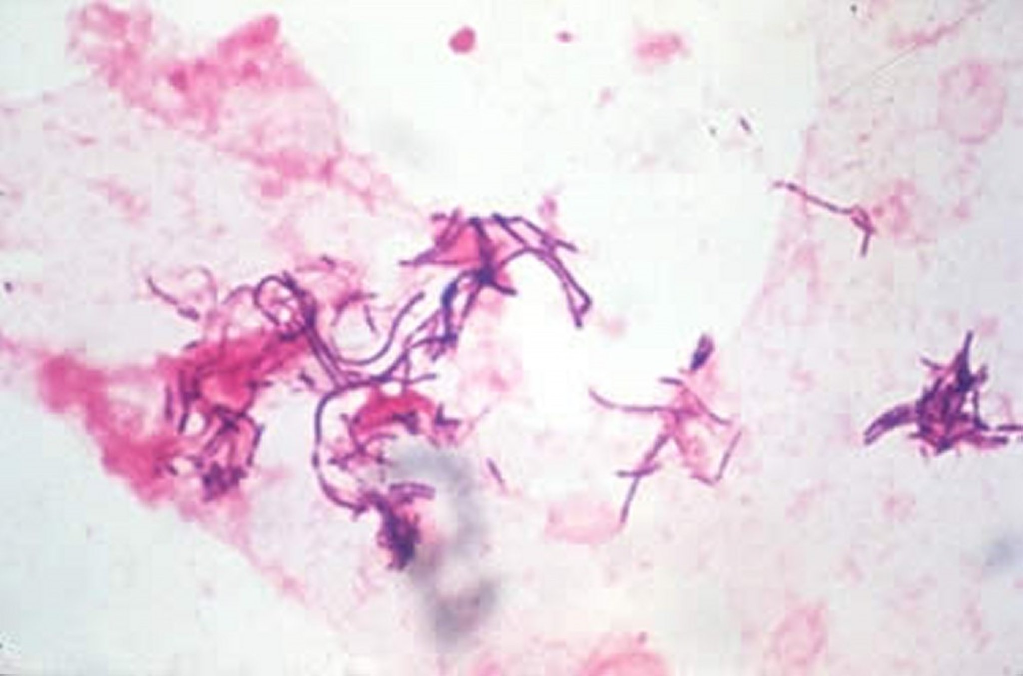
Isolation of E rhusiopathiae from blood of affected pigs, especially after enrichment, is possible in acute cases and helps establish a diagnosis. In addition, molecular methods capable of detecting E rhusiopathiae DNA in affected tissues or blood (ie, PCR assays) can also be used. Recently, immunohistochemical methods to demonstrate the organisms in formalin-fixed paraffin-embedded tissues have become available and are useful in cases when pigs have been treated with antimicrobials before sample submission. A rapid, positive response to penicillin therapy in affected pigs supports a diagnosis of acute erysipelas because of the sensitivity of the organism to penicillin.
Chronic erysipelas can be difficult to definitively diagnose. Arthritis and lameness, coupled with the presence of vegetative valvular endocarditis postmortem, may support a presumptive diagnosis of chronic erysipelas. However, these lesions can be caused by other infectious agents. A positive culture of valvular vegetations or demonstration of E rhusiopathiae DNA in the lesions by PCR is definitive for diagnosing chronic erysipelas.
Serologic tests cannot reliably diagnose erysipelas but can be useful to determine previous exposure or success of vaccination protocols, because antibody titers should increase after vaccination. For this purpose, ELISAs and complement fixation tests are available in selected laboratories.
Differential diagnoses to consider include conditions that can precipitate gross lesions suggestive of acute septicemia. Septicemic salmonellosis due to Salmonella Choleraesuis infection, classical swine fever due to pestivirus infection, and septicemia and endocarditis due to Streptococcus suis infection should be considered, based on similarity of lesions. Similar skin lesions can be found with porcine dermatitis and nephropathy syndrome caused by porcine circovirus, or infection with classical swine fever virus or Actinobacillus suis. Glasser’s disease due to Haemophilus parasuis infection and Mycoplasma hyosynoviae infection can precipitate similar changes in synovial tissues and joints of affected pigs.
Treatment of Swine Erysipelas
Preventive treatment through vaccination should be emphasized
Early treatment with appropriate antibiotics, particularly penicillin, generally leads to recovery
E rhusiopathiae is sensitive to penicillin. Ideally, affected pigs should be treated at 12-hour intervals for a minimum of 3 days, although longer durations of therapy may be necessary to resolve severe infections. On an economic basis, penicillin is the best choice for antibiotic therapy, but ampicillin and ceftiofur also yield satisfactory results in acute cases. When injecting large numbers of affected pigs is impractical, tetracyclines delivered in the feed or water may be useful. Fever associated with acute infections can be managed by administration of NSAIDs such as flunixin meglumine or by delivery of aspirin in the water. Erysipelas antiserum is described as an effective adjunct to antibiotic therapy in treating acute outbreaks but is not commonly available. Treatment of chronic infections is usually ineffective and not cost effective.
Prevention of Swine Erysipelas
Vaccination against E rhusiopathiae is very effective in controlling disease outbreaks on swine farms and should be encouraged. It may not be as effective in preventing chronic arthritis, however. Cessation of vaccination on some farms has been linked to disease outbreaks. Injectable bacterins and attenuated, live vaccines delivered via the water are available and provide extended duration of immunity. Optimal timing of vaccination may vary from farm to farm. When E rhusiopathiae is endemic in the production environment, vaccination should precede anticipated outbreaks. Susceptible pigs may be vaccinated before weaning, at weaning, or several weeks after weaning. Male and female swine selected for addition to the breeding herd should be vaccinated with a booster 3–5 weeks later. Thereafter, breeding stock should be vaccinated twice yearly. Vaccines should not be administered to animals undergoing antibiotic therapy, because antibiotics can interfere with the subsequent immune response to the vaccine.
Vaccination failures may occur in some herds due to management stresses that compromise the immune system of vaccinated pigs. The use of live vaccines may lead to clinical disease, particularly chronic erysipelas. Antigenic differences between serotypes in vaccines and serotypes circulating on farms could also result in incomplete immunity and disease outbreaks, but this is a rare event because there is thought to be good cross-protection among the major E rhusiopathiae strains infecting pigs.
In addition to vaccination, attention to sanitation and hygiene and elimination of pigs with clinical signs suggestive of erysipelas infection represent other viable methods that may help control the disease on swine farms.
Key Points
Erysipelas in swine is caused primarily by Erysipelothrix rhusiopathiae and has acute, subacute, and chronic manifestations.
Common clinical signs include characteristic diamond-shaped skin lesions, diffuse erythema, septicemia, and/or arthritis.
Pigs are often healthy carriers of the bacterium. Routine vaccination is effective at preventing acute disease, including animal losses and abattoir condemnations.


