


Most causes of dystocia in the mare are due to abnormal presentation, position, or posture. A dead or compromised fetus often is not properly positioned in the pelvic canal. Dystocia due to fetal–maternal disproportion or primary uterine inertia is rare in mares. A vaginal examination should be performed if the foal is not delivered within 30 minutes after rupture of the chorioallantois or if second-stage labor does not begin after >4 hours of obvious first-stage labor.
The initial examination may be performed in the standing mare. The perineum should be cleansed with povidone-iodine scrub and rinsed well with water. Efforts should be made to maintain hygiene at all times. A diagnosis of the presentation, position, and posture must be made, and a plan formulated to move the fetus into an anterior longitudinal, dorsosacral position with head, neck, and front limbs extended. Copious amounts of clean lubricant will ease repositioning and fetal expulsion. Excessive mechanical or manual traction should be avoided. Length of time spent on manipulations and any progress should be noted. Once the fetus is properly presented and positioned in the pelvic canal, if it cannot be delivered using traction exerted by two strong adults, the diagnosis should be reconsidered and plan of action altered. Compassion for the foaling mare must be considered in case management decisions.
Sedation can be provided by administration of xylazine (0.5–1 mg/kg, IV) and butorphanol (0.01–0.02 mg/kg, IV). If abdominal straining prevents adequate vaginal examination and fetal manipulation, epidural anesthesia may be administered using xylazine (0.17 mg/kg) and lidocaine (0.22 mg/kg) diluted to a total volume of 8 mL.
The front legs of the fetus should be identified and extended out the vulva. Obstetrical chains should be applied with one loop of the chain at the distal metacarpus and a second loop around the pastern to decrease trauma to the fetlock joint. The chains will aid manipulation and assist the application of traction. Once the head is exposed and the neck extended, it may be possible to intubate the fetus and administer low-flow oxygen using a portable oxygen tank or ventilate with a resuscitator (bag-valve mask). Proper rotation of the fetus may be facilitated by the mare getting up and down. After confirming that the fetus is in the proper orientation, manual traction can be applied, initially dorsally and then caudally. Once the foal’s chest is exposed, traction should be applied in a ventral caudal direction. Traction should be applied intermittently in rhythm with the mare’s abdominal contractions.
After the foal is delivered, an internal examination of the genital tract per vagina should be performed to identify any lacerations in the mare’s genital tract or the presence of a twin. A few low doses of oxytocin (5–10 IU, IV or IM) can be administered every 15–20 minutes to stimulate passage of the fetal membranes and uterine evacuation. Repair of primary perineal lacerations usually can be delayed until after foal heat, but the dorsal aspect of the vulvar lips should be temporarily sutured if pneumovagina/pneumometra develops.
Controlled Vaginal Delivery for Dystocia in Horses

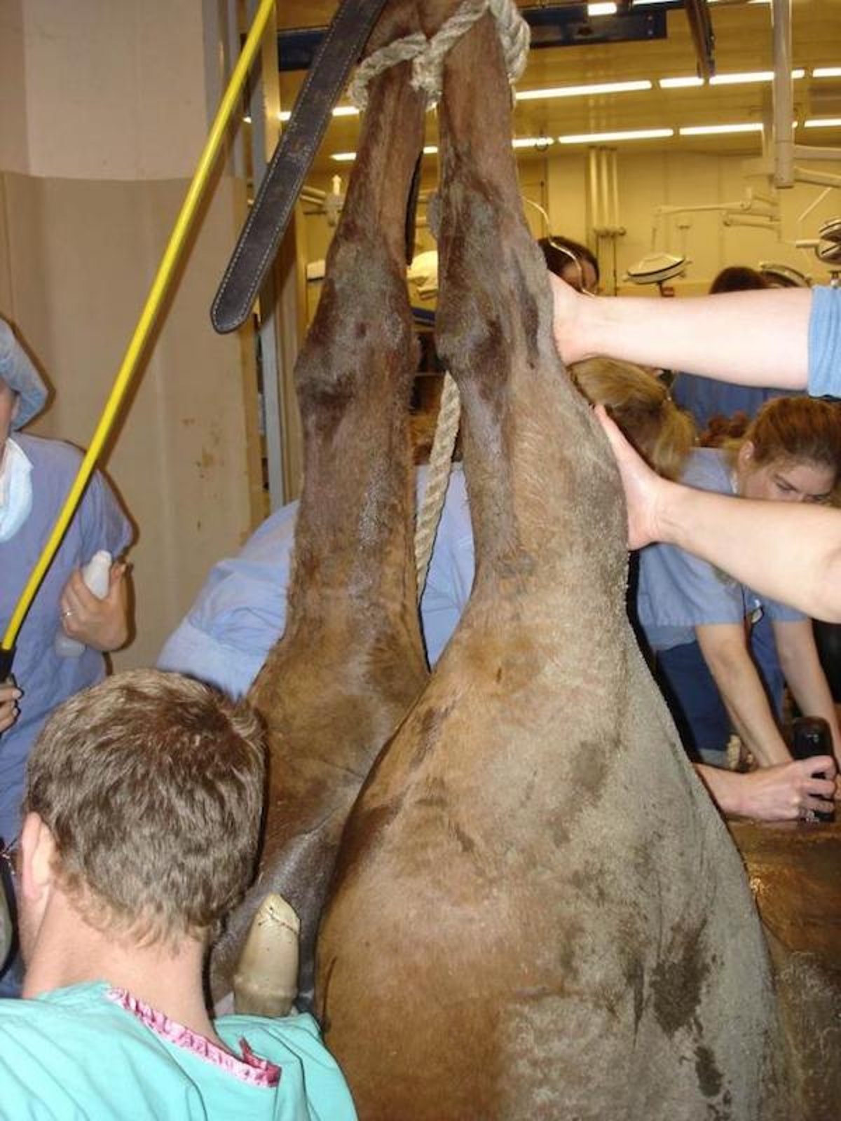
Courtesy of Dr. Patricia Sertich.
If resolution of the dystocia seems challenging or not possible in the standing mare because of the mare's straining or the orientation of the fetus, a controlled vaginal delivery should be considered. This is performed in an anesthetized mare. Field anesthesia can be accomplished by first heavily sedating with xylazine (1 mg/kg, IV) followed by diazepam (0.05–0.1 mg/kg, IV) and ketamine (2.2–2.5 mg/kg, IV). If gas anesthesia is available, routine induction and maintenance by inhalation will provide longer working time and greater relaxation. If general anesthesia and a controlled vaginal delivery are likely, epidural anesthesia should not be administered. The mare’s hindquarters are hoisted to allow the GI tract to move cranially in the abdomen, providing space to more readily perform fetal manipulations. It is prudent for managers of large breeding farms to have a designated location on the farm where a mare could be anesthetized and its hindquarters elevated (hobbles, winch, front end loader readily available) to hasten resolution of a dystocia.
Cesarean Section for Dystocia in Horses
If vaginal delivery attempts fail and surgical facilities are available, a decision should rapidly be made to deliver the foal by cesarean section to spare the mare's caudal genital tract from further trauma. Treatment for retained placenta should be initiated immediately after surgery. Barring complications, mares can usually be rebred 60 days after cesarean section.
Fetotomy for Dystocia in Horses
If surgical facilities are not available or the economic situation prevents referral to a surgical facility, a fetotomy may allow vaginal delivery of the fetus. In the mare, fetotomy is usually recommended only if fetal expulsion can be accomplished after one or two cuts. Care should be taken to avoid damage to the mare’s cervix and pelvic canal. Treatment for retained placenta should be initiated immediately after fetal delivery.
For More Information
Also see pet health content regarding the breeding and reproduction of horses.


