


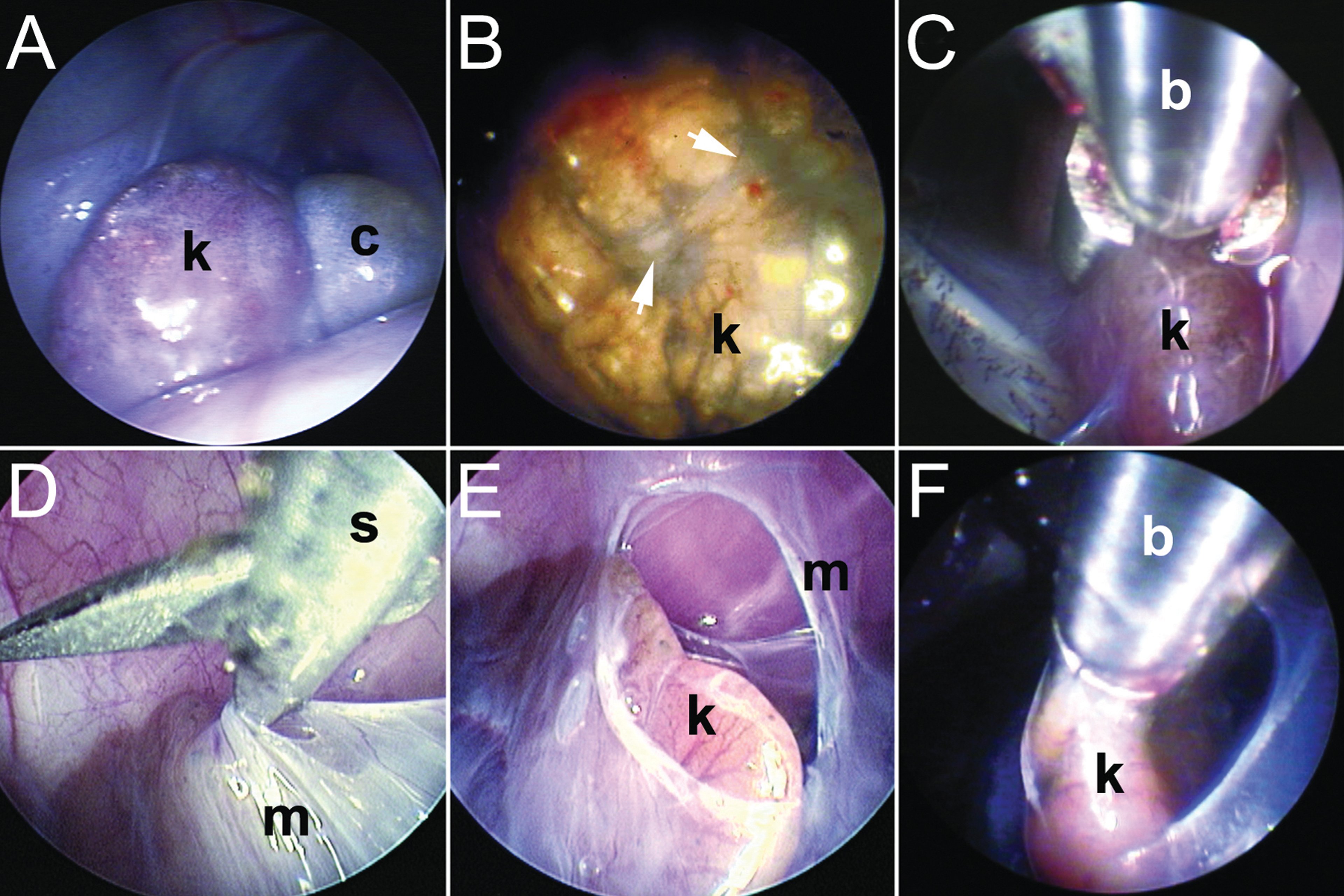
Endoscopic pathology and kidney biopsy. A) Enlarged kidney (k) with associated renal cyst (c) in a green iguana (Iguana iguana) that presented with anorexia and cachexia. Radiography and ultrasonography confirmed renomegaly, and plasma biochemistry revealed reversed calcium:phosphorus ratio and increased uric acid. Endoscopic biopsy confirmed the diagnosis of glomerulonephrosis with renal gout and mineralization (left lateral coelioscopy). B) Abnormal kidney (k) with fibrous bands (arrows) in a female Greek tortoise (Testudo graeca) that presented with anorexia, lethargy, and polyuria. Diagnostic imaging and clinicopathology were unremarkable; however, renal biopsy confirmed the diagnosis as severe tubulonephrosis (left prefemoral approach). C) Biopsy of an iguanid kidney (k) using 1.7-mm biopsy forceps (b) (right lateral coelioscopy). D) Incising the caudodorsal coelomic membrane (m) of a yellow-bellied slider (Trachemys scripta scripta) using 1.7-mm endoscopy scissors (s) to gain access to the retrocoelomic kidney (left prefemoral approach). E) View of the incised coelomic membrane (m) revealing the kidney (k) (left prefemoral approach). F) Biopsy forceps (b) being passed through the incision to collect a biopsy from the kidney (k) (left prefemoral approach).
Courtesy of Dr. Stephen Divers.