

Upper leg lameness is more common in beef cattle compared to dairy cattle. Both diagnosis and treatment are more difficult than in cases of lower limb lameness. In general, diagnosing the cause of upper leg lameness requires ancillary testing such as radiography and ultrasonography. Unless the animal is valuable breeding stock or a show animal, surgical treatment for upper leg lameness is typically not pursued as the overall prognosis is poor. The majority of surgical procedures for upper limb lameness are referral procedures or in-clinic procedures requiring advanced training. Conservative treatment consisting of rest and NSAIDS is typically considered the primary treatment. If the animal does not respond to conservative treatment, euthanasia or culling is usually recommended, depending on the ambulatory status of the animal.
Hock and Knee Injuries in Cattle

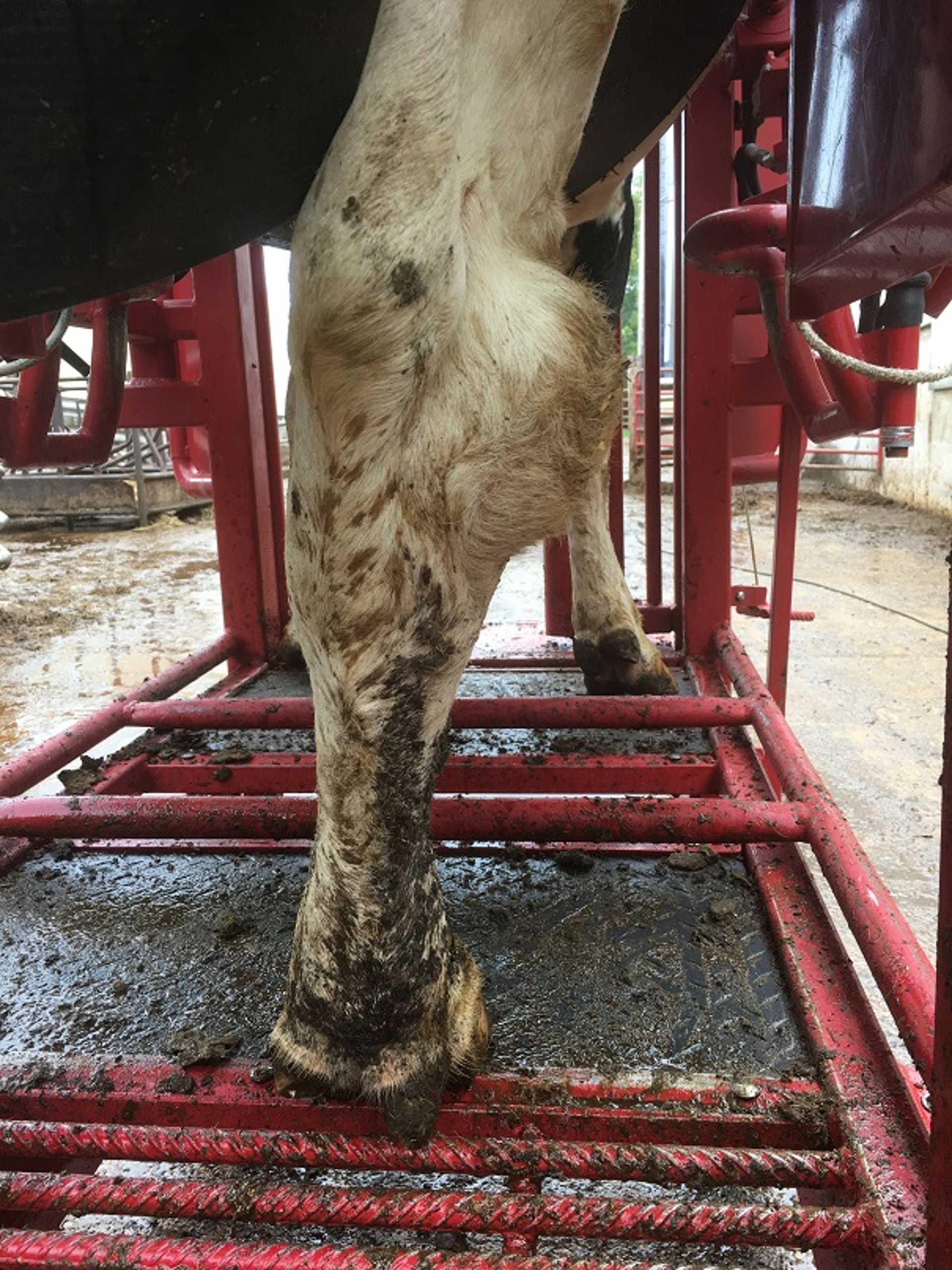
This photograph of the right hind leg of a cow shows severe lateral swelling of the hock.
Courtesy of Dr. Gerard Cramer.
Hock and knee injuries often present as hair loss, swelling, and/or broken skin in the region of the hock (tarsus) and knee (carpus). Severe lesions in these leg regions are associated with lameness. These injuries are more common in confined/housed dairy cattle, and their assessment at the herd level is part of welfare quality assurance programs.
Pathogenesis
Hock and knee injuries generally arise from animals rubbing on hard or abrasive surfaces in their resting area. These injuries are more common in cows housed in free-stall or tie-stall dairies, predisposed by suboptimal stall management and stall design, base, and bedding. Hard stall bases without sufficient bedding or with abrasive bedding, improper stall dimensions, and wet lying surfaces contribute to these injuries. The hocks and knees rub repeatedly as a cow moves in her stall or transitions between lying and standing, creating friction that results in injuries to these upper-leg regions over time.
Diagnosis
Hock and knee injuries vary in severity, ranging from areas of hair loss or broken skin, scabs, or minor swelling to areas with major swelling and/or open wounds with purulent discharge.
Prevention
Hock and knee injuries can be prevented by improving cow comfort. Cows should be housed in stalls that are appropriately sized and configured for the animals in the herd, and the stalls should be deeply bedded with nonabrasive bedding material.
Treatment
The best treatment for animals with hock and knee injuries is to remove the inciting cause of injury or move the animal to a deeply bedded lying area. Cows with open lesions may require supportive wound care; they will not recover, however, if the environmental cause of the injury is not addressed.
Stifle Injuries in Cattle
Stifle injuries are a common cause of upper-leg hind-limb lameness in cattle and often involve the cruciate ligaments, the meniscus, or the patella.
Pathogenesis
Most stifle injuries in adult cows and bulls result from traumatic events such as slipping, falling, mounting, or exertion in downer animals. In older animals, degenerative joint disease may also be a contributing factor.
Diagnosis
Stifle injuries typically manifest as nonspecific hind-limb lameness. A physical exam should be used to rule out lower-limb injuries or problems and to localize the issue to the stifle. With stifle injuries, joint effusion, pain, and crepitus in the joint are common signs. Hearing a clicking sound and eliciting a cranial drawer sign are diagnostic for cranial cruciate ligament injury. Meniscus injury presents as nonspecific stifle lameness and can accompany cranial cruciate injury. Patellar luxation presents similar to femoral nerve paralysis in calves; in both conditions, the animal cannot extend the stifle. Upward fixation of the patella results in a characteristic extension of the limb and is accompanied by locking and unlocking of the patella on the medial trocheal ridge. This condition is rare and is more common in bulls.
Prevention
To prevent stifle injuries, the focus is to provide secure footing within the animal’s housing environment to avoid issues with slipping and falling.
Treatment
Stifle injuries can be treated; usually, however, the prognosis is guarded to poor. Conservative treatment is comprised of confinement and prolonged NSAID administration. For valuable animals, surgical options are available.
Hip Injuries in Cattle
The most common injury to the hip is a coxofemoral luxation, in either the craniodorsal or the cranioventral direction. Traumatic fractures of the tuber coxae or femoral head can also occur.
Pathogenesis
Hip injuries result from traumatic events such as falling, slipping, or bumping into hard objects in the environment.
Diagnosis

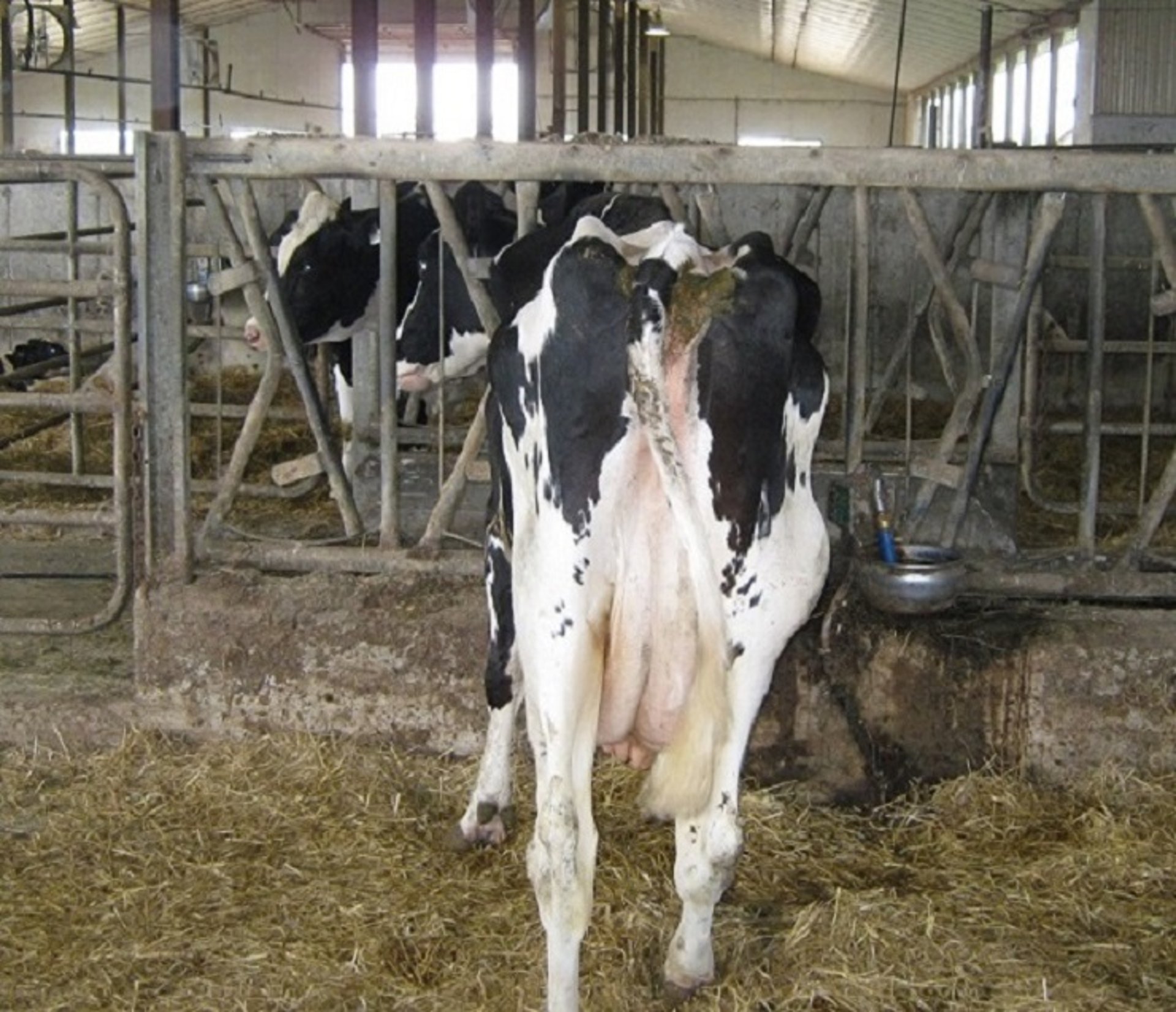
The cow in this photograph has a right hip injury, as the swelling and dropped hip appearance indicate.
Courtesy of Dr. Gerard Cramer.
Coxofemoral luxations can present as a dropped-hip appearance if the animal is standing. Cows suffering from cranioventrally displaced luxation usually present as down animals. Fractures can be palpated if they affect the tuber coxae; other fractures typically cannot be felt.
Prevention
Prevention of hip injuries focuses on preventing traumatic events, which typically result from aggressive handling practices, mounting, or slippery flooring.
Treatment
For most hip injuries, the prognosis is guarded; it is more favorable in younger animals. Usually, and especially for femoral head fractures, euthanasia is the most suitable course of action. However, some surgical approaches involving an open reduction can be successful for coxofemoral luxations. Conservative treatment of coxofemoral luxations has also been reported to be successful if done within 48 hours of the occurrence of the injury.
Noninfectious Joint Disease in Cattle
A variety of noninfectious joint diseases exist, including osteochondrosis, degenerative joint disease and osteoarthritis. In both beef and dairy cattle, noninfectious joint disease is rarely diagnosed and occurs infrequently.
Pathogenesis
Because of their infrequent occurrence, noninfectious joint diseases have not been studied extensively; however, the pathogenesis of these disorders is thought to be similar to the pathogenesis of the same types of disorders in other species.
Diagnosis


This photograph shows joint swelling around the fetlock of a heifer.
Courtesy of Dr. Gerard Cramer.
Most joint diseases present with a variable extent of lameness and joint effusion. Osteochondrosis and degenerative joint disease are commonly found in young, growing animals; osteoarthritis is more common in older animals. Further diagnostic tests, such as radiography and arthrocentesis, are required to differentiate these joint diseases more fully.
Prevention
Because of their unclear pathogenesis, strategies to prevent noninfectious joint diseases are limited.
Treatment
Conservative treatment for joint disease—restricting activity and administering NSAIDs—is often unrewarding. Surgical approaches, including arthroscopy, have been used successfully to treat some of these conditions.
Septic Arthritis in Cattle
Septic arthritis is the most common reason for swelling in one or more joints in younger animals.
Pathogenesis
Septic arthritis results when microorganisms enter the joint cavity either because of sepsis or through penetrating wounds. The resulting inflammatory process in the joint increases joint effusion. Persistent infections result in further damage to the joint.
Diagnosis
Septic arthritis typically causes severe, acute pain and lameness. The affected joint(s) are often swollen, warm, and painful to the touch. In distal limbs when a single joint is affected, the process is typically traumatic in origin. Polyarthritis typically has a systemic origin (ie, navel ill, mycoplasma), and the inciting cause should be determined. Arthrocentesis of synovial fluid can be used to confirm the diagnosis.
Prevention
Prevention of septic arthritis should focus on preventing sepsis and penetrating injuries near the joints. Common causes of sepsis in calves include mycoplasma and navel infections.
Treatment
Treatment of septic arthritis requires treating the sepsis, as well as managing the inflammation and pain. Extended administration of antimicrobials and NSAIDs, along with joint lavage, is necessary.
Muscular Ruptures in Cattle
Muscular ruptures are most commonly partial or complete tears of the gastrocnemius or fibularis tertius muscle.
Pathogenesis
Muscular ruptures are typically acute because of direct trauma or physical exertion when the animal attempts to get up while down for another condition.
Diagnosis
Animals with a gastrocnemius tear typically are unable to rise, and the hock touches the ground when the animal attempts to get up. Animals with a fibularis tertius tear typically can flex the stifle with the hock fully extended. Typically, the affected muscle is firm and swollen.
Prevention
Prevention of muscular injuries should focus on providing solid footing, especially for down animals.
Treatment
Treatment of muscular injuries, especially gastrocnemius tears, is unrewarding. Typically, euthanasia is the most appropriate course of action. In young animals with tears of the fibularis tertius, restricting activity may enable recovery.
For More Information
van Amstel SR. Corkscrew claw. Vet Clin North Am Food Anim Pract. 2017;33:351-364. doi:10.1016/j.cvfa.2017.02.010
Whay HR, Shearer JK. The impact of lameness on welfare of the dairy cow. Vet Clin North Am Food Anim Pract. 2017;33:153-164. doi:10.1016/j.cvfa.2017.02.008
Wilson JP, Randall LV, Green MJ, et al. A history of lameness and low body condition score is associated with reduced digital cushion volume, measured by magnetic resonance imaging, in dairy cattle. J Dairy Sci. 2021;104:7026-7038. doi:10.3168/jds.2020-19843
Schlageter-Tello A, Bokkers EAM, Koerkamp PWGG, et al. Manual and automatic locomotion scoring systems in dairy cows: a review. Prev Vet Med 2014;116:12-25. doi:10.1016/j.prevetmed.2014.06.006
Van Nuffel A, Zwertvaegher I, Pluym L, et al. Lameness detection in dairy cows: part 1. How to distinguish between non-lame and lame cows based on differences in locomotion or behavior. Animals 2015;5:838–860. doi:10.3390/ani5030387
Davis-Unger J, Schwartzkopf-Genswein KSG, Pajor EA, et al. Prevalence and lameness-associated risk factors in Alberta feedlot cattle. Transl Anim Sci. 2019;3:595-606. doi:10.1093/tas/txz008
Terrell SP, Reinhardt CD, Larson CK, Vahl CI, Thomson DU. Incidence of lameness and association of cause and severity of lameness on the outcome for cattle on six commercial beef feedlots. J Am Vet Med Assoc. 2017;250:437-445. doi:10.2460/javma.250.4.437
Solano L, Barkema HW, Pajor EA, et al. Prevalence of lameness and associated risk factors in Canadian Holstein-Friesian cows housed in freestall barns. J Dairy Sci. 2015;98(10):6978-6991. doi:10.3168/jds.2015-9652
Solano L, Barkema HW, Mason S, Pajor EA, LeBlanc SJ, Orsel K. Prevalence and distribution of foot lesions in dairy cattle in Alberta, Canada. J Dairy Sci. 2016;99:6828-6841. doi:10.3168/jds.2016-10941
Endres MI. The relationship of cow comfort and flooring to lameness disorders in dairy cattle. Vet Clin North Am Food Anim Pract. 2017;33:227–233. doi:10.1016/j.cvfa.2017.02.007
ICAR. ICAR Claw Health Atlas. Accessed March 21, 2023. https://www.icar.org/ICAR_Claw_Health_Atlas.pdf
Jacobs C, Beninger C, Hazlewood GS, Orsel K, Barkema HW. Effect of footbath protocols for prevention and treatment of digital dermatitis in dairy cattle: a systematic review and network meta-analysis. Prev Vet Med. 2019;164:56-71. doi:10.1016/j.prevetmed.2019.01.011
Plummer PJ, Krull A. Clinical perspectives of digital dermatitis in dairy and beef cattle. Vet Clin North Am Food Anim Pract. 2017;33:165-181. doi:10.1016/j.cvfa.2017.02.002
Desrochers A. Diagnosis and prognosis of common disorders involving the proximal limb. Vet Clin North Am Food Anim Pract. 2017;33:251–270. doi:10.1016/j.cvfa.2017.03.002
Shearer JK, van Amstel SR. Traumatic lesions of the sole. Vet Clin North Am Food Anim Pract. 2017;33:271-281. doi:10.1016/j.cvfa.2017.02.001
Guard CL, Peek SF, Fecteau G. Musculoskeletal disorders. In: Peek SF, Divers TJ, eds. Rebhun’s Diseases of Dairy Cattle. 3rd ed. Elsevier; 2018:553-604.
Anderson DE, Desrochers A, van Amstel SR. Surgical procedures of the distal limb for treatment of sepsis in cattle. Vet Clin North Am Food Anim Pract. 2017;33:329-350. doi:10.1016/j.cvfa.2017.02.011
Nichols, S. and H. Lardé. 2014. Noninfectious joint disease in cattle. The Veterinary clinics of North America. Food animal practice 30:205-223, vii. 10.1016/j.cvfa.2013.11.010
Stoddard GC, Cramer G. A review of the relationship between hoof trimming and dairy cattle welfare. Vet Clin North Am Food Anim Pract. 2017;33:365-375. doi.org/10.1016/j.cvfa.2017.02.012
Kofler J. Computerised claw trimming database programs as the basis for monitoring hoof health in dairy herds. Vet J. 2013;198:358-361. doi:10.1016/j.tvjl.2013.06.009
Raven ET, Haalstra RT, Peterse DJ, Lurvink A. Cattle Footcare and Claw Trimming: The Origin and Prevention of the Necrotising Inflammations of the Corium (Ulcerations of the Claw). Farming Press; 1985.
van Amstel SR. Corkscrew claw. Vet Clin North Am Food Anim Pract. 2017;33:351-364. doi:10.1016/j.cvfa.2017.02.010
Wilson JP, Randall LV, Green MJ, et al. A history of lameness and low body condition score is associated with reduced digital cushion volume, measured by magnetic resonance imaging, in dairy cattle. J Dairy Sci. 2021;104:7026-7038. doi:10.3168/jds.2020-19843
Jacobs C, Beninger C, Hazlewood GS, Orsel K, Barkema HW. Effect of footbath protocols for prevention and treatment of digital dermatitis in dairy cattle: a systematic review and network meta-analysis. Prev Vet Med. 2019;164:56-71. doi:10.1016/j.prevetmed.2019.01.011
Plummer PJ, Krull A. Clinical perspectives of digital dermatitis in dairy and beef cattle. Vet Clin North Am Food Anim Pract. 2017;33:165-181. doi:10.1016/j.cvfa.2017.02.002


