


Lameness in pigs is of increasing interest in North American swine production. Mycoplasma hyosynoviae in particular has received more attention, in part because of more intensive diagnostic efforts. Metabolic bone disease has also been an area of focus because problems have arisen after more complex formulations for gestation and growing-finishing diets.
Lameness Due to Infectious Arthritis in Growing-Finishing Pigs
Most of the bacteria discussed under Common Bacterial Causes can cause or contribute to arthritis in individual pigs at any age, including growing-finishing pigs and adults. Outbreaks of bacterial infections are more common when pig sources or groups are commingled, mixed, moved, or overcrowded; housed in cold and drafty or hot environments; or subjected to abrupt changes in management and feed or feeding.
With most endemic bacteria, the upper respiratory tract (also vagina, skin, environment) of sows may be a source of infection to a few suckling piglets that then may carry the organism and shed to cohorts after weaning or as maternal antibody wanes. Other sources of infection include older pigs, the commingling of pigs with other pig sources, and lateral introductions due to lax biosecurity.
Erysipelothrix rhusiopathiae
Outbreaks of lameness due to Erysipelothrix rhusiopathiae infection (erysipelas) are most common in the growing-finishing stage. Sudden deaths of a few peracutely affected pigs may precede acute clinical signs of fever, anorexia, and reluctance to move. Rhomboid lesions (diamond skin disease) occur 2–3 days after acute infection in some pigs.
Early aggressive treatment with injectable antimicrobials (eg, penicillin) and vaccination with a killed bacterin is warranted. Erysipelas antisera can be an effective adjunct where available.
See the Common Bacterial Causes discussion above; also see Swine Erysipelas.
Glaesserella parasuis
Glaesserella parasuis is another organism capable of causing acute outbreaks of infection in pigs, manifested as arthritis, fibrinous polyserositis, or meningitis. Risk factors and microecology are similar to those associated with other endemic bacteria.
See the Common Bacterial Causesdiscussion above; also see Glässer Disease.
Mycoplasma hyosynoviae
M hyosynoviae is commonly endemic in swine herds; however, it has emerged as an important infectious cause of lameness in pigs 10–30 weeks old. As colostral immunity wanes at 6–10 weeks of age, more shedding occurs and more pigs are susceptible to infection. Morbidity is usually low to moderate; however, it can be as high as 50% in naive or compromised pigs. Mortality is very low.
An acute, usually hind limb, lameness lasting up to 10 days develops in groups of growing-finishing pigs or selected replacement stock. Arthritis may be exacerbated by trauma or stress, and pigs exhibit pain in major joints (eg, elbows, stifles, and hocks) that may also develop soft, fluctuant swellings.
Necropsy in cases of M hyosynoviae infection in pigs reveals lesions restricted to the joints, especially the stifles and hocks. Affected joints contain excess, clear, yellow synovial fluid that may have fibrin flakes, as well as edematous, yellowish synovium with nonsuppurative synovitis and villous hypertrophy microscopically. Articular surfaces of bone and the periarticular tissues usually are unaffected.
M hyosynoviae can be detected in joints at slaughter or necropsy in pigs with degenerative joint disease (osteochondrosis); in such cases, however, it is considered a secondary infection or coinfection rather than a causative agent.
Diagnosis of lameness in pigs due to M hyosynoviae is based on the age of onset of typical clinical signs, including lameness in one or more legs that may be accompanied by fluctuating swelling around joints. Typically, pigs are afebrile and there is no evidence of pneumonia, pleuritis, or peritonitis.
Definitive diagnosis of M hyosynoviae infection that is based on detection of the organism and histological examination requires the collection of samples of synovium and synovial fluid from untreated pigs within 3–4 days of the onset of clinical signs. However, M hyosynoviae can be detected from healthy joints and is not always recovered from affected joints.
Lack of a response to penicillin in acute cases has been used to differentiate M hyosynoviae infection from erysipelas. Unlike polyarthritis caused by Mycoplasma hyorhinis, M hyosynoviae infection responds well to treatment with tylosin and lincomycin if they are administered promptly, and tiamulin or tetracycline may be effective where allowed.
Mycoplasmal arthritis may exacerbate clinical signs associated with degenerative joint disease and osteoarthrosis, and vice versa.
Lameness Due to Osteomyelitis in Growing-Finishing Pigs
Osteomyelitis can occur in pigs of any age. If the integument is damaged, sepsis develops and a suppurative lesion extends to the periosteum and bone. Alternatively, organisms can invade bone from the synovium of infected joints.
Many factors can increase susceptibility to osteomyelitis, including the following:
poor processing or injection techniques that initiate abscesses that may extend into adjacent bone
subclinical microfractures of bone or growth plates associated with metabolic bone disease
disruption of the integrity of the hoof wall that initiates cellulitis and osteomyelitis of a phalangeal bone
ear and flank biting wounds
tail biting that results in local infection that ascends the spinal canal and leads to epidural abscesses that can invade and affect vertebral bodies
Lesions and clinical signs of osteomyelitis may develop slowly. Depending on the site of infection, the pig may become ataxic and, ultimately, paralyzed in the pelvic limbs. If bones or joints of a limb are affected, the condition is usually chronic and the pig becomes three-legged lame. Young pigs cease to grow.
At necropsy, cream or green caseous pus is evident at the site of the osteomyelitic lesion. With infection by Trueperella pyogenes, there are abundant pockets of green, semiliquid pus.
Other organisms isolated from osteomyelitic abscesses may include streptococci, staphylococci, and enterobacteria. Treatment is not usually feasible, and pigs should be culled for humane reasons. When applicable, however, hygiene can be improved, and problems such as tail biting can be controlled or prevented.
Lameness Due to Osteochondrosis or Osteoarthrosis in Growing-Finishing Pigs
Lameness associated with osteochondrosis or osteoarthrosis may become clinically relevant by the time a pig is 4–6 months old. The major ramifications, however, are in gilts, sows, and boars (see Lameness in Breeding Gilts, Sows, and Boars).
Lameness Due to Rickets in Growing-Finishing Pigs
Rickets is a deficiency disease that affects growing animals during the period of skeletal growth. It is usually characterized by soft and deformed bones or growth plates that are the result of a failure to assimilate and use calcium and phosphorus normally, often associated with an inadequate amount of vitamin D.
Rickets affects rapidly growing, young pigs with a clinical onset of typical appearance (soft bones, bent limbs) at ~10 weeks old. However, histological lesions and effects of metabolic bone disease can be detected in pigs as young as neonates. Morbidity is high, and affected pigs become crippled, anorectic, and unthrifty.
In rickets cases, limbs are stunted and bowed, joints are swollen, and the head may seem disproportionately large. Long bones of the limbs can spontaneously fracture, making the pig severely lame and unwilling to move. Ribs may fracture. Some pigs develop posterior paresis and sit on the ground if vertebral bodies fracture and damage the spinal cord.
On necropsy, bones from pigs with rickets should be dissected to determine whether there are any fractures or healing fractures, particularly in the ribs, humeri, and femurs. The costochondral junctions of most ribs are enlarged to form a rachitic rosary (see rickets photograph), and ribs may bend with moderate manual force and break without a snap.

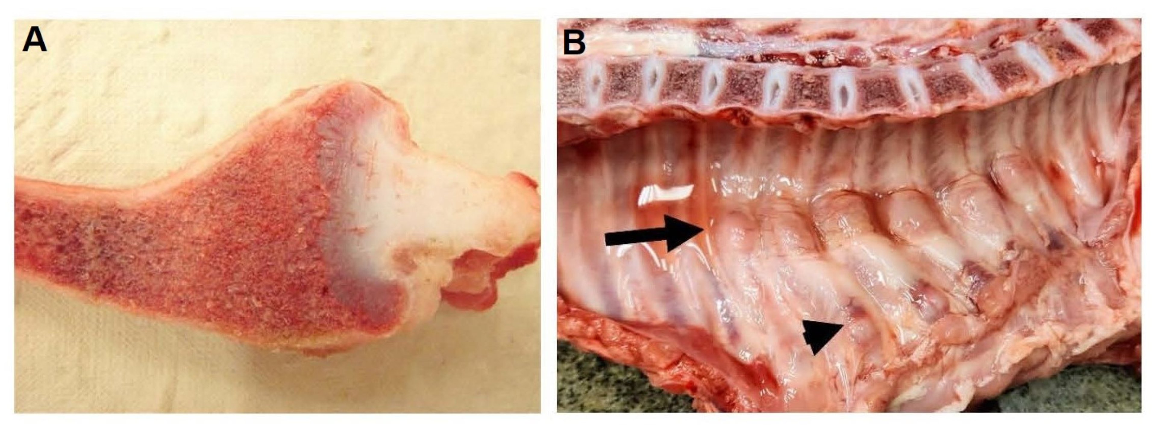
Courtesy of the Iowa State University Veterinary Diagnostic Laboratory photo archive.
Bone remodeling in rickets is inadequate, and radiographs show that long bones and ribs are poorly mineralized. Failure of calcification and endochondral ossification results in thickened, irregular growth plates and epiphyseal growth cartilages in which hemorrhages may be observed grossly if slab sections of the ends of long bones are cut on a band saw. In chronic cases of rickets, bones can be cut with a knife.
Bone analysis can confirm loss of normal density and ash (calcium, phosphorus) content. A sudden increase in carcass condemnations or partial condemnations because of fractured limb bones, ribs, or vertebrae at slaughter should trigger a nutritional investigation of the ration.
Ration analysis is useful in diagnosing rickets, but the nutritional error may have been corrected by the time testing is performed. Current batches of feed may have been mixed correctly or with different lots of ingredients, thus making it difficult to link cause and effect. Keeping frozen samples of each batch of feed for retrospective analysis is a good practice.
Although rations can be corrected and vitamin D given parenterally, there is no effective treatment for rickets, and attempts to rear large numbers of affected pigs have been economically disastrous. Culling affected pigs may thus be the most cost-effective alternative.
Lameness Due to Foot Disorders in Growing-Finishing Pigs
On occasion, growing-finishing pigs have overgrown claws or bruises and cracks in the wall or sole of the hoof. The floor type and condition are perhaps the most important factors affecting whether lesions develop or resolve.
Floors with wide spaces between slats allow digits to fall between the slats, causing damage.
Floors kept too wet can soften the hoof wall, making it more prone to trauma.
Floors that are too smooth destroy the balance between growth and wear of the horn.
Floors that are too rough damage the hoof wall, coronary band, or skin above the hoof, allowing infectious agents to penetrate the foot or adjacent joints, resulting in abscess formation.
An absolute or intermittent deficiency of biotin in pigs results in weak, flaky keratin that makes hoof walls susceptible to cracking. Flaky skin accompanies hoof lesions and generally leads to poor reproductive performance in the herd. As gilts are prepared for breeding, supplementing biotin may be helpful. Recommended inclusion rates of biotin are 250–400 mcg/kg complete feed.
Trace mineral deficiencies or imbalances can also contribute to compromised hoof wall and heel epidermis formation in pigs. Selenium toxicosis can cause coronary band swelling and necrosis, in addition to more generalized signs, such as anorexia or even paralysis. Selenium or ergot toxicoses can result in hoof sloughing in pigs.
Lameness Due to Nutritional Myopathy in Growing-Finishing Pigs
In contemporary systems with adequate ration preparation and storage, nutritional myopathy in pigs is rare. Unexpected deaths are more typical of selenium and/or vitamin E deficiency in swine than is nutritional myopathy; however, sometimes pigs are found recumbent and unable to rise and walk.
At necropsy, a variety of pathological changes may be evident in cases of nutritional myopathy in pigs, including pale muscle masses; however, epicardial hemorrhages (mulberry heart disease) and a pale, scarred liver with an uneven surface (hepatosis dietetica) are more common signs. Prevention includes supplementation of the ration with selenite to the legal limit of 0.3 ppm selenium, as well as providing supplemental vitamin E.
Key Points
As in all other stages of development, infectious arthritis due to agents such as Mycoplasma, Glaesserella, and Erysipelothrix is a common cause of lameness in growing-finishing pigs.
Osteochondrosis can manifest as swollen epiphyses or joints and cause lameness in rapidly growing pigs.
Rickets is a common, preventable nutritional bone disease of young pigs; feed and bone mineral analyses are required for diagnosis.
For More Information
Madson DM, Ensley SM, Gauger PC, et al. Rickets: case series and diagnostic review of hypovitaminosis D in swine. J Vet Diagn Invest. 2012;24(6):1137-1144. doi:10.1177/1040638712461487


