


The immediate effect of injury of a peripheral nerve is a variable degree of dysfunction, depending on the severity of the injury. The mildest form of injury is neuropraxia, which temporarily disrupts function with minimal morphologic alterations in the nerve. Axonotmesis is disruption of axons without disruption of the surrounding connective tissue of the nerve. The most severe form of injury is neurotmesis, which is complete severance of the nerve. With both axonotmesis and neurotmesis, there is subsequent degeneration of the axons distal to the injury site and in a portion of the nerve proximal to the injury site.
Diagnosis of peripheral nerve injuries is based on the history and clinical assessment of the motor and sensory function of the affected nerve(s). Electromyography often helps identify denervated muscles 5–10 days after injury. Nerve conduction studies may also be useful in diagnosis.
Prognosis is guarded. With neuropraxia, complete recovery usually occurs within 3 weeks. For function to return after axons are disrupted (axonotmesis, neurotmesis), the nerve must regenerate from the point of injury all the way to the innervated muscle. The growth rate of regenerating axons in the distal stump is 1–3 mm/day. Recovery is unlikely if the severed axons are substantially separated or if scar tissue interferes with axonal growth. Although various anti-inflammatory drugs have been recommended for traumatic nerve injuries, there is little evidence of benefit.
Surgery to appose the nerve stumps should be performed promptly in cases in which the nerve has been sharply transected. In instances of blunt trauma, surgical exploration and excision of scar tissue may help. Surgery is often successful in horses with fibrous compression of the suprascapular nerve. Long-term management consists of physical rehabilitation to minimize muscle atrophy and decreased mobility of joints. Bandages, splints, or various other methods of external coaptation may be necessary to help protect the affected limb.
Brachial Plexus Avulsion in Animals
Traumatic injury to the C6–T2 nerve roots that innervate the thoracic limb can lead to brachial plexus avulsion in dogs, cats, and birds. With severe extension or abduction of the limb, the nerve roots stretch or tear from their attachment to the spinal cord. Clinical signs vary with the extent of root involvement. Complete avulsion results in flaccid paralysis of the limb, anesthesia distal to the elbow, ipsilateral Horner syndrome, and ipsilateral loss of the cutaneous trunci (panniculus) reflex. The injured animal bears little or no weight on the limb and drags the dorsal surface of the paw on the ground. Sensation to the ventral surface of the paw is spared if only the cranial nerve roots are affected. Avulsion of the caudal nerve roots causes loss of sensation on the caudal surface of the limb, with variable loss on the cranial surface.
There is no treatment, and with complete avulsion, the prognosis is poor. Amputation of the limb may be necessary because of damage from dragging or self-mutilation. Recovery is possible in mild cases in which the roots are contused rather than avulsed.
Calving Paralysis in Cattle
Calving paralysis is seen in heifers with oversized fetuses. It has previously been attributed to bilateral compression of the obturator nerve, but damage to the sixth lumbar nerve root, which contributes to the obturator and sciatic nerve, likely accounts for most of the paralysis. Ischemic necrosis of muscles secondary to compression and rupture of muscles during attempts to rise also contribute to the paraparesis. Additionally, metabolic derangements, such as hypocalcemia, may complicate the clinical picture. (Also see Bovine Secondary Recumbency.)
Facial Nerve Trauma in Animals

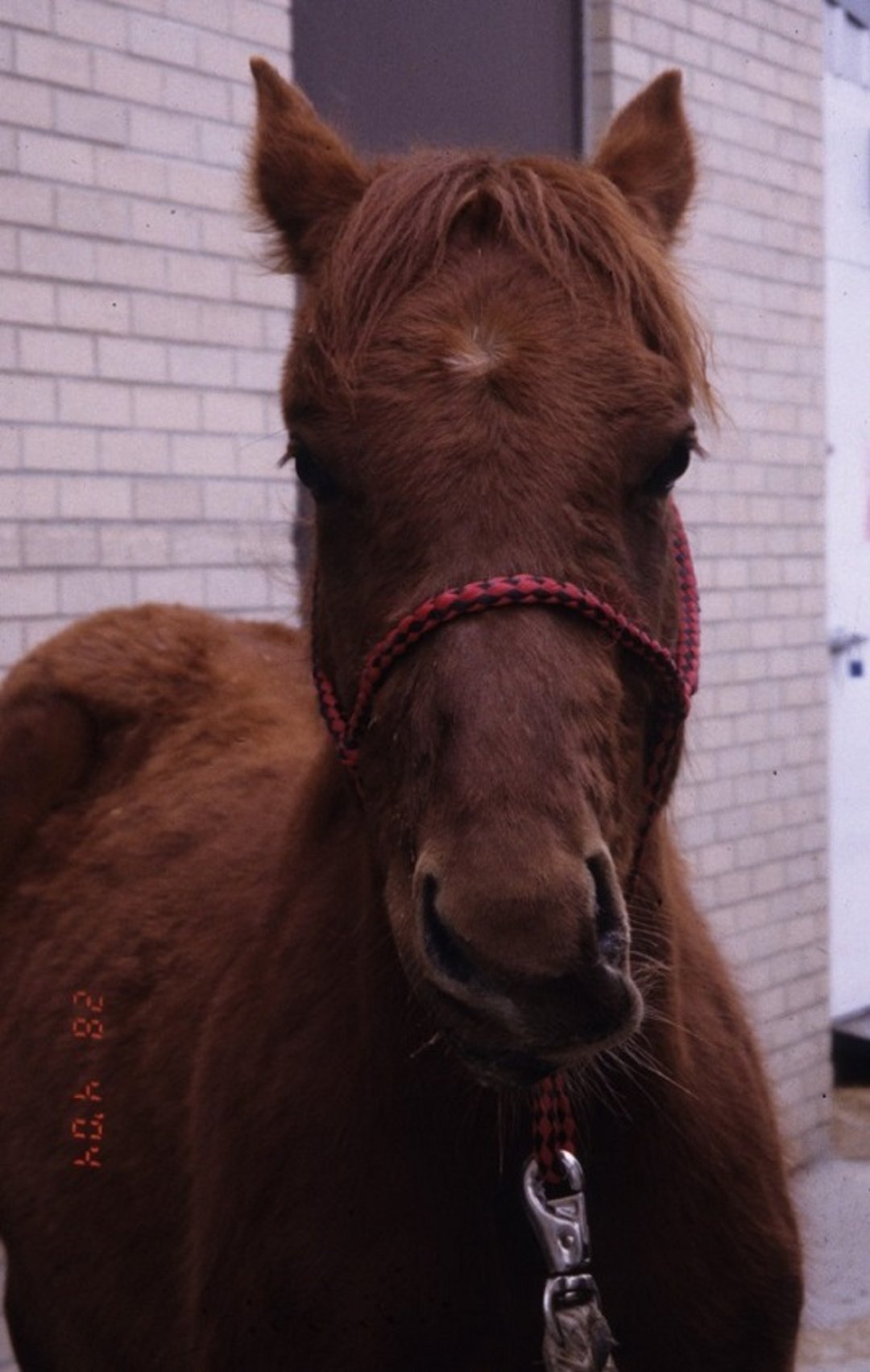
Courtesy of Dr. Sameeh M. Abutarbush.
Facial nerve trauma is most common in large animals that become recumbent, with subsequent compression of the side of the face. It can be caused by pressure from a halter that is left on horses during general anesthesia. There is ipsilateral lip paralysis, deviation of the muzzle to the contralateral side, and weak to absent palpebral reflex. A drooping ear can result from injuries to the proximal aspect of the nerve.
Peripheral Nerve Injuries in Animals
Peripheral nerve injuries are some of the most common neuropathies in animals. The sciatic nerve or its branches may be injured by pelvic fractures, during or after retrograde placement of intramedullary pins in the femur, or by injections of irritating substances in or near the nerve. Damage to the proximal aspect of the sciatic nerve causes monoparesis with inability to flex the stifle. The hock (tarsi) and digits (metatarsi) cannot flex or extend, and weight is supported on the dorsal surface of the foot with the hock excessively flexed. There may be loss of sensation below the stifle except for the medial aspect, which is innervated by a branch of the femoral nerve.
Injury to the tibial nerve results in inability to extend the hock or flex the digits as well as reduced sensation over the plantar surface of the foot.
Injury to the peroneal nerve results in inability to flex the hock or extend the digits as well as decreased sensation over the craniodorsal surface of the foot, hock, and stifle.
The femoral nerve may be injured in calves and foals during dystocia if excessive traction stretches or otherwise damages it. This results in an inability to bear weight on the limb because of an inability to extend the stifle. The patellar reflex is weak or absent. Sensation is lost along the medial surface of the limb (saphenous nerve).
The suprascapular nerve is most commonly injured in large animals secondary to trauma of the shoulder region. This results in atrophy of the supraspinatus and infraspinatus muscles and instability of the shoulder joint (sweeney, see Suprascapular Neuropathy in Horses). In horses, the nerve may be entrapped by connective tissue that develops in the region of the supraspinous fossa.


