


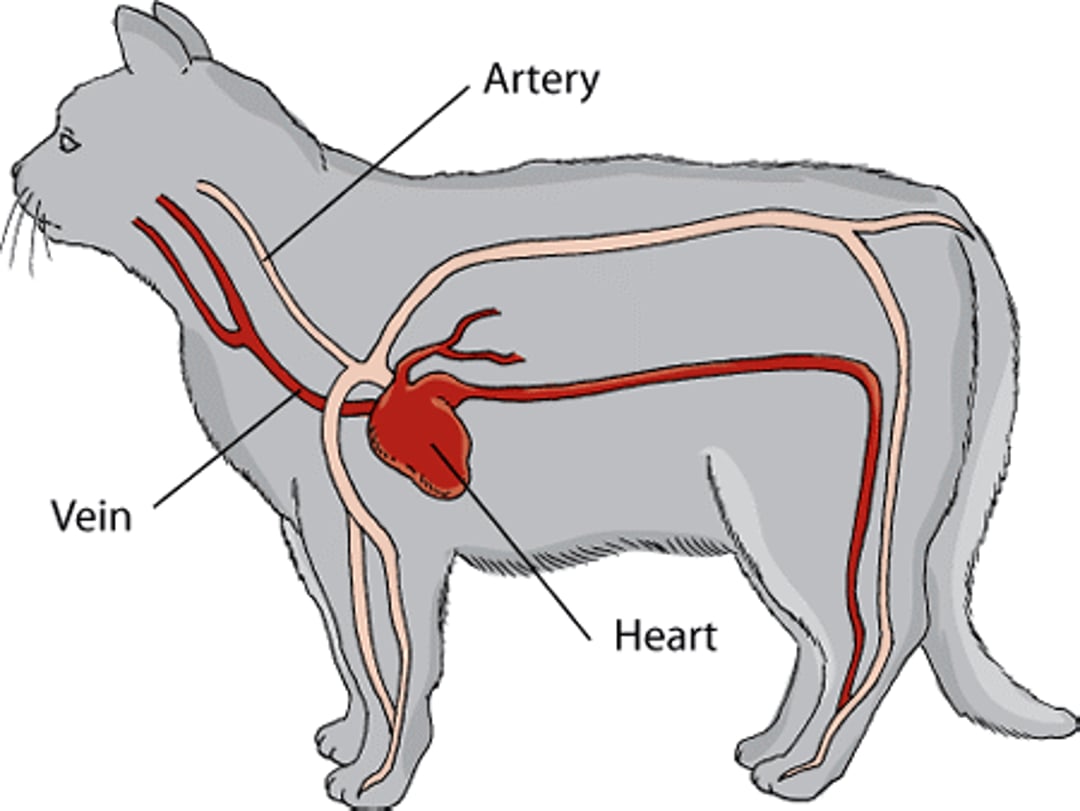
The cardiovascular system includes the heart and the blood vessels (the veins and the arteries). The function of the heart is to pump blood. The right side of the heart pumps blood to the lungs, where oxygen is added to the blood. The left side pumps blood to the rest of the body, where oxygen and nutrients are delivered to tissues, and waste products (such as carbon dioxide) are removed. The heart is a hollow, muscular organ which, in mammals and birds, is divided into 4 chambers. The muscular tissue is called the myocardium. There are upper chambers on both the left and ride sides of the heart called the left and right atria (the plural form of atrium). There are also 2 lower chambers called the left and right ventricles.
Cardiovascular system of a cat

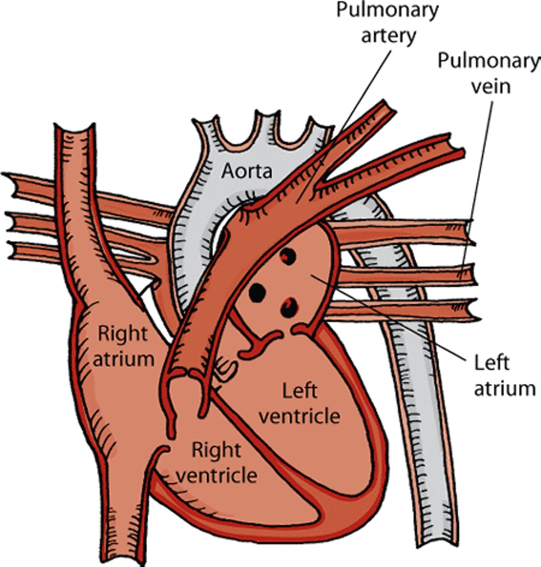
A series of valves keep blood flowing in one direction through the heart. The atrioventricular valves are valves between the atria and the ventricles. The semilunar valves are valves between the heart and the aorta and between the heart and the pulmonary artery. Each ventricle has an inlet and an outlet valve. In the left ventricle, the inlet valve is called the mitral valve, and the outlet valve is called the aortic valve. In the right ventricle, the inlet valve is called the tricuspid valve, and the outlet valve is called the pulmonary valve.
Blood from the body flows through the 2 largest veins, called the venae cavae, into the right atrium. When the right ventricle relaxes, blood in the right atrium pours through the tricuspid valve into the right ventricle. When the right ventricle is nearly full, the right atrium contracts, pushing additional blood into the right ventricle. The right ventricle then contracts, pushing blood through the pulmonary valve into the pulmonary arteries, which lead to the lungs. In the lungs, blood absorbs oxygen and gives up carbon dioxide. The blood then flows through the pulmonary veins into the left atrium. When the left ventricle relaxes, the blood in the left atrium pours through the mitral valve into the left ventricle. When the left ventricle is nearly full, the left atrium contracts, pushing additional blood into the left ventricle. The left ventricle then contracts, pushing blood through the aortic valve into the aorta, the largest artery in the body. This blood carried in the aorta distributes oxygen to all of the body except the lungs.
Inside a cat's heart

Each heartbeat consists of 2 parts: diastole and systole. One half of a heartbeat (diastole) is the sound of the mitral and tricuspid valves closing. The other half (systole) is the sound of the aortic and pulmonary valves closing. During diastole, the ventricles relax and fill with blood. During systole, they contract and pump blood out to the body.
The rate and force of contraction of the heart and the degree of narrowing or widening of blood vessels are controlled by several hormones and by the autonomic nervous system (the part of the nervous system that controls involuntary activity).
Heart Rate
The heart beats because of a tiny electrical current that begins in the sinoatrial node. The sinoatrial node is the heart’s natural pacemaker. Rhythmic electrical impulses or discharges from the sinoatrial node cause the contraction of muscle fibers in the heart. While an animal is at rest, the sinoatrial node discharges many times each minute; in a resting cat, it will discharge more than 120 times per minute.
Heart rate is also inversely related to blood pressure. When blood pressure increases, heart rate decreases; when blood pressure decreases, heart rate increases. In heart failure, nerve endings that are sensitive to blood pressure changes (called baroreceptors) report the lower blood pressure to the brain, resulting in an inappropriately elevated heart rate. Unfortunately, this further injures the heart.
Heart Sounds and Murmurs
Heart sounds are produced by the rapid acceleration and deceleration of blood and the resulting vibrations in the heart due to the circulation of blood. They can be heard using a stethoscope. In cats, 2 heart sounds can normally be distinguished.
Heart murmurs are vibrations that can be heard coming from the heart or major blood vessels and generally are the result of turbulent blood flow or vibrations of heart structures such as part of a valve. Murmurs are typically described by their timing (that is, whether they occur during diastole, systole, or continuously), their intensity (that is, whether they can be heard easily or with difficulty), and their location. Not every murmur indicates a heart disorder; for example, innocent murmurs are sometimes detected in healthy kittens less than 3 months of age.
Arrhythmias
Arrhythmias are abnormalities of the rate, regularity, or site of heartbeat formation. An arrhythmia does not necessarily indicate heart disease. Many arrhythmias are functionally insignificant and require no specific treatment. Some arrhythmias, however, may cause severe signs (such as loss of consciousness due to lack of blood flow to the brain) or lead to sudden death. Many disorders are associated with abnormal heart rhythms. Common findings in animals with an arrhythmia are a rate that is too slow (bradycardia), a rate that is too fast (tachycardia), premature beats (a beat that is heard too early), an irregular rhythm, and pauses in the rhythm. If an abnormal rhythm is heard with a stethoscope, your veterinarian may recommend other tests, such as an electrocardiogram or echocardiogram.
Pulse
A pulse is the rhythmic expansion of an artery that can be felt with the fingertips during physical examination. In cats, pulses are typically felt at the femoral artery (in the thigh). A jugular pulse in the lower neck can be noted in healthy animals. A pulse may be absent, increased (strong), or decreased (weak)—each of which may indicate a specific type of heart disease or defect.
For More Information
Also see professional content regarding heart and blood vessel disorders.


