


Common Bacterial Pathogens (Aeromonas, Vibrio, and Edwardsiella) in Aquaculture
Common bacterial pathogens in aquaculture include:
Aeromonas hydrophila
A salmonicida
Vibrio spp
Edwardsiella ictaluri
E tarda
Streptococcus spp
other related gram-positive cocci, can also infect aquaculture species
Organisms that may have previously been identified as E tarda have now been recognized as a new species of organism, with biochemical characteristics very similar to those of E tarda. Two new groups are also recognized, E piscicida and E piscicida–like bacteria, which have been associated with disease in channel catfish and freshwater game fish.
Treatments for food fish species are limited to the species and disease indicated on the label of approved drugs, although approved forms of the Veterinary Feed Directive drugs oxytetracycline, ormetoprim sulfadimethoxine, and florfenicol can be used extra-label under the supervision of a veterinarian. Florfenicol is the only drug approved for use against streptococcal infections in freshwater-reared, warmwater finfish; however, extra-label use is possible. Enrollment in an INAD program may also allow use of florfenicol for indications and species beyond the current label. Also see SRAC 4709 (Investigational New Animal Drug [INAD] Exemptions and the National INAD Program [NIP]). Appropriate withdrawal periods before harvest must be followed when used in food fish species ( see Table: Drugs Used in Aquaculture in the US – Approval Status and Withdrawal Times). Vaccines are available for a number of bacterial pathogens and species indications. Under supervision by a veterinarian, licensed commercial vaccines can be used off-label. In addition, autogenous vaccines, based on facility-specific pathogens, may help prevent recurring disease outbreaks (see Use of Vaccines in Finfish Aquaculture at ufl.edu).
Yersiniosis in Aquaculture
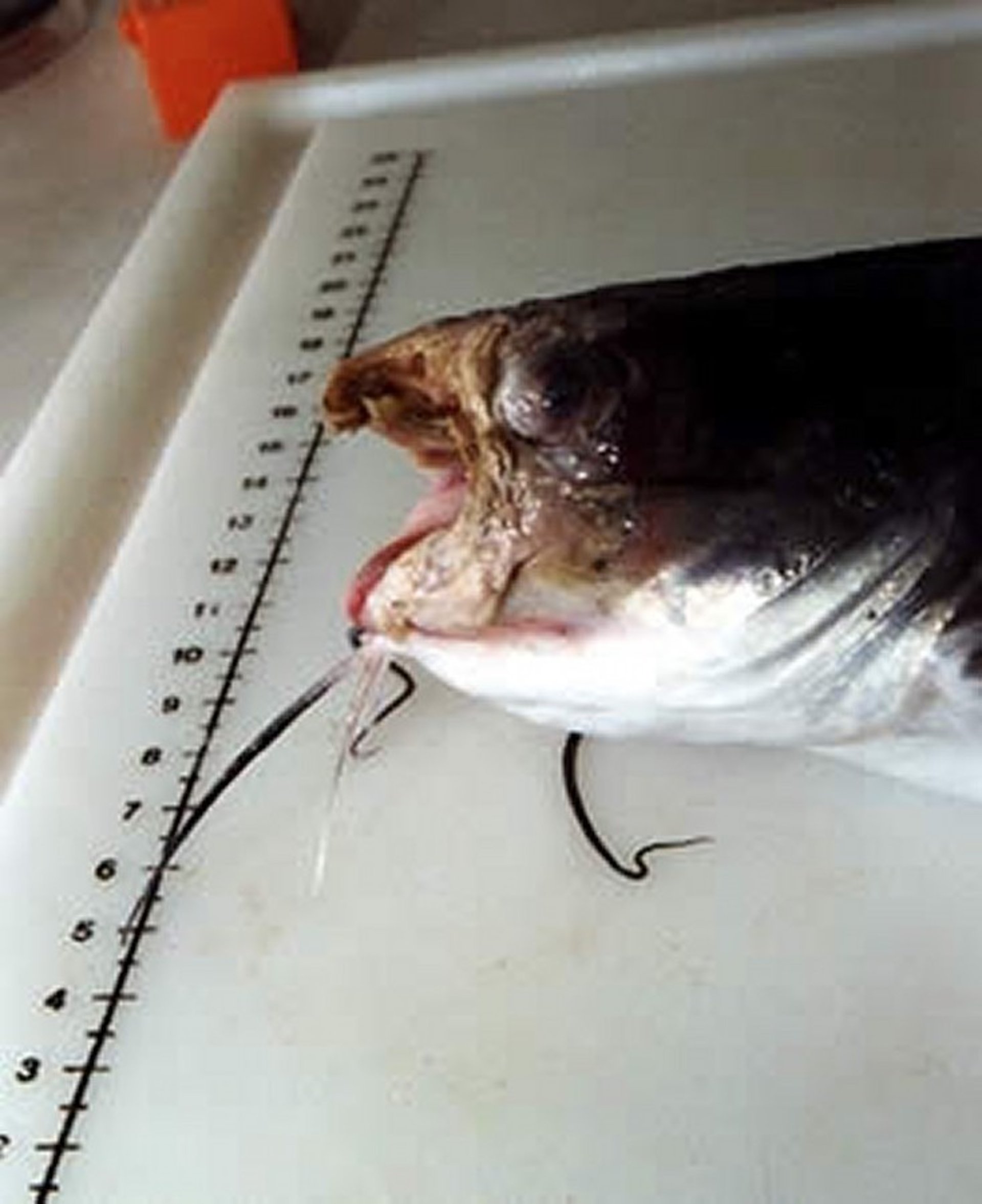
Yersiniosis (enteric redmouth disease) is a serious acute or chronic bacterial disease of intensively cultured salmonids. The etiologic agent is Yersinia ruckeri. Signs are darkening and hemorrhage of the mouth (red mouth), skin, anus, and fins. Chronic signs are associated with inappetence, exophthalmos, swelling, and degenerative changes of internal organs. Mortality rates are variable but are exacerbated by poor water quality and related stressors. Diagnosis is by isolation and identification of pure cultures of the organism obtained from the internal organs of infected fish. Fish that survive remain carriers and may cyclically shed bacteria, particularly when exposed to stressful conditions and water temperatures of 15°–18°C. Depopulation of infected fish and avoidance of introduction of infected fish can be recommended, but preventive vaccination is the usual procedure in endemic areas. Yersiniosis can be treated successfully with antibiotics, which should be selected based on a sensitivity test. Therapy should be continued for at least 14 days.
Edwardsiella piscicida and E piscicida-like Diseases in Aquaculture
Edwardsiella ictaluri causes enteric septicemia of catfish, the most important infectious disease in the channel catfish industry. Infection occurs in the spring and fall when water temperatures are 22°–28°C, and mortality may be exacerbated by handling stress, chemical treatment, or poor water quality. The disease occurs in two forms: the enteric (or intestinal) form and the meningeal form. In the enteric form, infected fish may develop skin lesions characterized by massive petechial hemorrhage around the mouth, operculum, and eyes, or they may develop measles-like, red, punctate lesions along the body wall. There is a hemorrhagic enteritis, and the intestine may be hemorrhagic and fluid- or gas-filled. Liver lesions are common and may be evident as multifocal areas of necrosis, abcessation, or hemorrhage.
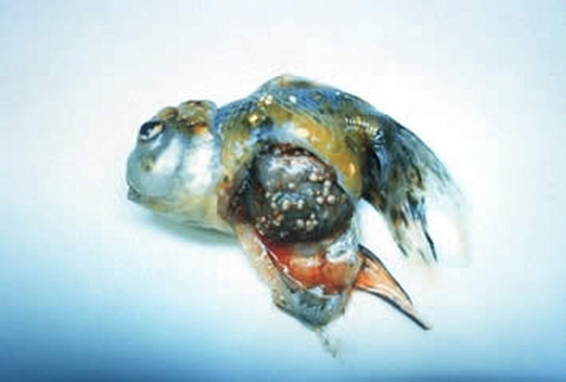
By contrast, in the meningeal form, few external signs may be seen in infected fish. The bacteria enter the CNS through the olfactory system, and affected fish develop severe meningitis. In fingerlings, the inflammation may be severe enough to erode the skull, resulting in the characteristic “hole-in-the-head” lesion. Fish affected with the meningeal form may demonstrate bizarre behavior, including spinning, erratic swimming, and general disorientation. Diagnosis is based on bacterial culture and isolation. Brain culture is indicated whenever E ictaluri is suspected. E ictaluri will grow on blood agar incubated at 25°C for 48 hours. Antimicrobial therapy should be based on results of sensitivity testing. Vaccination is available for channel catfish fingerlings.
There has been an important change in understanding of fish diseases caused by an organism historically identified as E tarda. This organism is the causative agent of emphysematous putrifactive disease of catfish. The name is descriptive of the most common lesions, an ulcerative dermatitis associated with a malodorous, gas-producing bacteria. E tarda is an enteric bacterium that is ubiquitous in terrestrial and aquatic environments. It has been associated with enteric disease in mammals, including humans, as well as birds, reptiles, and fish. Recent molecular work has demonstrated that several different organisms that may have been previously identified as E tarda are likely causing many of the disease outbreaks attributed to E tarda in fish.
These newly recognized taxa, E piscicida and E piscicida–like organisms, are considered important emerging diseases in the channel catfish industry. They can present not only as an ulcerative dermatitis but also can cause a systemic granulomatous disease with gross and microscopic lesions very similar to those seen in mycobacteriosis. Granulomatous disease attributed to E piscicida tends to result in hepatic lesions, which are not typical of mycobacteriosis. Histologically, the granulomas contain gram-negative and acid-fast negative rods. Molecular testing is required to confirm a diagnosis of E piscicida or E piscicida–like infection. The disease will respond to antibiotics, but it is unclear whether fish can clear bacteria sequestered in granulomas. These environmental bacteria thrive in organically rich environments, so sanitation may need to be addressed as part of the management strategy. For aquariums and recirculating systems, UV filtration may help decrease numbers of bacteria in the environment.
Columnaris and Related Diseases in Aquaculture
The taxonomic grouping of bacteria causing columnaris disease, coldwater disease, and bacterial gill disease has undergone notable revision in recent years based on genomic studies. The primary causative agent of each of these important diseases has been moved into the genus Flavobacterium, although other genera have also been implicated. These gram-negative, rod or filamentous bacteria have a distinctive gliding motion. Skin or gill lesions have slimy or cotton-like surface exudates, which usually cover surface necrosis, ulcerations, and marginal hemorrhages.
Flavobacterium columnare, the most prominent member of this group responsible for columnaris disease, is most common in warmwater species of fish. A presumptive diagnosis can be made from visualization of typical organisms on wet mounts of infected skin or gill tissue. Columnaris disease can be confirmed by isolation of the organism on Ordal’s or other cytophaga media. Sensitivity tests are difficult to perform, because F columnare will not grow on Müller-Hinton media. If the disease is diagnosed early in the course of infection, treatment with potassium permanganate or hydrogen peroxide may be effective. If the disease becomes chronic, it may have become systemic, in which case treatment with florfenicol or oxytetracycline is recommended. Columnaris disease can be prevented by reducing organic loading and avoiding traumatic injuries. A vaccine is currently available in the US for use in channel catfish and largemouth bass.
A similar organism affecting marine fish was previously grouped with F columnare but has been given its own genus and is now named Tenacibaculum maritimum. Potassium permanganate is not recommended in warmwater marine finfish because of its increased toxicity at higher salinities.

Courtesy of Dr. Ruth Francis-Floyd.

Courtesy of Dr. Ruth Francis-Floyd.
Flavobacterium psychrophilum causes coldwater (peduncle) disease, or bacterial coldwater disease, in salmonids and other coldwater species. Rarely, warmwater fish exposed to cold temperatures may be affected. Disease is most severe at water temperatures of 4°–10°C, and signs should not be seen at temperatures >18°C. Skin lesions usually begin on the dorsal and posterior surfaces of the fish but may be found on any part of the body. Advanced cases show necrosis and ulceration of the peduncle, and underlying musculature will be exposed. As the disease progresses, the infection becomes systemic, typically involving the spleen, kidney, and liver. Confirmed diagnosis is possible after isolation of F psychrophilum using cytophage media (15°–20°C for 3–6 days), or by immunohistochemistry or PCR assay. Outbreaks can be controlled with oxytetracycline.
Bacterial gill disease, caused by F branchiophilum, is most frequently reported in young cultured salmonids or fish cultured under conditions of high organic loading. It has been seen occasionally in aquarium fish. It may be initiated by crowding and poor water quality, particularly high organic loads, high ammonia levels, and silt. Gills appear swollen and mottled, with patchy areas of bacterial growth that can be confirmed by microscopic examination of direct gill smears. Hyperplasia, adhesions, and deformity of the gill lamellae can be seen. In young fish affected with the disease, mortality is high and morbidity sustained. Prevention efforts include improving water quality and avoiding overstocking. A single treatment with potassium permanganate, followed by addition of salt to the system (2–5 ppt) may help control losses, but sanitation is critical for longterm resolution of the problem. Antimicrobial therapy may be used as needed to control secondary bacterial problems.
Bacterial Kidney Disease in Aquaculture
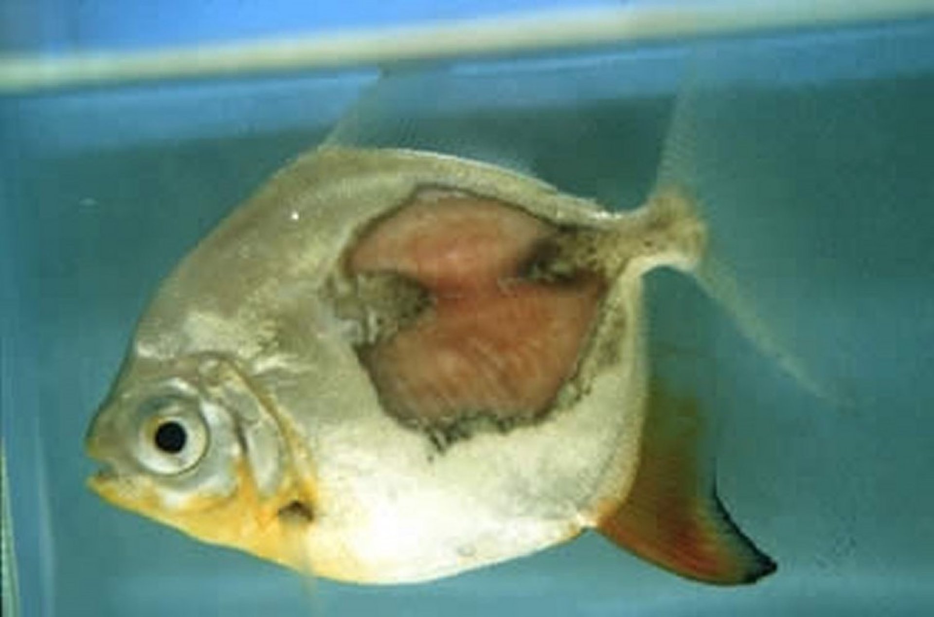
Bacterial kidney disease is economically important in cultured salmonids. The cause is Renibacterium salmoninarum, an obligate intracellular bacteria that is one of the relatively few gram-positive organisms that causes disease in fish. Bacterial kidney disease is a chronic disease affecting juveniles (6–12 months old) and prespawning adults, with clinical signs very similar to those of mycobacteriosis. Clinically, infected fish appear lethargic and darkened and may have coelomic distention, pale gills (anemia), and vent hemorrhages. Typical lesions include grayish, localized, or conglomerate granulomata in the viscera, especially the kidney or body wall; exophthalmos; blindness; cystic cavities in musculature; and emaciation.
A presumptive diagnosis can be based on visualization of small, gram-positive rods in kidney imprints. Definitive diagnosis requires isolation and identification of the bacteria by using a selective medium that contains cysteine and incubating at 15°C for 3–6 weeks. R salmoninarum is transmitted both horizontally and vertically, and fish that survive an epizootic remain carriers. Infected female fish should be injected with erythromycin (11–20 mg/kg, IM) 14–60 days before spawning to prevent vertical transmission. Erythromycin (100 mg/kg for 10–21 days) is effective when administered in feed early in the course of an outbreak; however, it is not FDA approved for this use. Erythromycin 200 injectable IM or IP is available through an FDA INAD for use against this disease. Obtaining disease-free stock and preventing contamination by infected wild fish are the best preventive measures.
Mycobacteriosis in Aquaculture

Courtesy of Dr. Ruth Francis-Floyd.
Mycobacteriosis is an important disease in aquaculture because of its typically chronic nature (although acute and subacute presentations can occur) and the lack of effective antibiotics. External or internal masses, nodules, or granulomas are a common presentation in aquaculture species. Important differential diagnoses for a suspect case of mycobacteriosis, based on identification of granulomas in tissue, are Edwardsiella piscicida and francisellosis. Further, although granulomatous lesions are typical of mycobacteriosis, they are not always seen. In some species, and under some conditions, granulomas may not be present. Instead, caseous (“pus-like”) lesions may be seen, or histologically, only a histiocytic inflammatory response (increased macrophages/tissue phagocytes without granuloma formation) may be apparent. Acid-fast stains and/or culture are necessary to exclude mycobacteriosis in cases with chronic morbidity/mortality and chronic inflammation but no granulomas. In addition, processing differences (including the decalcification method, if necessary, and the type of acid-fast stain used) may affect staining characteristics of mycobacteria.
Rickettsia and Rickettsial-like Diseases in Aquaculture
Rickettsial disease associated with Piscirickettsia salmonis has been described in salmonid species from Chile, Norway, Ireland, and Canada. Rickettsial-like organisms have been reported in tilapia, sea bass, and blue-eyed plecostomus. Rickettsial disease can result in acute mortality, affecting up to 95% of fish with few gross signs. In tilapia, acute mortality may be triggered by sudden drops in temperature. Chronic disease is manifest by nonspecific external lesions, including anorexia, pale gills, and skin lesions. Internally, lesions are more typical, with granulomatous lesions possible throughout the viscera. The most characteristic lesions may be found in liver and kidney tissue and appear as gray to yellow mottled areas with ring-shaped foci. Visceral lesions are grossly similar to those seen in advanced cases of mycobacteriosis, and differentiation is important. Histologically, intracellular organisms may be seen in macrophages and hematopoietic tissue in the liver, spleen, and kidney. Blood or tissue smears stained with Giemsa or acridine orange may reveal the intracellular organisms, often appearing as paired, curved, gram-negative rods in macrophages or hepatocytes. Rickettsia-like organisms can be isolated using a variety of cell lines; however, confirmation of a suspect case may also be based on serology.
Transmission of rickettsial-like diseases in fish is not understood. In terrestrial species, a vector is often required; however, R salmonis has been demonstrated to survive for 14 days in seawater, suggesting that horizontal transmission in the absence of vectors may be possible in aquatic species. Oxytetracycline is the treatment of choice, although it is unclear whether an advanced case can be completely resolved with antimicrobial treatment. Rickettsia-like organisms do not appear to be a zoonotic threat, because they do not seem able to survive at mammalian body temperatures.
Francisellosis in Aquaculture
Francisellosis, an important emerging bacterial disease caused by Francisella noatunensis, has been identified as the primary cause of disease in a variety of aquacultured and wild-caught species, including tilapia, hybrid striped bass, Atlantic cod, Atlantic salmon, various grunt species, ornamental cichlids, fairy wrasses, and blue-green damselfish. In tilapia, francisellosis is often associated with environmental stressors, especially cooler temperatures and poor water quality, and so increasing temperatures (for indoor facilities) may help mitigate the disease. Many signs and necropsy findings are grossly similar to those seen in mycobacteriosis. The disease can be acute, with few clinical signs and high mortality, or subacute to chronic, with fish demonstrating erratic swimming, spiraling, buoyancy control problems, anorexia, lethargy, exophthalmia, anemia, petechiation, and darkening. Severely infected fish may have necrotic or nodular gills with red and white patchiness, and histologically, portions of the gill may also be severely hyperplastic with consolidation of secondary lamellae.
On necropsy, the spleen and kidney typically have a granular or nodular (granulomatous) appearance, and most other organs can also be affected. Francisellosis should be another differential diagnosis for granulomatous disease in fish. Identification is supported by history, clinical signs, species affected, and presence of non-acid-fast bacterial organisms that are Giemsa positive and weakly gram negative. Because Francisella does not grow on routinely used media (such as blood agar), confirmation requires PCR assay of affected tissue and/or culture on various special media, all of which include increased levels of cysteine (or cystine) and glucose. Culture and sensitivity should be performed by a microbiologist and laboratory with expertise in this pathogen for identification and to determine sensitivity patterns. Because Francisella is intracellular and causes granulomatous disease, chemotherapy may be less effective, unless the disease outbreak is identified very early in its course. Optimization of husbandry, including increasing temperatures when possible, may help reduce morbidity and mortality.


