


Courtesy of Dr. Stephen Divers.
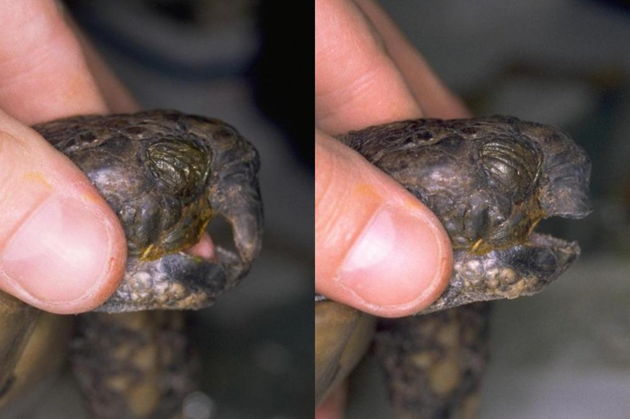
Beak anomalies in chelonians inhibit feeding and are often associated with trauma or secondary nutritional hyperparathyroidism leading to hypocalcemia, distortion of the skull, and abnormal occlusion and wear. Increased levels of dietary protein may contribute to accelerated growth of these tissues, while a lack of abrasive food items limits natural wear in captivity. Treatment consists of trimming and reshaping the beak into a more normal conformation. The condition usually recurs because of primary malocclusion, and longterm maintenance may be required.
Males of many species can be highly territorial and exhibit aggression toward other males or toward females during mating periods. Injuries to cagemates can be severe and are best avoided by separating animals at feeding and reducing the number of animals in a breeding group. When separated individuals are placed together for breeding, they should be carefully monitored. If reptiles must be kept together, it is vital the enclosure is large enough to avoid competition for resources, especially basking areas, and retreats. Food and water is best placed in multiple locations to prevent dominant cohabitants from intimidating the others.
Fractures due to trauma are commonly seen in all species. They are often associated with secondary nutritional hyperparathyroidism in chelonians and lizards. Long bones may be repaired with lightweight external coaptation. A simple way of splinting the legs of lizards is to tape the injured leg to the body (front legs) or the tail (rear legs). These splints are tolerated well and protect the injured limb from further injury. Fractures unaffected by metabolic bone disease can be repaired using established fixation techniques.
Injury to the spinal column must be assessed individually; when clear displacement is not evident, radiographic evaluation should be performed. Spinal injuries caudal to the vent may be well tolerated, but injuries cranial to the vent frequently result in constipation and retention of urates, with variable limb movement. Environmental changes (eg, low branches, shallow water dish, nonabrasive substrates) may permit the lizard to survive with an acceptable quality of life. Because these fractures are often pathologic (secondary to secondary nutritional hyperparathyroidism or chronic osteomyelitis), a thorough investigation is typically required.
Aggressive iguanids may frequently lash out with their tails and damage them against the vivarium glass or other furniture. Continual damage can lead to ischemic necrosis of the tail. Secondary infection may follow and progress to osteomyelitis. In some cases, septic emboli may lead to tail infection. In either scenario, tail amputation, in addition to investigation and correction of predisposing factors, is required. Radiography should precede surgery, because osteomyelitis may appear more cranial than the external lesion. Tail amputation in iguanids should be performed under anesthesia, taking advantage of the lizard’s ability to perform autotomy. The tail is simply bent and twisted sharply to cause a fracture through a fracture plane. Muscle fibers are trimmed, but the tail is left unsutured to encourage regeneration. Clean postoperative conditions are essential, but antibiotics are not. Tail necrosis in reptiles that do not exhibit autotomy requires a more traditional surgical amputation with primary closure of the wound.
Burns are generally associated with unscreened incandescent lights or other heat sources. Fluid loss, dehydration, and secondary bacterial or fungal infections are common sequelae. However, many can be treated by cleansing the site, applying topical antimicrobial agents, providing analgesics, and placing the reptile in a clean, dry environment. In uninfected burns, sterile skin protectants can be applied to the area to act as a “second skin.” These products allow access to the water and help keep contaminants out. In severe burn cases, fluids are given to offset losses, and systemic antibiotics may be indicated, especially if surgical debridement is required.
Courtesy of Dr. Stephen Divers.
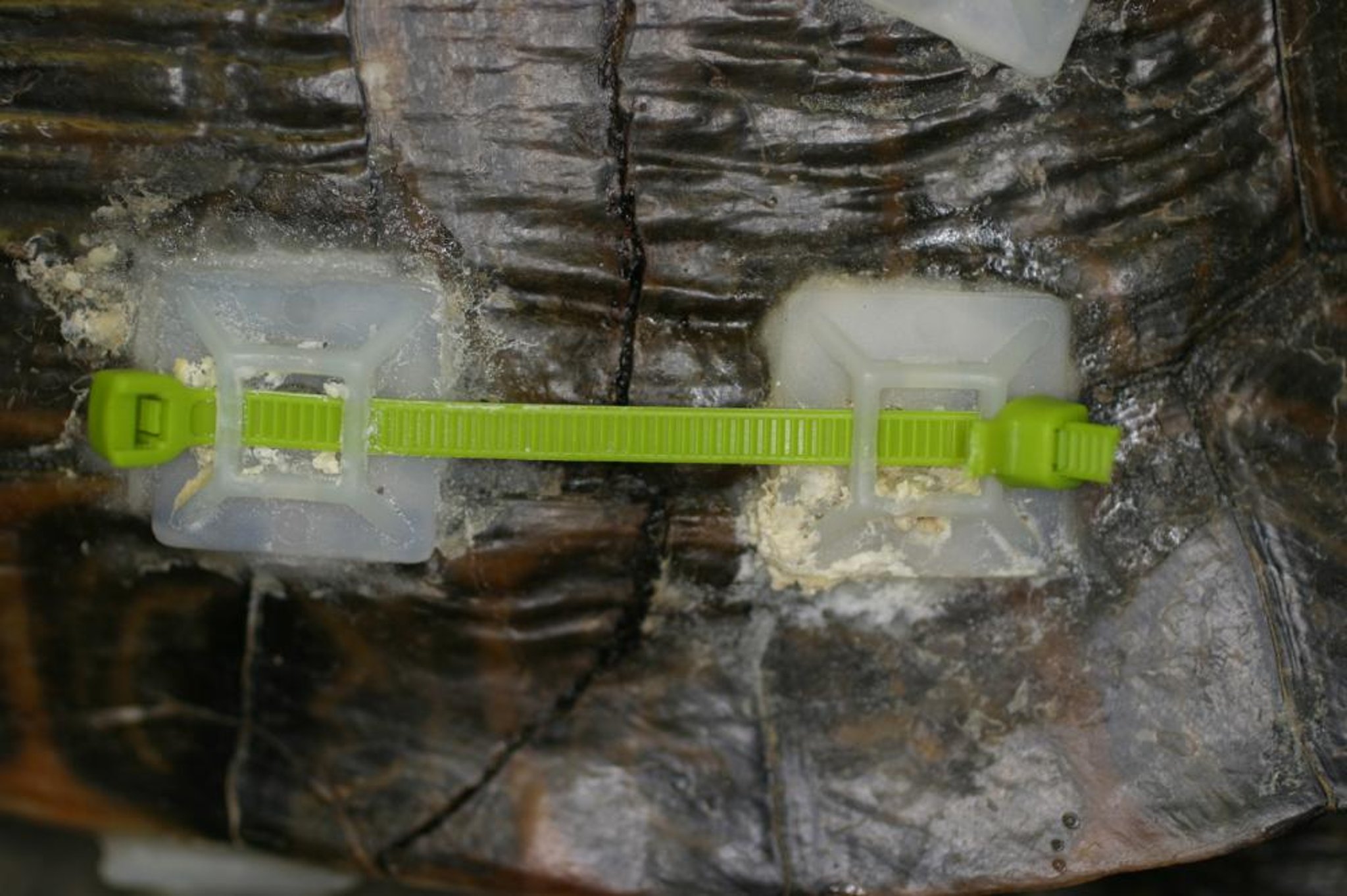
Traumatic injuries to turtles may result in fractures to the plastron, the carapace, or both. Repairs should be delayed in anything other than fresh wounds. Contaminated tissues should be gently debrided, flushed, and appropriately bandaged using a wet-to-dry technique. Holes in the bandages can be created to allow the legs to remain exposed. If obvious infection is present, samples should be submitted for microbiology before systemic antibiotics are started. Once stable, the wounds should be debrided, and the fractures should be realigned under general anesthesia and repaired using zip ties, or a similar fastener. These injuries also can be repaired using epoxy resin or a quick-setting, epoxy glue layered over fiberglass screen. Dental and orthopedic cements have also been used to stabilize fractured tissues. Healing is slow and may require 4–6 months or longer.
Courtesy of Dr. Stephen Divers.
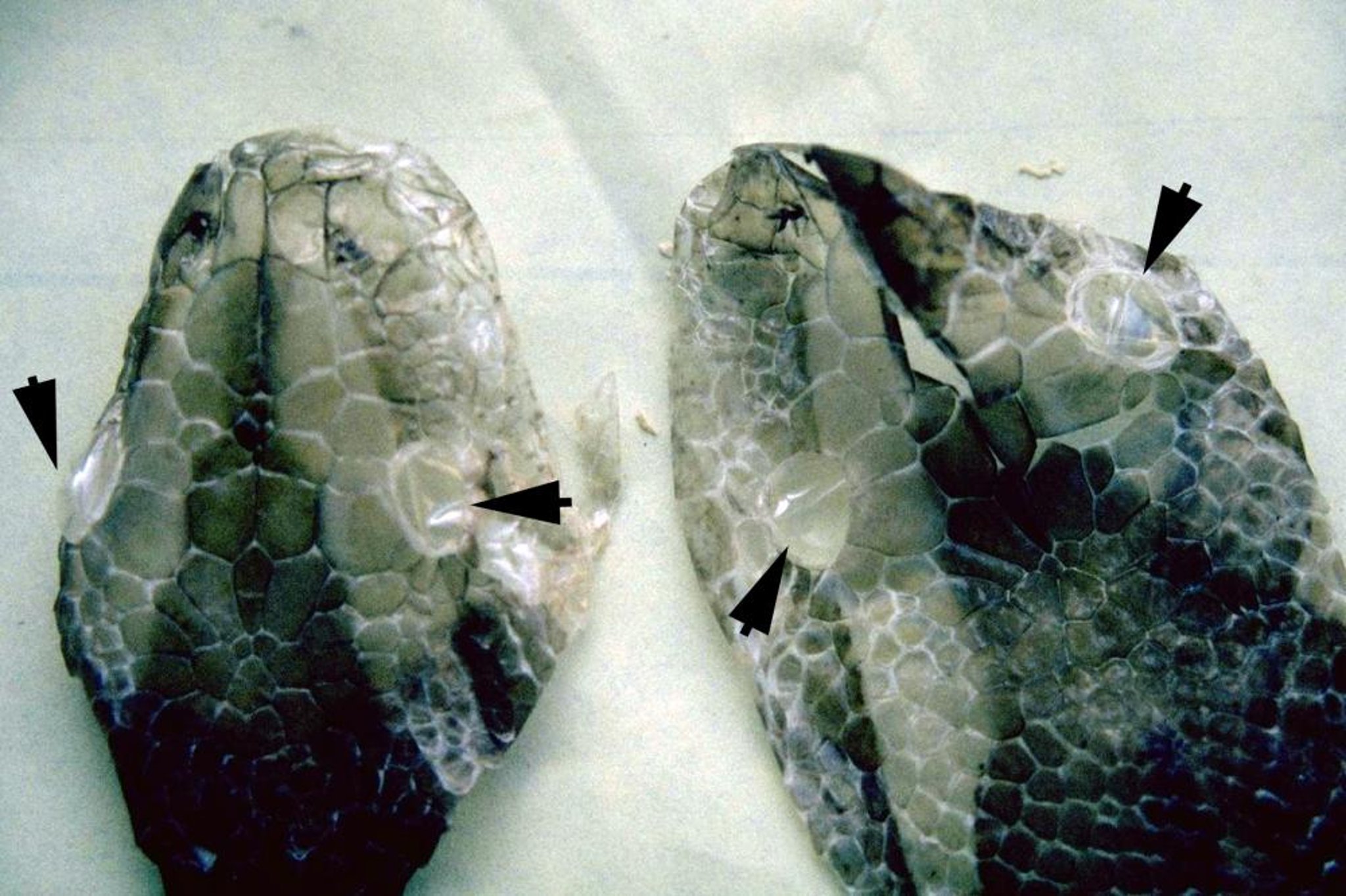
Dysecdysis, or incomplete or inadequate skin shedding, may be caused by low humidity, ectoparasitism, nutritional deficiencies, infectious diseases, lack of suitable abrasive surfaces, or even decreased thyroid function. Often, eyecaps or annular bands on the tail or digits are retained. Eyecaps are best treated by application of an ophthalmic ointment for several days or by placing the snake on wet towels in a ventilated, warm box overnight. If the spectacles do not fall off they can be carefully removed using magnification and fine forceps. Patience is advised—eyecaps should never be forced off because of the possibility of damaging the spectacle and exposing the cornea.
Courtesy of Dr. Stephen Divers.
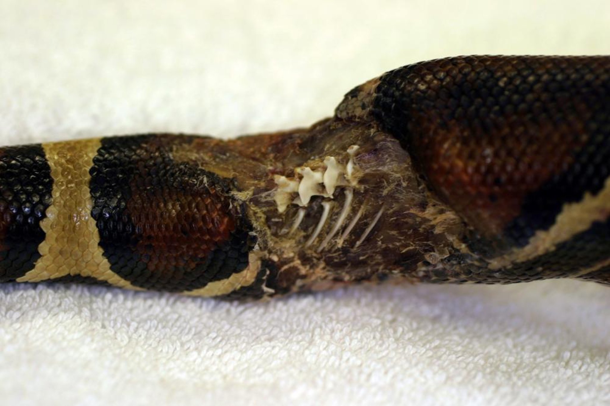
Prey-induced trauma, caused by live, uneaten invertebrate and vertebrate prey, can cause severe trauma, with secondary infection and abscessation. Whenever possible, rodents that have been freshly killed or frozen and thawed should be offered to prevent injury to the reptile (dead prey should be discarded after 12 hours if uneaten). The feeding of live prey is illegal in many countries, but even where legal it should be actively discouraged. Fresh bite wounds may be treated by cleansing with povidone-iodine (diluted 1:10). Topical antibiotics are indicated, but parenteral antibiotics, based on results of culture and sensitivity tests, may be necessary. Untreated wounds frequently abscess and are seen as a soft or hard swelling. The abscess, including the fibrous capsule, should be removed surgically. Open or draining abscesses should be curetted and flushed with povidone-iodine until closed by second-intention healing. ( See also Abscesses.)


