


Courtesy of Dr. Ronald Green.
Hip dysplasia is a multifactorial abnormal development of the coxofemoral joint in dogs that is characterized by joint laxity and subsequent degenerative joint disease. It is most common in large breeds. Excessive growth, exercise, nutrition, and hereditary factors affect the occurrence of hip dysplasia. The pathophysiologic basis for hip dysplasia is a disparity between hip joint muscle mass and rapid bone development. As a result, coxofemoral joint laxity or instability develops and subsequently leads to degenerative joint changes, eg, acetabular bone sclerosis, osteophytosis, thickened femoral neck, joint capsule fibrosis, and subluxation or luxation of the femoral head.
Clinical signs are variable and do not always correlate with radiographic abnormalities. Lameness may be mild, moderate, or severe and is pronounced after exercise. A “bunny-hopping” gait is sometimes evident. Joint laxity (Ortolani sign), reduced range of motion, and crepitation and pain during full extension and flexion may be present.
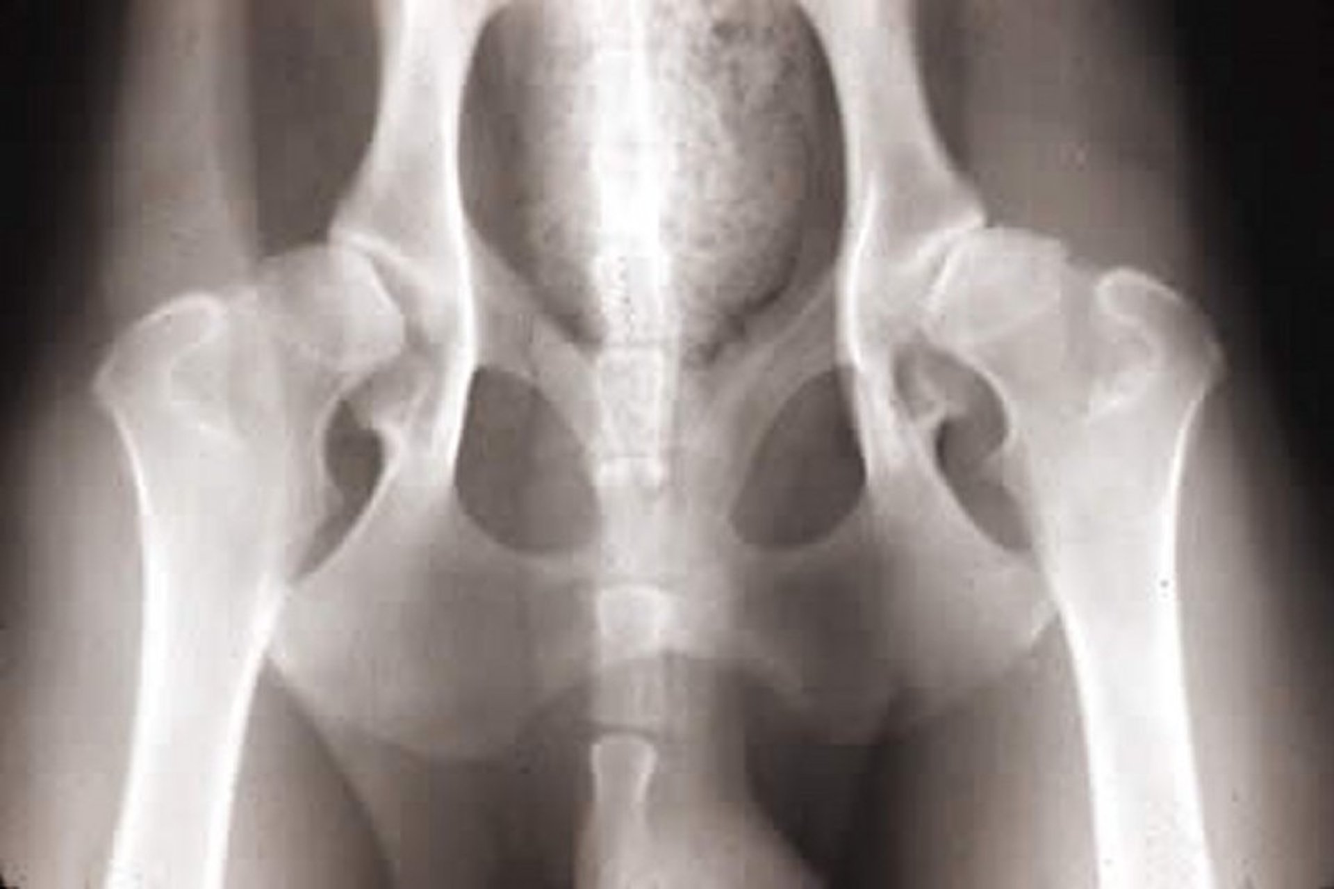
Radiography is useful in delineating the degree of arthritis and planning of medical and surgical treatments. Standard ventrodorsal views of sedated or anesthetized animals can be graded by the Orthopedic Foundation for Animals, or stress radiographs performed and joint laxity measured (Penn Hip). A dorsal acetabular rim view is used by some surgeons to evaluate the acetabulum before reconstructive surgery. Modified ventrodorsal and dorsoventral projections have also been proposed in an effort to mimic the normal standing posture of dogs. Recent reviews of American and international radiographic screening programs have failed to identify a "gold standard." An evaluation shift toward genome screening may yield more promising results in the future.
Treatments are both medical and surgical. Mild cases or nonsurgical candidates (because of health or owner constraints) will benefit from weight reduction, restriction of exercise on hard surfaces, controlled physical therapy (eg, hydrotherapy) to strengthen and maintain muscle tone, and anti-inflammatory drugs (eg, aspirin, corticosteroids, NSAIDs). Joint fluid modifiers (glucosamines) and acupuncture may be useful in some patients, although most reports are anecdotal.
Surgical treatments include pectineal myotenectomy to reduce pain, triple pelvic osteotomy to prevent subluxation, pubic fusion to prevent subluxation, joint capsule denervation to reduce pain, dorsal acetabulum reinforcement to reduce subluxation, femoral head and neck resection to reduce arthritis, and total hip replacement for optimal restoration of joint and limb functions. Additionally, femoral corrective osteotomies can be performed to reduce femoral head subluxation, although degenerative arthritis may persist.
Prognosis is highly variable and depends on the overall health and environment of the animal. In general, if surgery is indicated and performed correctly, it is beneficial. Animals on which surgery is not performed may require a change in lifestyle to live comfortably.


