


Significant trauma can result in breakage of any type, but the most commonly encountered fracture of the equine elbow affects the olecranon of the ulna. Stress fractures affect the craniodistal metaphysis of the humerus in racehorses.
Olecranon fractures in mature horses occur as a result of external trauma and may be incomplete (rare), complete but non- or minimally displaced, or complete and significantly displaced. A number of fractures are also open, as a result of the trauma that caused them. In cases of incomplete or nondisplaced fractures, conservative therapy can be rewarding, although some will displace during convalescence. Most authorities recommend cross-tying the horse to prevent it from getting up and down during the first 6–8 wk of box stall rest. The use of splints and Robert Jones bandages is more controversial—a “pendulum effect” can result from increased weight on the lower limb, possibly doing more harm than good. Many olecranon fractures displace under the influence of the triceps muscle and require internal fixation to repair. The use of a tension band plate is most common, with reported success rates for return to athletic function of ~75%.

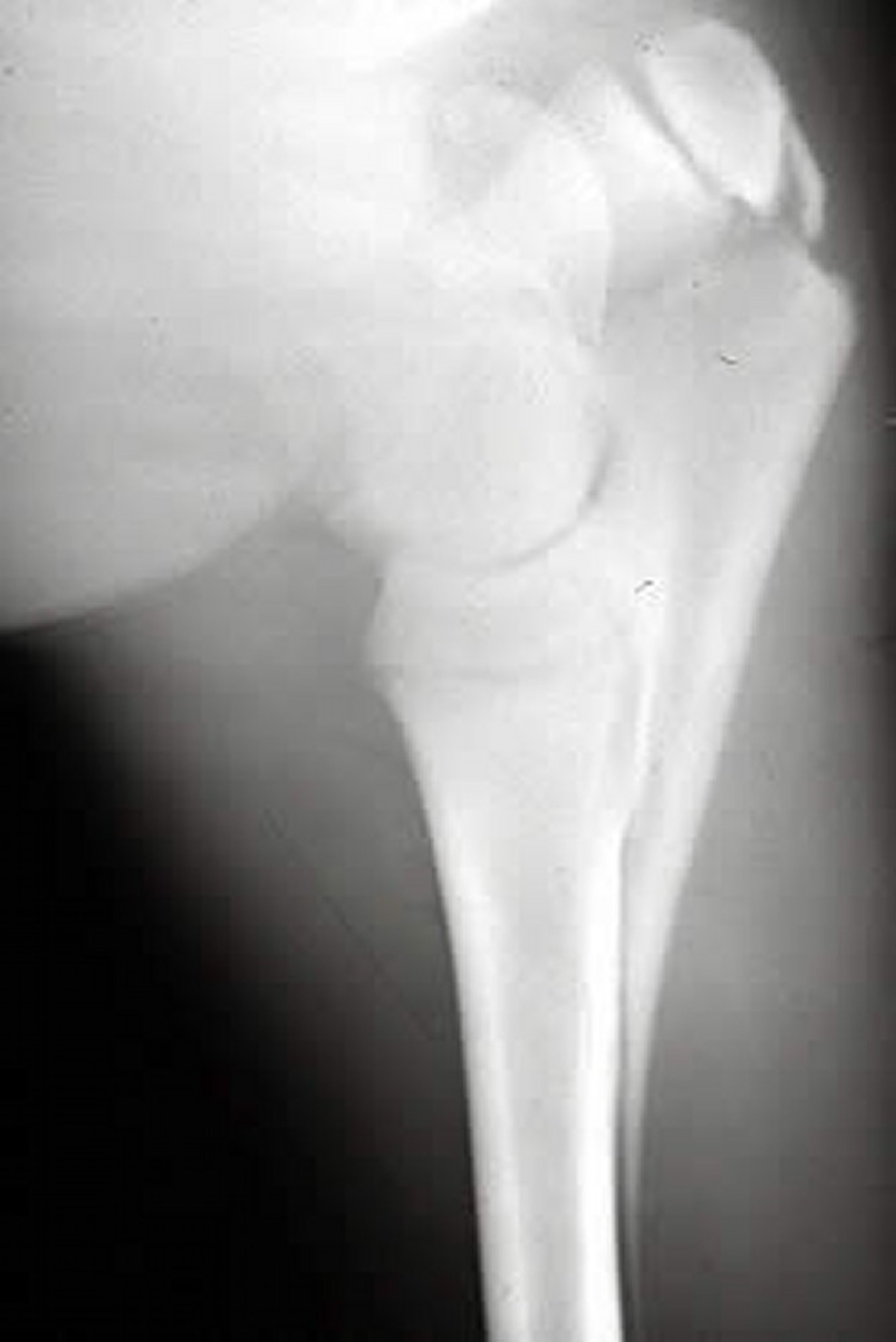
Courtesy of Dr. Ronald Green.
Fractures of the ulna in foals are less liable to displace and may therefore be treated more commonly with rest alone. If such fractures are displaced, tension band plates can be applied, but their use must be monitored carefully because they will interfere with growth of the limb (the proximal radial physis fuses at 11–24 mo of age) and must be removed as soon as satisfactory healing has been achieved. An unusual but potentially difficult injury to diagnose and treat occurs when the proximal ulnar epiphysis is avulsed due to Salter Harris type 1 or 2 injury of the physis, which fuses at 24–36 mo of age. In some cases, the epiphysis is retracted so far proximally that it does not appear on the standard mediolateral radiograph and can be missed. Less dramatic injuries of this type can be managed conservatively, but significant displacement requires surgical intervention.
Treatment and prognosis may be influenced by whether the fracture enters the elbow joint or not. To determine this, careful assessment of high-quality radiographs is needed.
Stress fractures at the craniodistal metaphysis of the humerus, just above the elbow, occur in racehorses. The history is often similar to that for fractures of the proximal humerus and other stress fractures. Mediolateral radiographs will often detect periosteal and endosteal reaction at the predilection site. This can also be documented ultrasonographically. Scintigraphy is a sensitive method to detect fractures that cannot be seen on radiographs. Management is tailored to the comfort of the horse and severity of the initial injury but proceeds as for other injuries of this type with a careful return to exercise, balancing structural integrity with biomechanical requirements for healing.


