


Fractures of the first/proximal phalanx (P1) may occur in any type of horse used for performance. They may be small osteochondral “chip” fractures along the dorsal margin of the proximal joint surface, sagittal (complete or incomplete), or comminuted. Another category involves fragments of the palmar or plantar proximal aspect of P1, which may be associated with osteochondrosis.
“Chip” fractures of the dorsoproximal aspect of P1 typically involve the medial aspect of the joint and occur in horses that exercise at speed. These fractures are normally traumatic in origin and result from hyperextension of the fetlock joint. Acute lameness and increased effusion in the fetlock joint along with sensitivity to firm flexion of the fetlock are clinical signs that a fracture may be present and radiographic examination indicated. In nonracing breeds, a chip fracture may be present on radiographs, but its clinical significance should be determined with diagnostic analgesia before being implicated as a source of lameness.

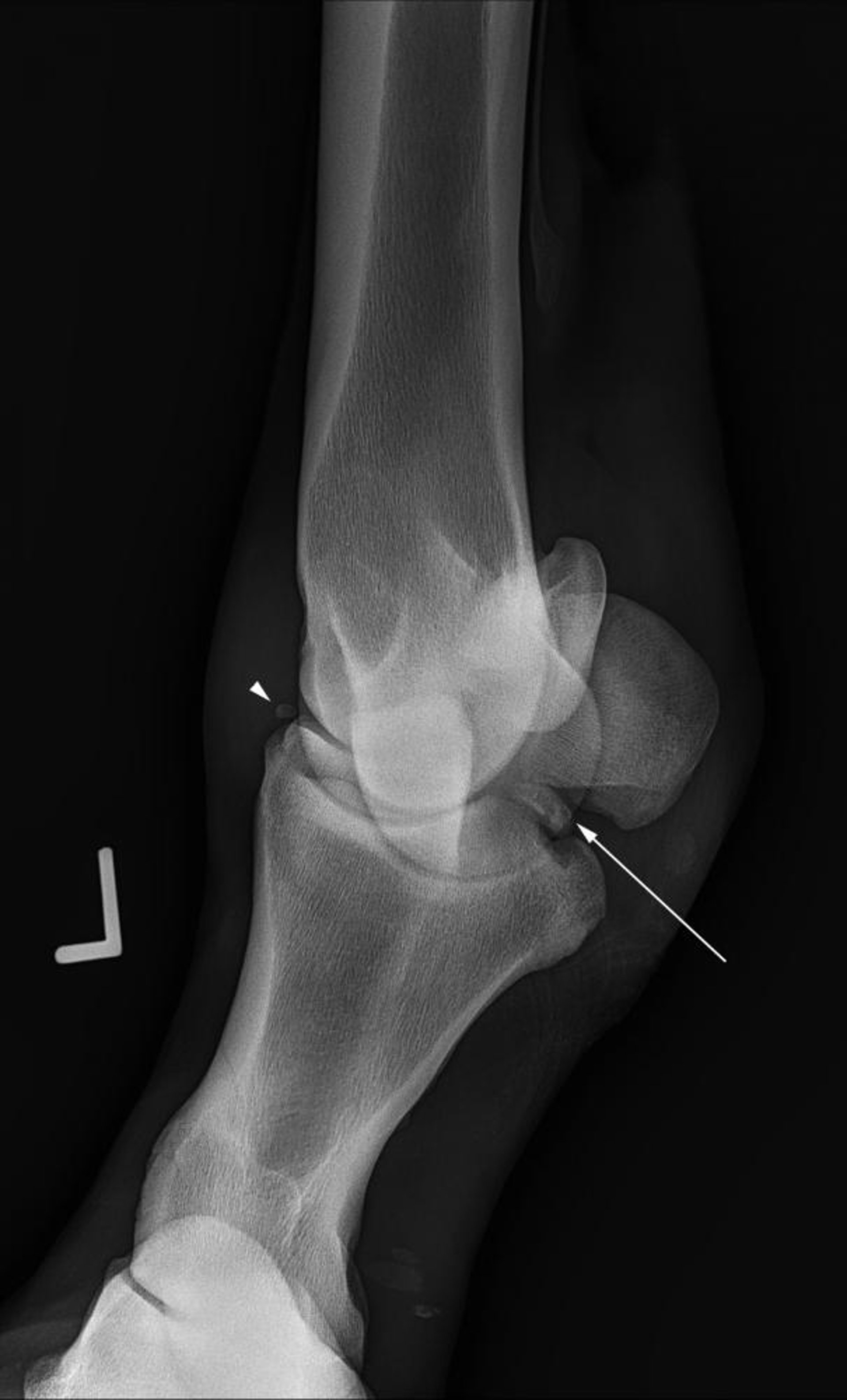
Courtesy of Dr. Matthew T. Brokken.
The cause of proximopalmar and proximoplantar osteochondral fractures is questionable; one thought is that they are from osteochondrosis, the other is that they are fractures. Axial fractures are classified as type I fractures and are generally articular. Type II fractures are located abaxially and typically have minimal articular cartilage present. Type I fractures are generally associated with lameness at speed with clinical signs similar to those of dorsoproximal P1 fractures. These fractures are more common in the hindlimb, and intra-articular diagnostic analgesia is often needed to implicate these fractures as a cause of lameness.

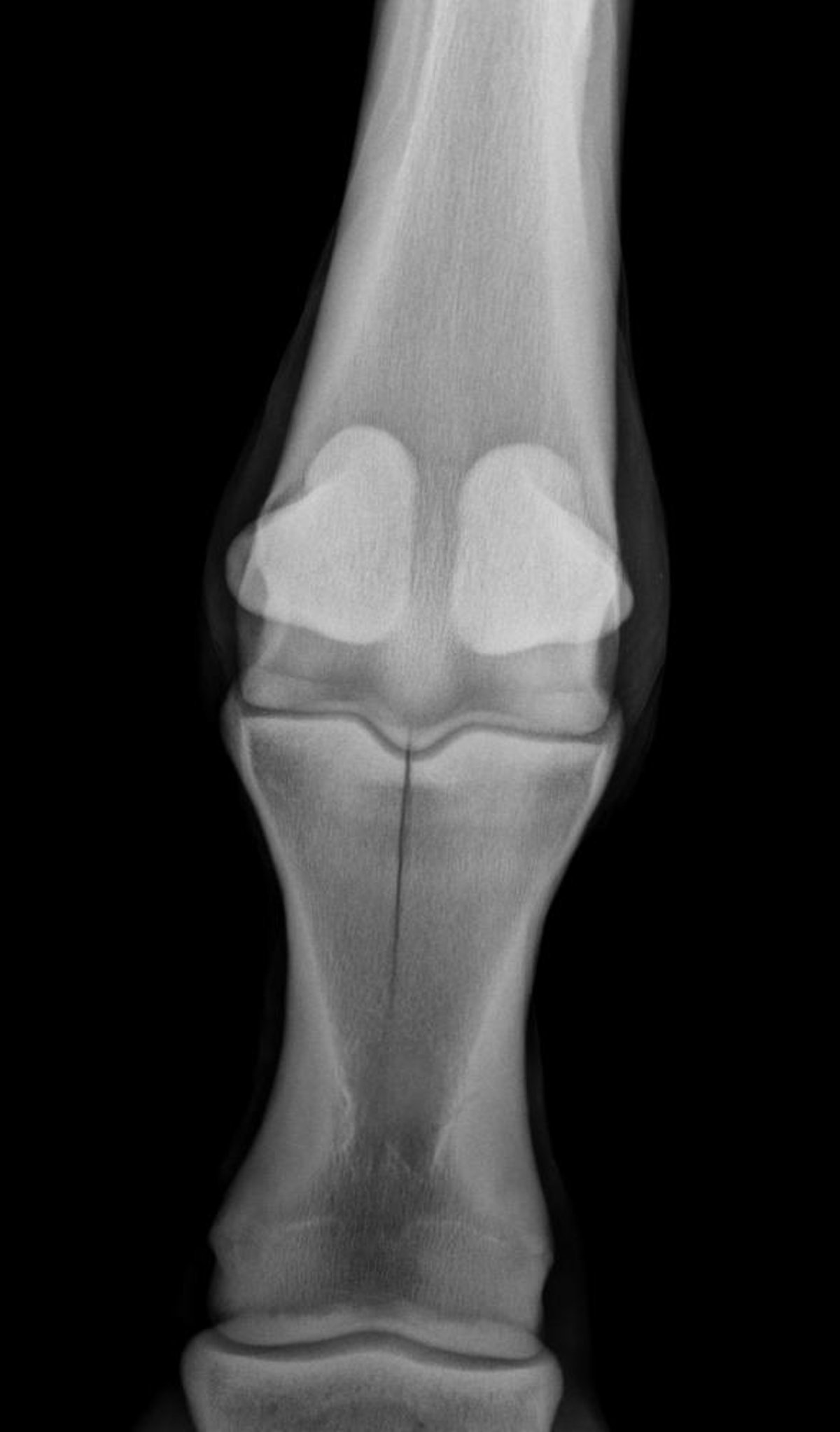
Courtesy of Dr. Matthew T. Brokken.
Diagnosis is confirmed by radiography. A number of oblique radiographic views may be necessary to ensure visibility of the fractures. For palmar or plantar osteochondral fractures, oblique radiographic views with the beam raised ~20° from horizontal may be more helpful for identification. The diagnosis of sagittal or comminuted fractures of P1 is typically straightforward, with marked lameness and swelling. Incomplete, short sagittal fractures can be more difficult to diagnose and may require special radiographic views off of dorsopalmar/plantar and/or nuclear scintigraphy in the initial stages, because the fracture line may be difficult to detect radiographically.
Osteochondral “chip” fractures can be removed arthroscopically with an excellent prognosis if no other abnormalities within the joint exist. Routine, nondisplaced sagittal P1 fractures can be repaired by internal fixation using screws placed in lag fashion via stab incisions. More complex P1 fractures typically require open reduction and repair via lag screws to allow accurate realignment of the articular surface of the fetlock to limit postoperative arthritis. Careful attention should be paid to the fracture configuration to ensure that all components are incorporated in the repair. In some circumstances, CT may aid an accurate diagnosis and reconstruction of the fracture. Conservative treatment of severely comminuted fractures involves immobilization with a plaster or fiberglass cast for up to 12 wk, with or without the use of transfixation pins through the third metacarpal/tarsal bone. Complications of P1 fracture repair include implant failure, poor alignment at the fracture site leading to secondary arthritis, and contralateral limb laminitis.

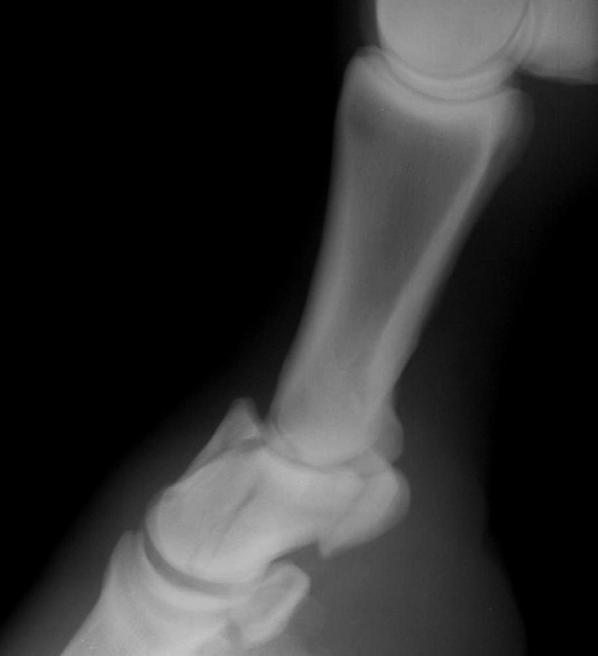
Courtesy of Dr. Matthew T. Brokken.
Fractures of the second/middle phalanx (P2) are most common in Quarter horses and typically affect the hindlimbs. Whereas osteochondral “chip” fractures are common off of proximal P1, osteochondral fractures are relatively uncommon off of P2. The most common fractures of P2 are either palmar/plantar eminence fractures of proximal P2 or comminuted fractures. Treatment of most P2 fractures is either with internal fixation with a combination of plate(s) and screws and/or a transfixation pin cast. Residual lameness typically is present and depends on the degree of osteoarthritis that develops in the distal interphalangeal joint and, to a lesser extent, the proximal interphalangeal joint (if not arthrodesed in the fracture repair). Prognosis depends on how comfortable the horse will be after fracture stabilization to limit the risk of contralateral limb laminitis.


