

Abnormalities of the leukogram include quantitative or numerical concentration abnormalities and WBC morphologic abnormalities.
Numerical Leukogram Abnormalities in Animals
WBC concentration values are interpreted by comparison with species-specific reference values. Interpretations should be made only by considering the absolute numbers. For reference values for total WBC and differential WBC concentrations in absolute numbers for common domestic species, see Table: Hematology Reference Ranges. The total WBC concentration is more variable and often higher in neonates than in adults. Age-related reference values should be used to evaluate leukograms in young animals, especially species in which lymphocytes are more numerous (and neutrophils less numerous) in adults, such as ruminants. Generally, differential WBC patterns of adults are reached at about the age of sexual maturity.
Abnormality in the total WBC concentration is useful only to alert the clinician to look for and interpret abnormalities in the cell distributions in the differential. When the total WBC is abnormal, one or more distributional abnormalities in the differential are likely. When the total WBC is normal, there still may be one or more distributional abnormalities in the differential. As a result, evaluation of the differential absolute values is the most important component of the leukogram.
Leukocytosis is an increase in the total WBC concentration, whereas leukopenia is a decrease in the total WBC concentration. Changes in the concentrations of specific leukocyte types are more important for clinical interpretation purposes.
Neutrophilia or neutrophilic leukocytosis is an increase in neutrophil concentration. Lymphocytosis is an increase in lymphocyte concentration. Monocytosis is an increase in monocyte concentration. Eosinophilia refers to an increase in eosinophil concentration, and basophilia is an increase in basophil concentration. Metarubricytosis or rubricytosis is an increase in nucleated RBCs (nRBCs) in blood. Mastocytosis is an increase in mast cells in blood.
White Blood Cell Terminology
WBC Type | Increased Concentration | Decreased Concentration |
|---|---|---|
Neutrophil | Neutrophilia, neutrophilic leukocytosis | Neutropenia |
Lymphocyte | Lymphocytosis | Lymphopenia |
Monocyte | Monocytosis | |
Eosinophil | Eosinophilia | Eosinopenia |
Basophil | Basophilia |
Decreases in concentration of a cell type are indicated by the suffix “penia.” This is applied only to cell types in which a decrease is possible. It does not apply to cell types for which the concentration may be 0, such as monocytes, basophils, nRBCs, and any other abnormal cell type. Hence, neutropenia is a decrease in neutrophil concentration, lymphopenia is a decrease in lymphocyte concentration, and eosinopenia is a decrease in eosinophil concentration. Cytopenia is a nonspecific term indicating a decrease in cell concentration(s), but the cell type is not specified. Pancytopenia indicates all cell types are decreased, often to a severe degree.
Terms used to describe or qualify abnormalities most often associated with inflammatory responses include various left shifts and leukemoid response. A left shift is an increase in concentration of immature, nonsegmented neutrophils, typically bands, but may also include metamyelocytes or even more immature forms. A regenerative left shift describes leukocytosis characterized by the combination of neutrophilia and a left shift. In this situation, the segmented neutrophils will be greater in concentration than bands and less mature forms. A degenerative left shift describes a neutrophil pattern characterized by normal to decreased total neutrophil concentration, but with a left shift in which the concentration of bands and less mature forms is greater than segmented neutrophils. This is an indication of maximal release from bone marrow in response to inflammation and signifies the presence of an acute, severe lesion.
Morphologic Leukogram Abnormalities in Animals
Abnormalities of WBC morphology may be associated with either acquired or inherited disease.

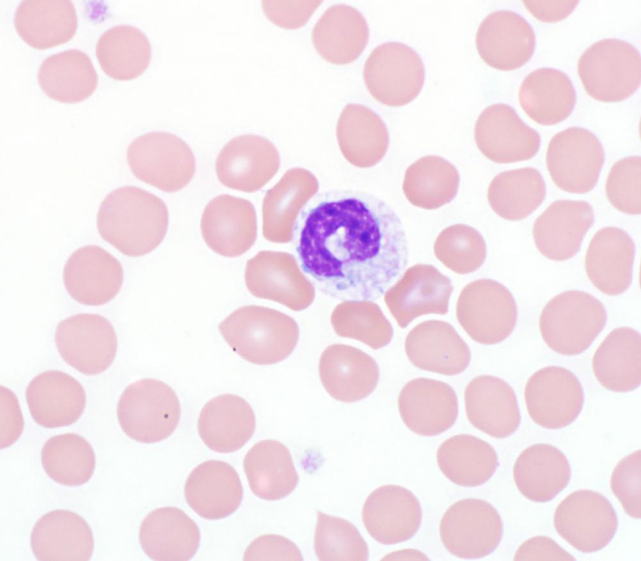
Photomicrograph of a blood smear from a dog showing a neutrophil with cytoplasmic basophilia, vacuolation and small Döhle bodies (Wright's stain, 1,000X).
Courtesy of Dr. Darren Wood.
Toxic changes are identified only in neutrophils. The term originates from historical observation that certain cell features were associated with general, usually overwhelming, toxic states, such as systemic bacterial infections and severe, acute inflammatory lesions. The term is misleading in that it implies neutrophil injury. The cells are not injured and have normal function. Toxic change is best defined as a set of morphologic changes observed on the blood smear that occur as a result of accelerated bone marrow production of neutrophils. The accelerated production is in response to relatively severe inflammatory states that maximally stimulate the bone marrow. Morphologic changes include (in order of frequency) diffuse cytoplasmic basophilia, Döhle bodies, and fine cytoplasmic vacuolation. Rarer changes include increased prominence of cytoplasmic azurophilic granules and nuclear immaturity.
Toxic changes are almost always associated with the concurrent presence of a left shift. It is graded as mild, moderate, or severe by subjective evaluation of the more common changes noted on examination of a blood smear. Döhle bodies, blue-gray cytoplasmic inclusions, are aggregates of endoplasmic reticulum. They are unique in that they may be found in clinically healthy cats and therefore are not interpreted as toxic change in this species unless excessively frequent and accompanied by other features.

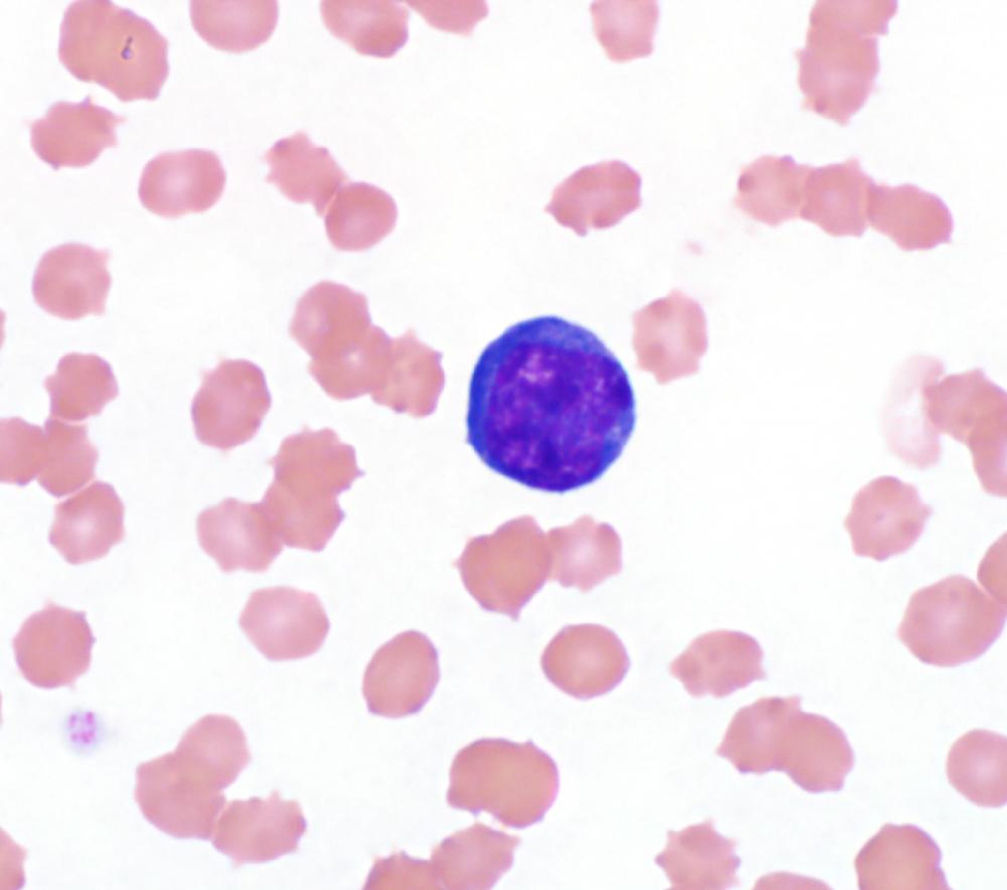
Photomicrograph of a blood smear from a dog with a reactive lymphocyte, a nonspecific indicator of immune stimulation. Note the large size and deeply basophilic cytoplasm. (Wright's stain, 1,000X).
Courtesy of Dr. Darren Wood.
Reactive lymphocytes have increased, distinctly basophilic cytoplasm and may have irregular or clefted nuclei. They may vary considerably in diameter. They have condensed chromatin and therefore are not blasts. They are interpreted as immunologically stimulated B cells.
Granular lymphocytes have condensed chromatin and increased pale blue-gray cytoplasm that contains several small pink or azurophilic granules. The nucleus may be round to clefted. These are large granular lymphocytes and may be either natural killer (NK) lymphocytes or T lymphocytes.
Blast cells are usually an indication of hematopoietic cell neoplasia if they are reproducible or present in large numbers. Their lineage may be tentatively identified by morphologic criteria, but flow cytometric analysis is required to definitively identify lineage.
Many of the following morphologic changes are uncommon.
Chédiak-Higashi syndrome, described in Persian cats, people, mink, foxes, Hereford and Brangus cattle, mice, and killer whales, is an autosomal recessive defect involving lysosomal granules. There is hyperfusion of granules resulting in large, eosinophilic cytoplasmic inclusions. Susceptibility to bacterial infections is increased, as is the tendency to bleed because of both neutrophil and platelet function abnormalities, respectively. Partial oculocutaneous albinism due to abnormal melanin granule formation may occur.
The mucopolysaccharidoses are a group of lysosomal storage disorders in which there is a defect in degradation of glycosaminoglycans. Both neutrophils and lymphocytes may contain accumulated mucopolysaccharide product in the form of purple or metachromatic intracytoplasmic granules. Lymphocytes may also be vacuolated. These disorders are associated with a variety of systemic clinical abnormalities and are seen in dogs and cats.
Another group of lysosomal storage disorders recognized in dogs and cats may result in cytoplasmic vacuoles predominantly in lymphocytes and occasionally in neutrophils. These disorders include gangliosidoses, alpha-mannosidosis, Niemann-Pick disease variants, acid-lipase deficiency, and fucosidosis. Most of these disorders result in severe, progressive neurologic disorders resulting from accumulated product in neuronal tissue.
Locoweed toxicosis is regarded as an acquired form of lysosomal storage defect in large animals. It is due to toxins from the plant that inhibits one or more enzymes of oligosaccharide metabolism. This may result in vacuolation in lymphocytes.

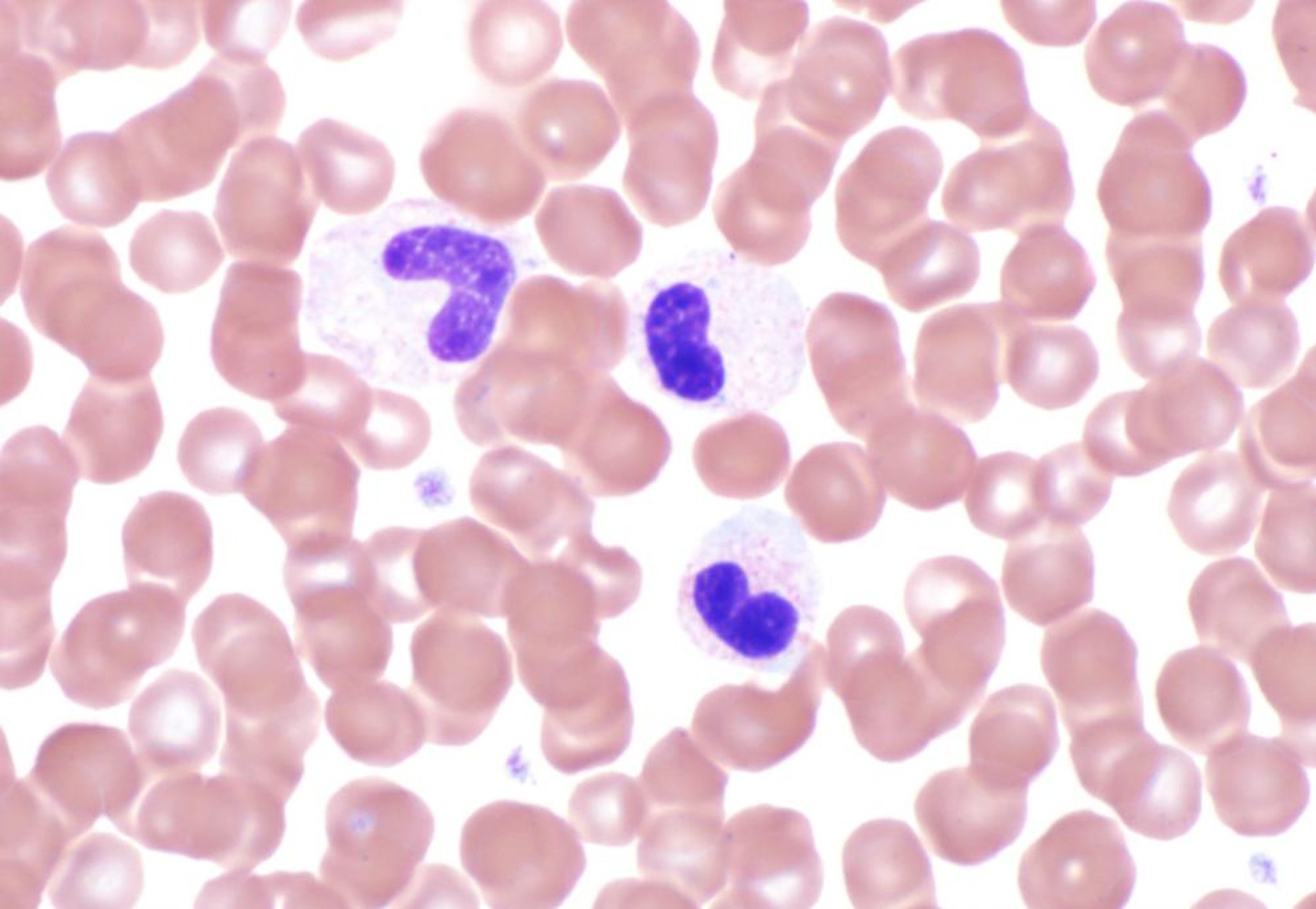
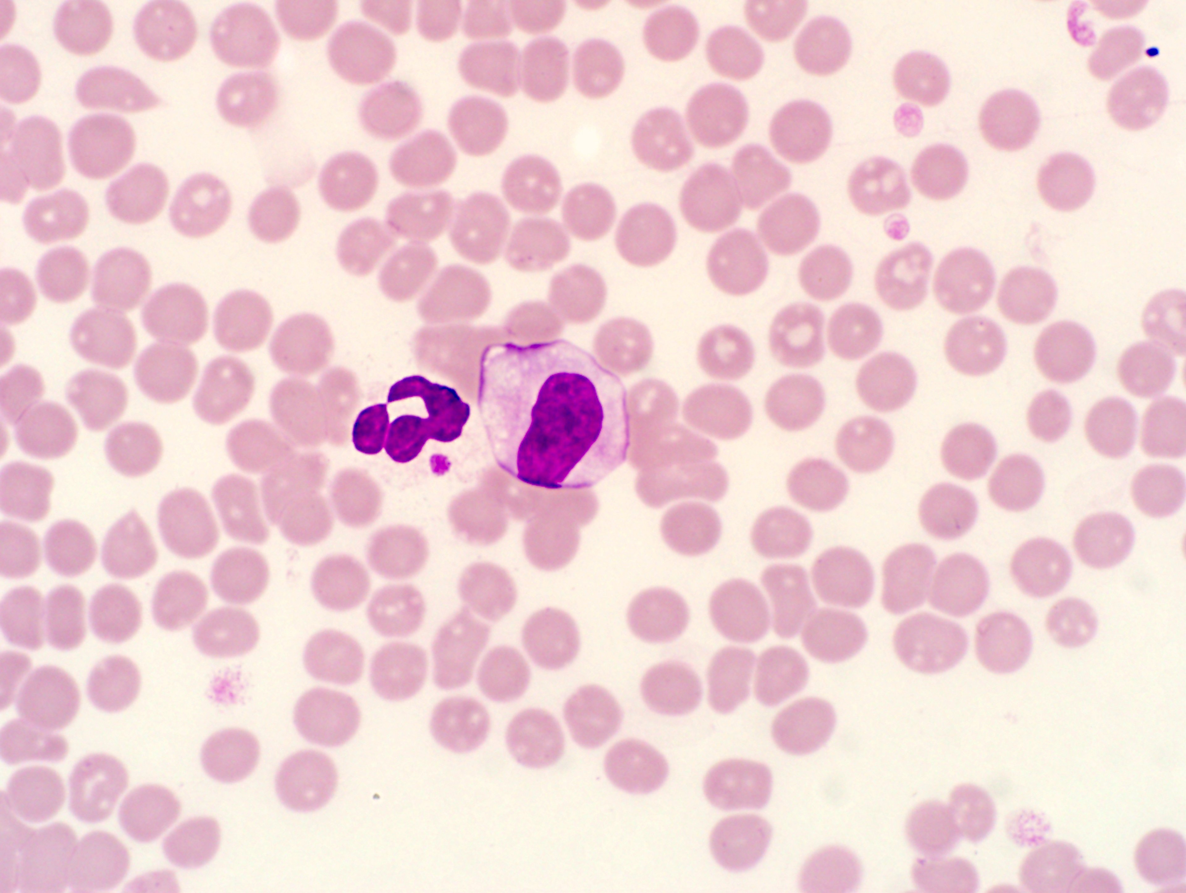
Photomicrograph of a blood smear from a dog. There are three neutrophils with mature clumped chromatin and hyposegmented nuclei present; (Wright's stain, 1,000X).
Courtesy of Dr. Darren Wood.
Pelger-Huët anomaly is a nuclear hyposegmentation defect of granulocytes in people, cats, rabbits, horses, and dogs that are heterozygous for the anomaly. Neutrophils have normal function but a near absence of segmented nuclear morphology. Most or all of the neutrophils appear as bands and metamyelocytes and may appear as a marked left shift in an otherwise normal leukogram. Eosinophils and basophils, if present, also exhibit nuclear hyposegmentation. Affected heterozygote animals are clinically normal; the homozygous inheritance of the trait is lethal.
Hypersegmentation is an increased degree of nuclear segmentation resulting in multiple lobes connected by nuclear filaments. It is a nonspecific indication of increased time in circulation and is normal aging of the cell. This may be seen with stress leukograms or corticosteroid administration.

Photomicrograph of a blood smear from a cat showing several mature lymphocytes clumped together (Wright's stain, 1,000X).
Courtesy of Dr. Darren Wood.
Leukocyte agglutination may occur with either neutrophils or lymphocytes. This is seen on low magnification as aggregates of 5–15 tightly clumped leukocytes. Avid agglutination may result in a markedly false low total WBC concentration on some cell counting instruments. This is likely due to the presence of a naturally occurring cold agglutinin that is operative only in vitro at laboratory temperature. There is no known clinical significance.

Photomicrograph of a blood smear from a dog showing the morula of a rickettsial organism in the cytoplasm of a neutrophil (cell on left stained purple). The most likely infectious agent is Anaplasma phagocytophilum.
Courtesy of Dr. Darren Wood.
Infectious disease inclusions are occasionally recognized. Canine distemper inclusions may be seen in neutrophils, monocytes, and lymphocytes, as well as in newly produced erythrocytes. The ehrlichioses of various animal species and canine hepatozoonosis may have cytoplasmic inclusions of respective organisms of these tickborne diseases.
Specific Interpretative Leukogram Responses
The abnormal leukogram is typically interpreted into one of several responses, each of which may consist of one or more abnormalities in the differential. Some may also be associated with concurrent changes in erythrocytes and platelets. Important species differences in leukogram responses are described below.
Corticosteroid-induced or Stress Response in Animals
In this very common leukocyte response, endogenous steroid release from stress or treatment with exogenous corticosteroids results in a leukogram with multiple changes. Lymphopenia is the most consistent change, and mature neutrophilia is usually present. Monocytosis and eosinopenia are expected changes in dogs but are more variable in other species. Eosinopenia cannot be determined when the lower reference value is zero. Neutrophilia is due to decreased adherence to the vascular endothelium, which inhibits margination and increases circulating time. As a result, neutrophils may also become hypersegmented. There may also be increased marrow release of neutrophils. Lymphocytes become redistributed to lymphoid tissues instead of remaining in circulation. This response may be misinterpreted as inflammation, but a left shift and toxic changes are not usually present.
Excitement or Epinephrine Response in Animals
Leukocytosis may occur as a result of exercise or excitement; this response is mediated by increased epinephrine concentration and may be thought of as a transient physiologic response. Epinephrine flushes cells from the marginal to the central pool. The effect may double the total WBC concentration within minutes. In addition, splenic contraction releases WBCs and RBCs into the peripheral circulation. The leukocytosis is usually due to a mature neutrophilia without a left shift or toxic changes. Lymphocytosis may be present, especially in young horses or cats. The effect in cats is often recognized as a prominent lymphocytosis—as much as two times the upper reference value. The excitement response is relatively uncommon in dogs.
Inflammatory Response in Animals
The concentration of neutrophils in blood in response to inflammatory disease is highly variable and dynamic. It is best viewed as a balance between tissue demand and bone marrow production at all phases of the response. There are important species differences in this balance that are related to bone marrow storage reserve and proliferative capacity.
At the beginning of an inflammatory process, the bone marrow responds by delivery of its reserve of late-stage maturing neutrophils, including band cells. If consumption exceeds marrow delivery during this acute stage, neutropenia with a prominent left shift will develop. In dogs and cats, this is an indication of severity of the inflammatory lesion.
Subsequently, it takes 2–4 days for the marrow to accelerate neutrophil production by increased stem-cell entry and expansion of proliferative stages that feed the maturation stages and amplify neutrophil delivery to blood. In dogs, the acute stage of the inflammatory response is usually characterized by mild to moderate neutrophilia, with the left shift being somewhat proportional to severity of demand.
After a few days, accelerated bone marrow production adds to the picture. Neutrophilia may increase along with a left shift and toxic changes. As the process becomes chronic, the balance between increased marrow output and consumption may favor the development of even higher magnitudes of neutrophilia. The most chronic form, present for weeks or even months, is described as a “closed cavity” inflammatory process in which a lesion becomes somewhat "walled off" and therefore consumes fewer neutrophils, yet still stimulates maximal bone marrow production. Examples of closed cavity processes are pyometra in dogs and traumatic reticuloperitonitis (hardware disease) in cattle. In these conditions, the magnitude of total WBC concentration, consisting of neutrophilia, may be as high as 100,000/μL (100 × 109/L) in dogs. Extreme neutrophilia, exceeding upper reference limits usually seen in inflammation, may be associated with leukemia, Hepatozoon canis infections, and rarely other neoplasms that produce colony-stimulating factors.

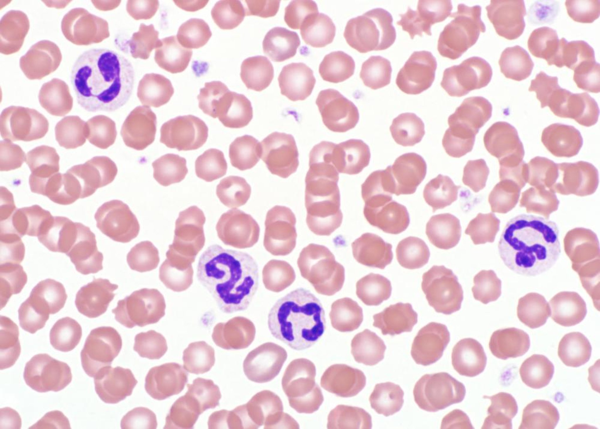
Photomicrograph of a blood smear from a dog with neutrophilia. The cells are mature, segmented and lack toxic changes. (Wright's stain, 1,000X).
Courtesy of Dr. Darren Wood.
In contrast, cattle and most other ruminants have a relatively low reserve of bone marrow neutrophils and a lower capacity for accelerating granulopoiesis. This is reflected in the relatively low neutrophil concentration in the blood of healthy ruminants. As a result, acute inflammation in cows is characterized by neutropenia that can be profound and neutropenia in cattle does not reveal the severity of inflammation. After several days, the bone marrow response may establish a return of blood neutrophils in modest concentration, characterized by a marked left shift and toxic changes. Chronic, closed cavity inflammatory lesions are associated with magnitudes of neutrophilia that rarely exceed 25,000/mcL (25 x 109/L) of blood. Cats and horses are intermediate in these responses, with cats being more like dogs and with horses being more like cattle. Pigs have an inflammatory leukogram similar to that of dogs.
Bovine leukocyte adhesion deficiency is a lethal, autosomal recessive disorder of Holstein cattle. It is associated with marked neutrophilia; the neutrophils have a deficiency of the glycoproteins (integrins) that are essential for normal leukocyte adherence and emigration from the vasculature. Recurrent bacterial infections, persistent neutrophilia (often >100,000/mcL [100 × 109/L]), lymphocytosis, and death (usually between 2 weeks and 8 months of age) are characteristic. Calves often are stunted and have recurrent pneumonia, ulcerative stomatitis, enteritis, and periodontitis. On histologic examination of tissues, there are few neutrophils, except within vessel lumens, because they persist in the circulation and have impaired entry into the tissues. Testing is available to detect carriers. A similar defect has also been reported in some Irish Setter dogs.
Neutropenia may develop because of excessive tissue demand for neutrophils or reduced granulopoiesis. It may be occur in the presence of overwhelming bacterial infections, especially gram-negative sepsis or endotoxemia, in all species. Immune-mediated destruction of neutrophils is diagnosed by exclusion of other consumptive processes. Stem-cell injury may occur from many causes, such as various viral infections ( see Table: Viral Infections that May Cause Transient Neutropenia), chemical injury, and idiosyncratic drug reactions, eg, caused by sulfonamides, penicillins, cephalosporins, and chloramphenicol in cats. These reactions typically affect all bone marrow cell lines but are recognized initially as neutropenia because of the relatively short lifespan of this cell type.
Viral Infections that May Cause Transient Neutropenia
Species | Infection |
|---|---|
Dogs | Parvovirus, canine distemper (acute phase) |
Cats | Panleukopenia (parvovirus), feline leukemia virus |
Horses | Equine influenza, equine viral arteritis (acute phase), equine herpesvirus |
Cattle | Bovine viral diarrhea virus |
Pigs | Classical swine fever virus, African swine fever virus |
Neutropenia is seen in the now rare cyclic hematopoiesis syndrome of gray Collie dogs, also known as canine cyclic neutropenia. It is an inherited, autosomal recessive disease characterized by a profound recurrent neutropenia, associated overwhelming recurrent bacterial infections, bleeding, and coat color dilution. The defect is due to a mutation in a protein that may regulate neutrophil elastase activity. Neutrophil maturation is arrested at regular intervals of 11–14 days; the peripheral blood neutropenia lasts 3–4 days and is followed by neutrophilia. All other hematopoietic cells, including lymphocytes, also have cyclic production that is minimally evident because of the relatively long circulation time of other cell types. Affected puppies often die at birth or during the first week of life, and rarely live longer than a year. Surviving dogs may be weak with stunted growth, and develop serious recurrent bacterial infections during periods of neutropenia. Monocytosis may be seen in the inflammatory pattern at any stage of its progression. Monocytosis is more likely and tends to be of greater magnitude when the process becomes chronic.
Combined Corticosteroid-induced and Inflammatory Response in Animals
Inflammatory disease processes commonly induce a concurrent endogenous corticosteroid response, recognized by the presence of lymphopenia in conjunction with an inflammatory neutrophil response (left shift). The neutrophil response to inflammation overrides and may be additive to the corticosteroid influence on neutrophils.
Lymphocytosis in Animals
Mild lymphocytosis and reactive lymphocytes may occur after vaccination. Modest lymphocytosis, between 7,000–20,000/mcL (7–20 × 109/L), should prompt consideration of a possible physiologic excitement response, particularly in cats. If that is excluded, then a lymphoproliferative disorder should be considered. If examination of lymphocyte morphology reveals prolymphocytes and/or blast cells, then lymphocytic leukemia is the working interpretation. If the cells are all small with normal-appearing chromatin, then chronic lymphocytic leukemia is a consideration requiring further evaluation. Chronic ehrlichiosis may result in lymphocytosis of this magnitude in dogs. At higher concentrations, the lymphocytosis may be regarded as conclusive evidence of leukemia.
Persistent lymphocytosis in cattle is defined by lymphocyte concentrations consistently >7,500/mcL (7.5 × 10⁹/L). It is due to a B-cell proliferation that occurs in a subset of animals infected with bovine leukemia virus (BLV). Affected cattle are usually asymptomatic. The finding of persistent lymphocytosis is regarded as a positive indication of BLV infection in individual animals. A smaller subset of BLV-infected cattle, either with or without lymphocytosis, may progress to develop lymphoma or lymphocytic leukemia.
Lymphopenia in Animals
Lymphopenia is a common leukogram abnormality most commonly associated with stress (endogenous) or corticosteroid administration (exogenous). The most likely cause is steroid-induced apoptosis of lymphocytes. Lymphopenia also rarely occurs due to other causes, such as extravasation of lymph (eg, lymphangiectasia, chylous effusion), some viral infections with tropism for rapidly dividing cells (eg, parvoviral infections), and hereditary immunodeficiency disease (eg, combined immunodeficiency disease of Arabian foals).
Stem-cell Injury and Pancytopenia in Animals
A number of factors may cause reversible or irreversible stem-cell injury. These injuries may affect erythrocyte, platelet, lymphocyte, and/or granulocyte production. Because of short circulating lifespan, neutropenia is often the first abnormality seen. When chronic or irreversible, these injuries result in decreases in all three major blood cell lines, with the hemogram demonstrating leukopenia, nonregenerative anemia, and thrombocytopenia. General causes include 1) overdoses of radiation and antineoplastic drugs, 2) drug or plant toxicities (eg, estrogen toxicity in dogs, bracken fern toxicity in cattle, phenylbutazone toxicity in species other than horses), 3) hematopoietic cell neoplasia involving bone marrow (myelophthisis), and 4) viral infections that injure rapidly dividing cells and may cause transient neutropenia ( see Table: Viral Infections that May Cause Transient Neutropenia).
Eosinophilia and Basophilia in Animals

Photomicrograph of a blood smear from a dog. Note several eosinophils with bright orange-red cytoplasmic granules are present (Wright's stain, 1,000X). This can be observed with parasitism or allergies.
Courtesy of Dr. Darren Wood.
Eosinophilia, or the combination of eosinophilia and basophilia, prompts the consideration of the following processes: allergic-based inflammation, parasitic infestation, subepithelial (skin, respiratory, GI) inflammation that is likely allergic in nature, and less commonly, paraneoplastic induction. Eosinophilia is seen in most dogs with heartworm disease and may be seen in dogs and cats with flea infestation. Decreased concentration of these cell types in blood has no pathologic relevance.
Hypereosinophilic syndrome has been reported in cats, dogs, and ferrets. This poorly understood syndrome is characterized by persistent marked eosinophilia and eosinophil tissue infiltration with associated organ dysfunction.
Metarubricytosis in Animals

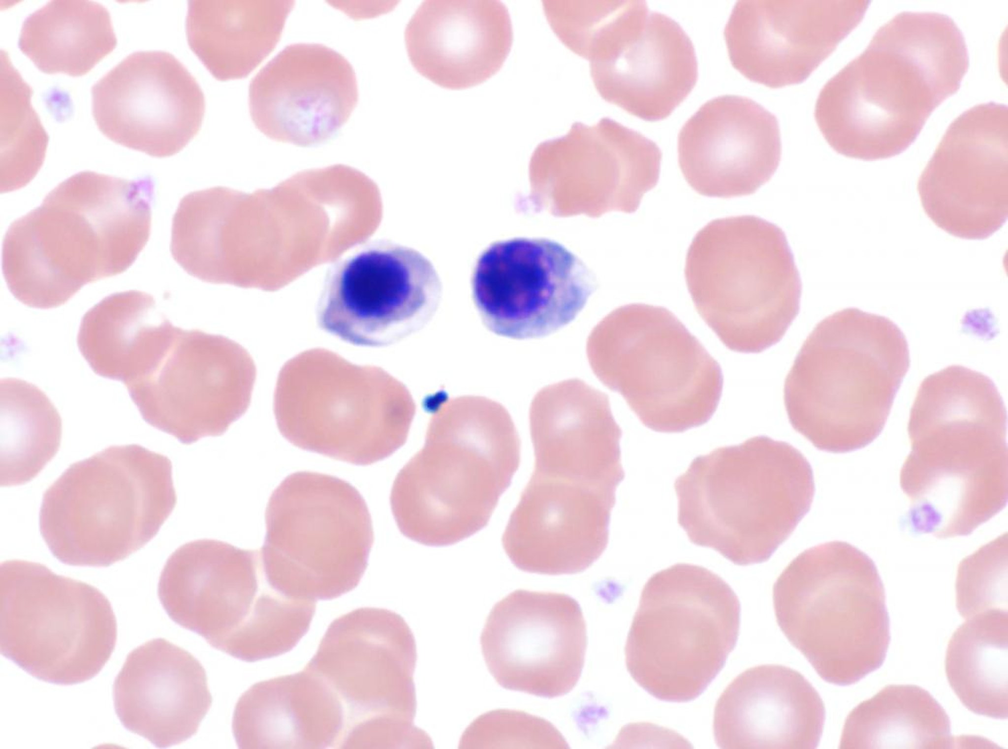
Photomicrograph of metarubricytes in a blood smear from a dog. The cell on the left is slightly more mature than the one on the right, with more condensed nuclear chromatin and less basophilic cytoplasm. (Wright's stain, 1,000X).
Courtesy of Dr. Darren Wood.
Although typically absent, metarubricytes occasionally become a major component of the total nucleated cell count. The magnitude may be 10%–50% of the nucleated cell population or more, with absolute numbers reaching 5,000–10,000/mcL (5–10 × 109/L). This may occur rarely in early phases of an intense regenerative response to anemia. It may also be associated with endothelial injury (eg, heat stroke) resulting in abnormal release rate of nRBCs from bone marrow. Most nRBCs will be counted as lymphocytes on cell counters with differential capability. This may result in a preliminary result of lymphocytosis being corrected later only by examination of the blood smear.
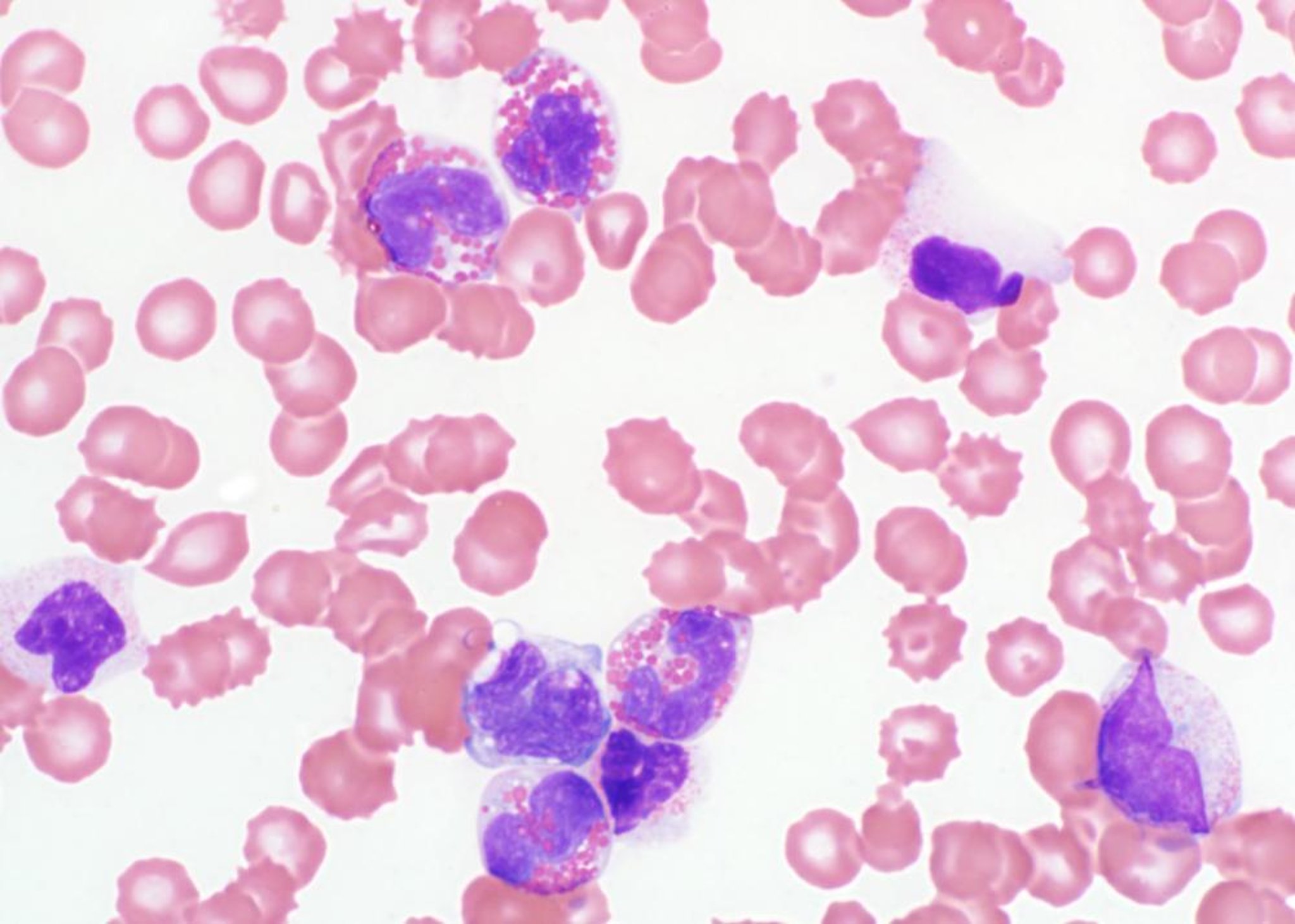
Hematopoietic Cell Neoplasia and Leukemia in Animals

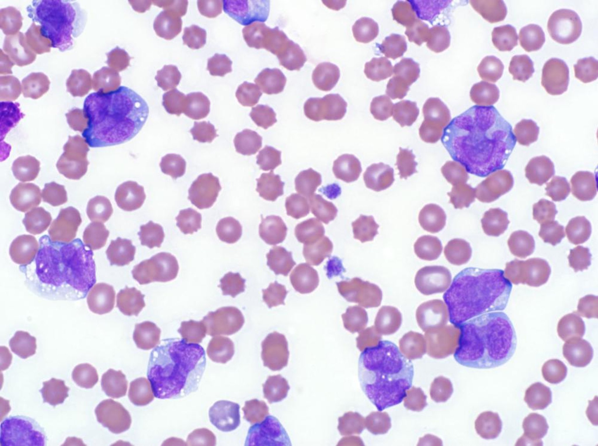
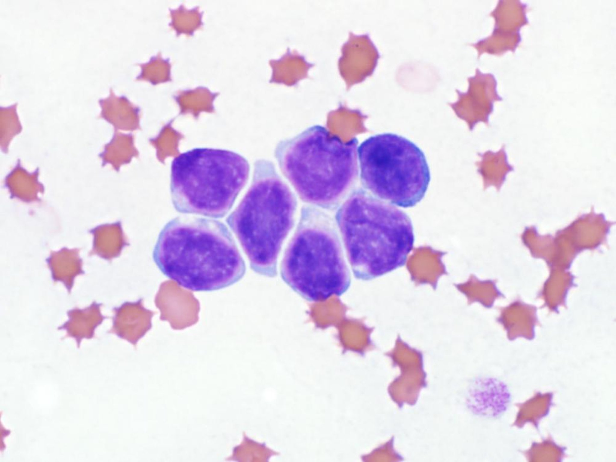
Photomicrograph of a blood smear from a dog with leukemia. Several atypical large cells of hematopoietic origin with convoluted nuclei are present.
Courtesy of Dr. Darren Wood.
Most cases of hematopoietic cell neoplasia of either lymphocytic or bone marrow origin will have some abnormal cells in blood. Sometimes, neoplastic cells are present in low numbers and are detected only by scanning the blood smear under low magnification. Finding abnormal hematopoietic precursor cells in blood in small numbers prompts investigation of bone marrow and/or other hematopoietic tissues (such as the spleen) for possible neoplastic disease.
The opposite extreme is marked leukocytosis with a predominance of the abnormal (neoplastic) cell population. In this situation, the blood sample is diagnostic for leukemia. If poorly differentiated, the cells are classified as blasts, with possible cell lineage determined based on morphologic appearance. If well differentiated, the cell lineage is usually more clear, because the cells are mature. Definitive diagnosis can be made using specialized techniques such as flow cytometry.
Lymphocytic leukemias are more common in domestic animals than are myeloproliferative neoplasms (ie, leukemias of granulocytes, erythrocytes, or platelets). Myelodysplasia is a term used to describe peripheral blood cytopenias, a hyperplastic response in bone marrow, and dysplastic features in developing cells. This can occur with a toxic insult to the bone marrow but may also indicate a preleukemic phase (myelodysplastic syndrome), and serial hemograms should be monitored.
A distinction is often made between an "acute" or "chronic" leukemia. The terms are more in reference to the clinical course of the disease rather than the duration of the tumor. An acute leukemia of leukocytes often causes systemic signs of illness and a poor short-term prognosis. These animals have variable numbers of poorly differentiated blast cells in circulation, as well as cytopenias in other cell lines. In contrast, a chronic leukemia of leukocytes often causes few to no clinical signs, may be discovered incidentally, and can have a long clinical course. These animals usually have large numbers of well-differentiated cells in circulation and lack other cytopenias except perhaps anemia.
Considerable progress is being made in the use of monoclonal antibody labeling and flow cytometric analysis to better establish cell lineage, particularly when the morphology is equivocal. This is particularly useful for poorly differentiated leukemias, in which morphology alone is unreliable. The distinction between well-differentiated or chronic myelogenous leukemia and extreme neutrophilic leukocytosis can be difficult. The latter is much more frequent, and a source of inflammation should be sought before concluding the changes indicate neoplasia.
For More Information
Also see pet health content regarding white blood cell disorders of dogs, white blood cell disorders of cats, and white blood cell disorders of horses.


