

Coccidia are single-celled obligate intracellular protozoan parasites in the class Conoidasida within the phylum Apicomplexa. The main clinical sign of coccidiosis is diarrhea. Oocysts can be identified in feces by use of salt or sugar flotation methods, direct intestinal smears, or a McMaster counting chamber. Antiprotozoal treatment can shorten the length of illness, decrease discharge of oocysts, alleviate clinical signs, and reduce likelihood of secondary infections and death.
Coccidia are obligate intracellular protozoan parasites in the class Conoidasida within the phylum Apicomplexa. The genera Eimeria (sporulated oocyst contains four sporocysts) and Isospora (sporulated oocyst contains two sporocysts) contain many species that infect a wide variety of birds, mammals and reptiles, but almost all are species host-specific. Infections occur throughout the world. Other genera cause besnoitiosis, cryptosporidiosis, sarcocystosis, and toxoplasmosis.
Infection is oral from ingestion of infective sporulated oocysts from a contaminated environment. Most animals are exposed to infection early in life.
Clinical signs of infection usually only occur in the young. The parasites mainly invade the intestinal epithelial lining and underlying connective tissue of the mucosa, sometimes leading to catarrhal inflammation and hemorrhage. In rabbits, the liver is also affected. During their lives most animals become infected but show no illness, and the disease is self-limiting. However, when clinical signs occur, they range from diarrhea and decreased growth in mild cases to dysentery, dehydration, and tenesmus in severe cases, usually occurring with widespread hind gut involvement. Mortality rate is generally low. Chronic infections can occur, leading to pasty feces, staring coats, poor growth, and variable sizes within an age group. In production animals, infection can have serious economic consequences. In cats, dogs, and horses, infection is mainly from Isospora spp, and clinical signs are less common and usually less severe than with Eimeria spp.
Various treatments are available but those approved vary in different countries. Control depends on decreasing environmental contamination and the stresses to which susceptible animals are subjected. Coccidiostats can also be used to decrease oocyst production and disease.
Etiology and Pathogenesis of Coccidiosis in Animals
All species of Eimeria complete their life cycle within a single host (monoxeny), as do most Isospora spp. However, some Isospora spp can use optional transfer or paratenic hosts. Development does not occur in these hosts, but allows the parasite to be dispersed to new areas. A new genus name, Cystoisospora, has been proposed for those species of Isospora with paratenic properties. Otherwise, coccidia are host-species specific and there is no cross-immunity between different coccidial species in the same host. Intercurrent infections are common and may be exacerbated because coccidiosis can be immunosuppressive.
The life cycle is complex but is all completed within the one host producing oocysts. There are three stages in the life cycle: sporogony, merogony and gametogony.
Sporogony/sporulation: Oocyst passed in the host’s feces are not immediately infective. They have a thick, resistant wall but require moisture, oxygen, and warmth to sporulate. In sporulation, the oocyst nucleus divides a number of times, dependent on the species, to produce sporocysts. The divided nucleus is surrounded by protoplasm forming a conical body, a sporoblast, which in turn secretes a wall around itself and divides into two sporozoites to become a sporocyst. The development of this infective sporulated oocyst can occur in hours but it may take days or weeks. These infective oocysts are resistant in the environment and can survive long periods, including over winter.
Merogony/schizogony (asexual stage): Once the infective sporulated oocyst is ingested, it releases the sporozoites in the small intestine and, after activation by the bile or trypsin, they invade the intestinal mucosa. Here, they become rounded trophozoites, which then divide into nucleated elongated merozoites that together form a meront. Once developed, the meront ruptures, as does the containing intestinal cell wall, thereby releasing the merozoites. Depending on the coccidial species, the merozoites may repeat the process of meront production in one or more cycles before they ultimately progress to the gametogony stage.
Gametogony (sexual stage): The last wave of merozoites enters other intestinal cells where they develop into large cells (macrogametocytes), which are female, or a large number of small flagellated male cells (microgametocytes). The male gametocytes are released by the rupture of the containing intestinal cell. One microgametocyte penetrates a macrogametocyte, resulting in their nuclei fusing. The resultant cell develops a thick wall and becomes an oocyst, which is ultimately released into the feces. With this life cycle, depending on the coccidial species, one ingested oocyst after two asexual generations could produce 24 × 106 merozoites and many million oocysts.
Infection results from ingestion of infective sporulated oocysts. When shed, oocysts are unsporulated and so are not infective. Under suitable environmental conditions of moisture, oxygen, and warmth, sporulation occurs within several days. The oocysts have a thick, resistant shell and can remain in the environment ≥1 year. However, they survive poorly at temperatures <~30°C or >40°C.
Of the numerous species of Eimeria or Isospora that can infect a particular host, many are not pathogenic. In general, those species that only invade the small intestine have fewer pathogenic effects on the host as the absorptive function more or less can be compensated for in the large intestine. More and more severe clinical signs (tenesmus, bloody feces, severe diarrhea, anemia, weakness) tend to occur in those species invading the large intestine and cecum because there is no subsequent region to undertake absorption and because the rate of cell replacement after damage is much slower in this area. Anemia is a rare clinical sign. Concurrent infections with two or more Eimeria spp are common; some of these may not normally be considered pathogenic, but also influence clinical disease. Within pathogenic species, strains may vary in virulence.
Coccidial infection tends to be immunosuppressive, allowing other diseases to become potentially more severe or of longer duration.
The life cycles of Eimeria and Isospora are self-limiting and end spontaneously within a few weeks, unless reinfection occurs. After Isospora infections, immunity is good and prevents further infection. Immunity after Eimeria exposure is usually lifelong, although small numbers of oocysts may occur in the feces at times, thereby maintaining immunity. Protective immunity appears to be both humoral and cellular but is mainly mediated by T-lymphocytes.
Epidemiology of Coccidiosis in Animals
Coccidiosis occurs universally, and most coccidial species are ubiquitous. Usually young mammals, particularly herbivores, are infected from an oocyst-contaminated environment either indoors or outdoors. Illness occurs more often indoors because of lower exposure rates outdoors with lighter exposure levels, allowing the host to be exposed to small numbers of oocysts over an extended period, thereby allowing natural immunity to develop without illness.
Infections tend to be seasonably variable. In the northwestern and midwestern US, cattle infection is higher in the summer, fall, and spring, and lower in midwinter and early summer. In Canada, winter coccidiosis occurs after a prolonged cold spell or after a sudden change to severe weather. In Queensland, Australia, weaned cattle tend to be more affected in dry years, possibly partly due to stress. In the UK, most cattle infections occur in the summer followed by the spring, fall, and winter. Most cases appear in the month of June and the fewest in January (US) and February (UK).
Coccidia are opportunistic pathogens. In general, for most farm animals, the infection rate is very high, but typically without any clinical disease. Clinical coccidiosis normally develops because of high oocyst exposure or various stressors on the hosts such as poor nutrition, nutritional deficiencies, overcrowding, poor sanitation, or intercurrent disease. Problems can also be related to stresses such as weaning, grouping, shipping, sudden feed changes, or weather changes, especially when severe. Usually, when clinical signs develop, they only develop in a few animals (5%–10%), although serial fecal sampling over a period shows most or almost all the others shedding oocysts.
The age of disease onset depends on the coccidial species and its prepatent period. Thus, most disease occurs from about 1–3 months old (piglets from under a week), although in animals born outside, it may be up to 1 year. Usually clinical signs are less severe in older animals, provided they are not exposed to new farm environments and are not immunosuppressed. During initial infection, there is usually some extent of immunosuppression which can result in outbreaks of other diseases. Older animals are usually highly resistant to clinical disease, but not totally resistant to infection, especially around parturition. Such events in clinically normal animals allow infection to persist on premises and act as an infection source for young susceptible animals.
Further illness after previous exposure occurs rarely, usually due to other problems in specific animals such as immunosuppression or intercurrent illness. Corticosteroid injections allow coccidia to complete their life cycle and shed oocysts.
Clinical Findings of Coccidiosis in Animals
Most infected animals are young when exposed to oocysts in the environment and show no observable clinical signs. Animals with subclinical infection may have decreased growth rates and decreased feed conversion, which can be important in production animals. For example, the initial decrease in weight gain in cattle may remain for at least one grazing season.
The clinical signs of coccidiosis are due to destruction of the intestinal epithelium and, frequently, the underlying connective tissue of the mucosa. This may be accompanied by hemorrhage into the lumen of the intestine, catarrhal inflammation, and diarrhea. The main clinical sign is diarrhea. Dehydration may result. Mainly when there is hind gut involvement, hemorrhage and tenesmus may occur. Typically, mortality rate is low. Serum protein and electrolyte concentrations (typically hyponatremia) may be altered. Anemia with decreased blood hemoglobin or PCV occurs uncommonly and then only in severely affected animals, which may die.
Chronic infection occurs in some animals, causing economic loss due to decreased feed intake, feed conversion efficiency, and weight gain. Some of these animals have poor-quality coats and are unthrifty. Infection can lead to nutritional deficiencies and digestive problems. In some cases, it can result in exacerbation of any intercurrent infections, causing animals to have more severe clinical signs and lengthening their duration or recovery period, either due to immunosuppression or for other reasons.
Postmortem Lesions
Postmortem findings typically show damage to various parts of the mucosal lining with some or many parts of the small intestine. Depending on the species of coccidia, lesions may be found in the small or large intestine. Most coccidial species have preferred development sites. There may be diffuse inflammation of parts of the small intestine, cecum, and large intestine. The mucosa is thickened, congested, and edematous with petechial or larger hemorrhages. The intestinal surface, hemorrhage, and diphtheritic fibromembranous lesions may show visible pinpoint or larger white areas of meronts. Mucosal scrapings or smears show large numbers of the various stages of the parasitic life cycle, especially gamonts and oocysts. Histologic examination shows loss and shedding of the epithelial surface, villous atrophy, oocysts, and gamonts; inflammatory cells are seen in the lamina propria and submucosa.
Diagnosis of Coccidiosis in Animals
Presence of fecal oocysts
History (age of affected animals, environment, and management)
Clinical signs (diarrhea in several young animals is indicative)

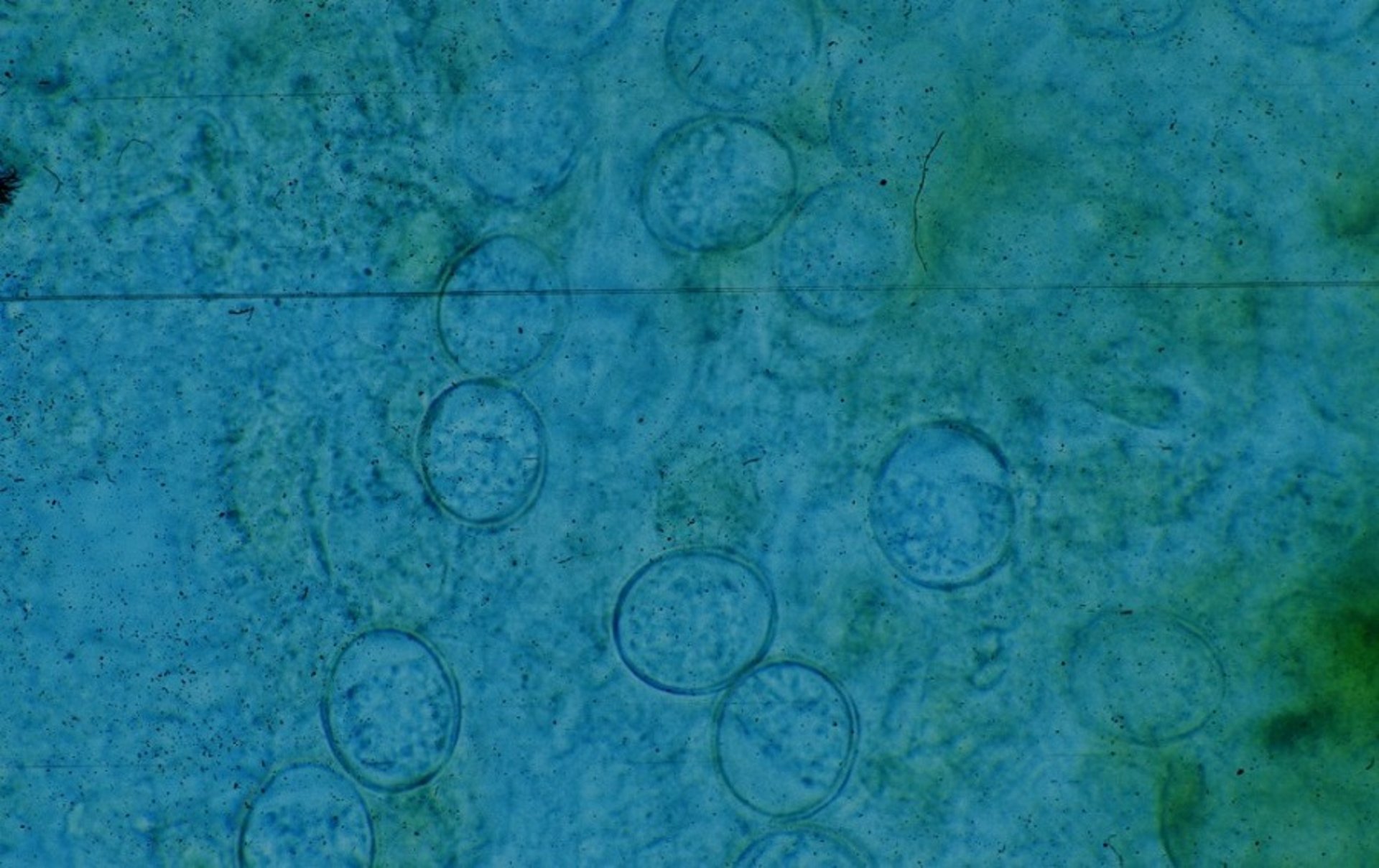
Photomicrograph of oocysts of Eimeria zuernii in a fecal smear from a calf.
Courtesy of Dr. Sameeh M. Abutarbush.

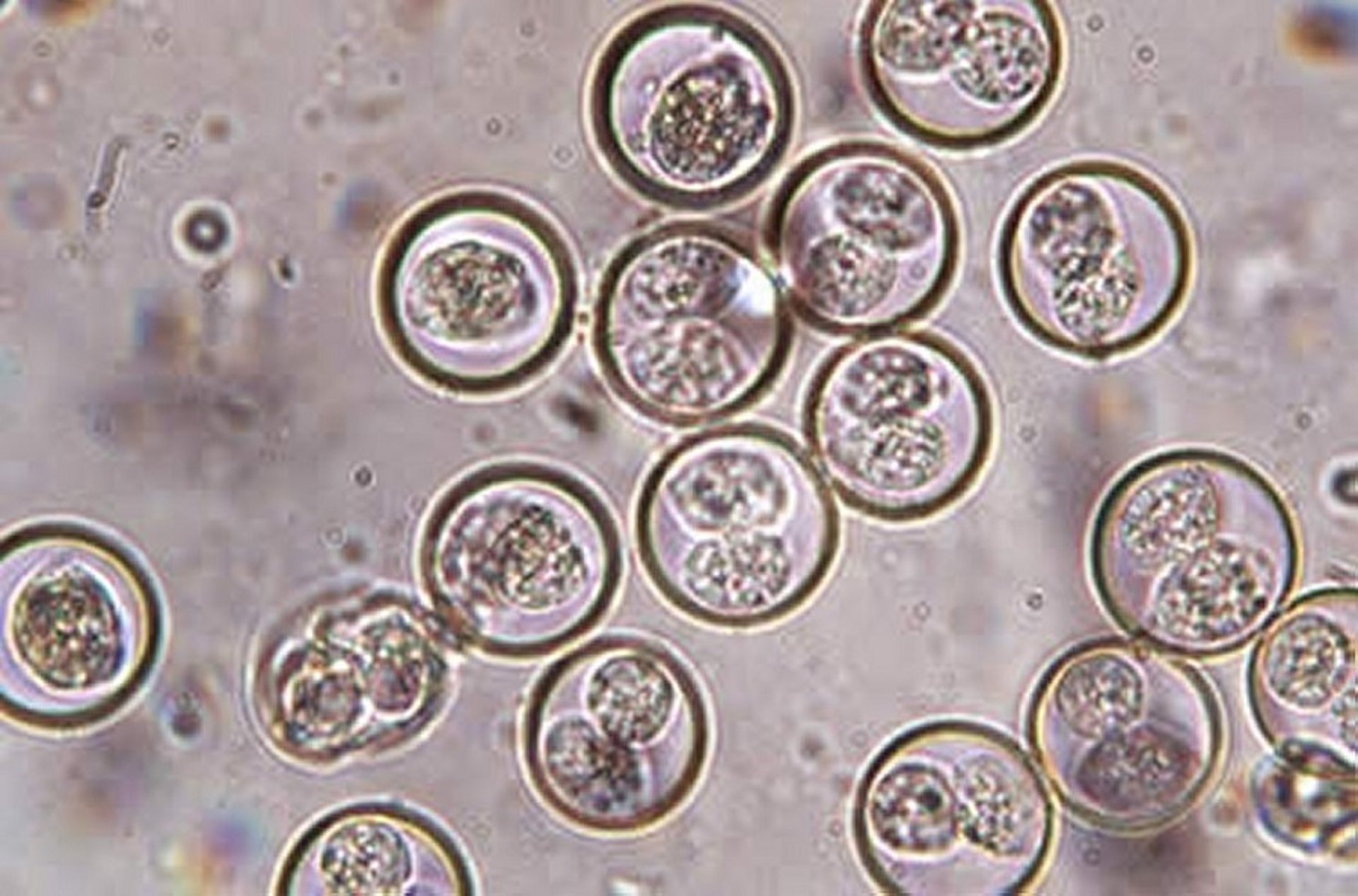
Photomicrograph of Isospora amphiboluri oocysts from a bearded dragon. Original magnification, 400×.
Courtesy of Dr. Roger Klingenberg.
Fecal samples should be taken from several animals in a group with and without (recorded) clinical signs of coccidiosis. They usually indicate oocyst presence. Oocysts can be identified in feces by use of salt or sugar flotation methods, direct intestinal smears, or a McMaster counting chamber. However, the severity of diarrhea may not correlate with the peak period of oocyst shedding. Finding notable numbers of oocysts of pathogenic species in the feces is diagnostic (>100,000 oocysts/g of feces in severe outbreaks). Usually a count >5,000 oocysts/g with clinical signs is suggestive of infection with coccidia. However, in some host species, oocyst counts >2,500 oocysts/g may be important depending on number of animals affected with and without clinical signs and may indicate potential problems. The number of oocysts in feces is influenced by the genetically determined reproductive potential of the species, the number of infective oocysts ingested, stage of the infection, age and immune status of the animal, prior exposure, consistency of the fecal sample (free water content), and method of examination. Therefore, the results of fecal examinations must be related to clinical signs and, when deaths occur, the intestinal lesions (gross and microscopic).
Speciation should be undertaken even if treatment is required before results. Although there is some overlap, many of the species in a particular host can be identified by oocyst size, shape, and wall thickness. Artificial sporulation of the oocyst facilitated with potassium dichromate may be required to confirm identification of some species.
When a diagnosis of coccidiosis is established, the possibility of intercurrent disease should be considered. Coccidial infection may need to be differentiated from other causes of diarrhea that occur at the same age, which include those due to bacteria, viruses, other protozoal parasites, helminths, nutritional deficiencies, and feeding and management faults.
Treatment of Coccidiosis in Animals
Stress reduction
Anticoccidial treatment
During outbreaks, ill animals should be separated and provided with good-quality feeds and a nonstressful environment. This also reduces reinfection pressures on others within the group that are less affected. If possible, in-contact animals should be moved to a less contaminated and stress-free environment with a lowered stocking density. Vacated areas should be thoroughly cleaned and suitably disinfected. The new accommodation should be regularly cleaned, generously bedded, and disinfected as necessary.
Those animals outside should also be moved to new areas and the stocking density decreased. All feed and water supplies should be sited in well-drained areas and fecal contamination avoided. Any medications used should be administered individually to the affected animals to ensure that they receive the correct full dose. It is not sufficient to place the anticoccidial in the feed or water as consumption of both is decreased in the ill animals. Provision of anticoccidial treatment to the rest of the group can usually be justified to prevent further illness and decrease oocyst environmental contamination.
There are wide variations between different countries in the licensing procedures and authorized or approved use of medications used for coccidial treatment and prevention. These medicines should always be used under veterinary supervision. Many of the medicines used in treatment of coccidiosis are only licensed in some countries or not at all. None are licensed for usage in all the species that can be infected. Use of unlicensed products in a particular country depends on the circumstances, regulations, and codes of practice in that country. For example, in parts of Europe, many anticoccidial medications are administered under the cascade principles for use of drugs in the absence of the availability of an authorized veterinary medicine. The rules for extralabel drug use (ie, when a drug is used in a manner not in accordance with approved label directions, such as a different dosage, interval, route, indication, or species) in each country need to be known and observed. Owners of affected animals should consult their veterinarian before beginning treatment.
Prompt administration of medication may slow or inhibit development of stages resulting from reinfection and thus can shorten the length of illness, decrease discharge of oocysts, alleviate clinical signs, and lessen the likelihood of secondary infections and death. However, the efficacy of treatment for clinical coccidiosis has not been well demonstrated for most drugs, although it is widely accepted that treatment is effective against reinfection and should therefore facilitate recovery. Most coccidiostats have a depressant effect on the early, first-stage schizonts and are therefore more appropriately used for control rather than treatment.
Because coccidiosis often reduces feed and water intake, individual nursing is often necessary to ensure sufficient intake. Affected animals should be kept in a warm, dry environment. If there is any doubt about the appetite and water consumption, it is best to ensure that all medications are individually administered. If animals are severely affected, electrolyte solutions or other fluid therapy may be needed.
Sulfonamides in soluble forms are commonly administered orally to calves and other animals with clinical coccidiosis. They appear to be more effective than in-feed or intestinal sulfonamide formulations (boluses) given that, if administered individually, an adequate dose can be achieved even when drinking or eating is decreased. Sulfonamides are also used in cats, dogs, goats, pigs, and sheep. Potentiated sulfonamides are occasionally prescribed by injection or orally, mainly in cats, dogs, and pigs, but the trimethoprim component is ineffective against coccidia. They are also used in cattle, goats, and sheep.
The thiamine (vitamin B1) antagonist amprolium has also been administered orally in water to calves with clinical coccidiosis and, on an extralabel basis, in goats, pigs, and sheep. It is approved in the US for 5-day use to treat clinical coccidiosis; it is best administered by drench or in the drinking water. It may be advisable to provide vitamin B1 after treatment. At times, amprolium is used in dogs, but this is an extralabel use.
The 4-hydroxyquinolone coccidiostat decoquinate is licensed in some countries, including the US, for in-feed treatment of cattle and sheep. Decoquinate is used in an extralabel fashion in goats.
Triazine antiprotozoals are used for coccidiosis in different species. Diclazuril is approved in some countries for oral use in cattle and sheep and is used in cats, dogs, goats, and pigs. Toltrazuril is approved for oral use in cattle, goats, pigs, and sheep and is used in cats and dogs. Ponazuril is used in horses and, mainly in an extralabel fashion, in cats, dogs, goats, and sheep.
As a result of US FDA Guidance for Industry #209 and #213, the marketing status of medically important antimicrobial drugs (ie, drug classes important in human medicine) that are used in food-producing animals (eg, the sulfonamides) has changed from over-the-counter to veterinary prescription for water medication or Veterinary Feed Directive (VFD) for feed medication. Drugs that are not used in human medicine (eg, the ionophores) continue to have an over-the-counter marketing status.
Control and Prevention of Coccidiosis in Animals
Preventive management is based on limiting intake of sporulated oocysts by young animals so that immunity is induced but not clinical signs. Good feeding practices and good management, including sanitation, contribute to this goal. With some Eimeria species, it may at times be necessary to treat pregnant dams to decrease oocyst shedding.
Neonates should receive adequate amounts of good-quality colostrum. Young, susceptible animals should be kept in clean, dry quarters. Feeding and watering devices should be clean and must be protected from fecal contamination; this usually means using troughs raised above the ground and positioning them to minimize fecal contamination. Stresses (eg, weaning, grouping, sudden changes in feed, and shipping) should be minimized. Any management alterations should not take place in extreme weather conditions or when weather is very variable.
Oocysts are resistant to most disinfectants. Some ammonia-based disinfectants kill oocysts but can only be used in areas vacated by animals. Infected areas should be cleaned, disinfected, and then steam cleaned. Animals should only occupy the areas once dry and after a fallow period.
Ideally, coccidiostats should not be administered prophylactically. However, when animals under various management regimens can be predictably expected to develop coccidiosis, then medication may be necessary to prevent clinical disease. This does not mean that management procedures to improve matters can be ignored. In virtually all cases of clinical coccidiosis, Eimeria spp are implicated. Isospora spp tend to mainly involve the small intestine, and so are less likely to produce illness except in piglets and very occasionally in kittens and puppies, mainly in catteries and kennels. Sulfonamides in the drinking water or in the feed can be helpful in preventing clinical disease in cattle, goats, sheep, and pigs but they are not licensed for such a purpose in many countries.
Ionophores (ie, lasalocid or monensin) are widely used for prevention in young ruminants. Continuous low-level feeding of lasalocid or monensin during the first month of cattle feedlot confinement is reported to be preventive. These treatments have been reported effective in goat kids, piglets, and lambs.
Amprolium is used in water or in feed for 21 days as a preventative for cattle in some countries. While not approved, it is used in goats and sheep. In pigs. it is administered to sows to decrease environmental oocyst contamination for young piglets. It is used as a drench in puppies.
Continuous low-level feeding of decoquinate during the first month of cattle feedlot confinement is reported to be preventive. It also has approved used in sheep in some countries, and is used in an extralabel fashion for goats.
Diclazuril and toltrazuril are approved in many countries for oral administration in cattle and sheep; in some countries, toltrazuril is approved for use in goats and pigs. Both are also used for goats and pigs where not licensed. In some cases a second dose is administered about 3 weeks later. These drugs are reported to have been used in catteries and kennels. Ponazuril is used in an extralabel or experimental fashion in cats, dogs, goats, and sheep. It is licensed in the US for equine protozoal myeloencephalitis.
Some plants, such as oregano oil, may assist in coccidiosis control. Feed pellets containing Lespedeza cuneata have decreased fecal signs and oocyst counts in lambs and kids.
Although commercial vaccines are available for poultry, vaccines are not available for other production animals. This is partly because of the wide range of coccidial species and a lack of knowledge of the immunobiology.
Key Points
Coccidiosis is a gastrointestinal illness caused by a protozoan.
Clinical signs include diarrhea (with or without mucus), hematochezia, lethargy, weight loss, vomiting, signs of abdominal pain, pallor, and anorexia.
Oocysts can be identified in feces by use of salt or sugar flotation methods, direct intestinal smears, or a McMaster counting chamber.
Antiprotozoal treatment can shorten the length of illness, decrease discharge of oocysts, alleviate clinical signs, and reduce likelihood of secondary infections and death.
For More Information
Also see pet health content regarding coccidiosis in dogs and cats.
Companion Animal Parasite Council. Coccidia in cats and dogs.


