


Abomasal displacement and volvulus are common disorders of high-producing dairy cattle. Clinical signs include anorexia and decreased milk production. With abomasal volvulus, clinical deterioration is rapid. The most important diagnostic finding is a ping upon simultaneous percussion and auscultation of the abdomen. Conservative (ie, medical) treatment for left abomasal displacement may be successful, especially if the diagnosis is made early, but prompt surgery is always required for right abomasal displacement due to high risk for volvulus. Improved management techniques may help reduce the frequency of all three conditions in dairy herds.
Because the abomasum is suspended loosely by the greater omentum and lesser omentum, it can be moved from its normal position on the ventral part of the abdomen to the left or right side (LDA and RDA, see below), or it can rotate while displaced to the right and lateral to the liver (abomasal volvulus). The abomasum can shift from its normal position to left displacement or to right displacement over a relatively short period (pendulous LDA). Abomasal volvulus can develop rapidly or slowly from an RDA. LDA and RDA result in partial ileus; abomasal volvulus leads to complete ileus and abomasal wall ischemia.
Left displaced abomasum (LDA) is displacement of the gas-filled, distended abomasum to the left side of the abomasum, trapping it between the rumen and the abdominal wall
Right displaced abomasum (RDA) is displacement of the gas-filled, distended abomasum from the ventral abdominal wall into the craniodorsal right abdominal cavity
Abomasal volvulus is displacement of the gas-filled, distended abomasum from the ventral abdominal wall into the craniodorsal right abdominal cavity, secondarily creating a volvulus by vertical and horizontal rotation (abomasum, wrapped in the greater omentum)
Etiology of Displaced Abomasum and Abomasal Volvulus in Cattle
Although LDA, RDA, and abomasal volvulus (incorrectly referred to in the past as right torsion of the abomasum) are often considered separately, there is evidence of a common underlying etiology; they may be different manifestations of the same or a similar disease process.
The etiology of displaced abomasum and abomasal volvulus is multifactorial, although decreased abomasal emptying by abomasal hypomotility and/or dysfunction of the intrinsic nervous system are thought to play an important role in development of displacement or volvulus. Important contributing factors include abomasal hypomotility associated with hypocalcemia and hypokalemia, as well as concurrent diseases (mastitis, metritis) associated with endotoxemia and decreased rumen fill, periparturient changes in the position of intra-abdominal organs, and genetic predisposition, particularly in deep-bodied cows. Genetic predisposition is correlated with milk production, indicating that current selection practices for milk production increase the incidence of abomasal displacement.
Hypomotility is also related to ingestion of high-concentrate, low-roughage diets, which reduce abomasal motility through a poorly defined mechanism that may involve hyperinsulinemia or increased concentrations of volatile fatty acids. Whether one of the mechanisms of excessive gas accumulation in cattle with abomasal displacement is the reticulum-mediated inflow of ruminal gas into the hypomotile abomasum is debatable but seems less likely. In addition, high-concentrate diets may result in increased gas production in the abomasum (mostly carbon dioxide, methane, and nitrogen). Finally, subclinical and clinical ketosis increase the risk of abomasal displacement through an unknown mechanism that may be associated with decreased rumen fill. Mechanical effects—eg, lying on one side, thus pushing the abomasum to the other side and contributing to the displacement—are considered less likely.
Approximately 80%–85% of displacements occur within 1 month after parturition; however, they can occur at any time, including during late pregnancy. RDA and abomasal volvulus cases are also commonly seen in the postparturient period; however, the association with calving is not as strong as with LDA. For most species, LDA is much more common than RDA (30 LDA to 1 RDA); cases of abomasal volvulus are also more common than RDA (10 LDA to 1 abomasal volvulus). It is thought that abomasal volvulus is preceded by RDA.1
References
Doll K, Sickinger M, and Seeger T. New aspects in the pathogenesis of abomasal displacement. Vet J. 2009;181(2):90-96.
Pathogenesis of Displaced Abomasum and Abomasal Volvulus in Cattle
In LDA, as a result of abomasal hypomotility and gas production, the partially gas-distended abomasum becomes displaced, buoying upward along the left abdominal wall lateral to the rumen. The corpus of the abomasum and greater curvature of the abomasum are primarily displaced, which in turn causes displacement of the pylorus and duodenum. The omasum, reticulum, and liver are also rotated to varying extents. The abomasal obstruction is partial, and although the segment contains some gas and fluid, some can still escape, and the distention rarely becomes severe. Because there is minimal interference with blood supply unless the gas distention is marked, the effects of displacement are entirely due to interference with digestion and passage of ingesta, which lead to decreased appetite and dehydration.
Left abdominal viscera, cow
A) Normal topography of left abdominal viscera, cow. B) Left displacement of abomasum. Illustration by Dr. Gheorghe Constantinescu. Adapted, with permission, from DeLahunta and Habel, Applied Veterinary Anatomy, W.B. Saunders, 1986. |

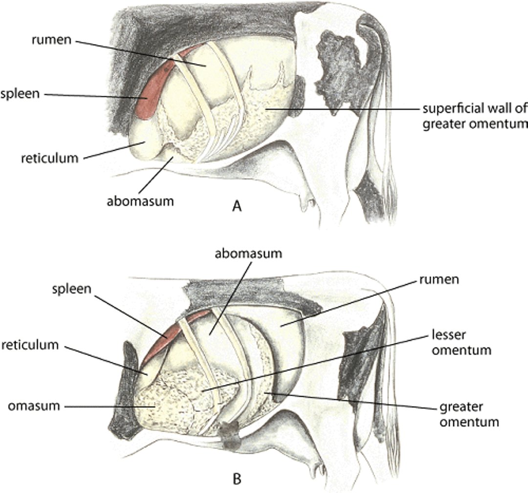

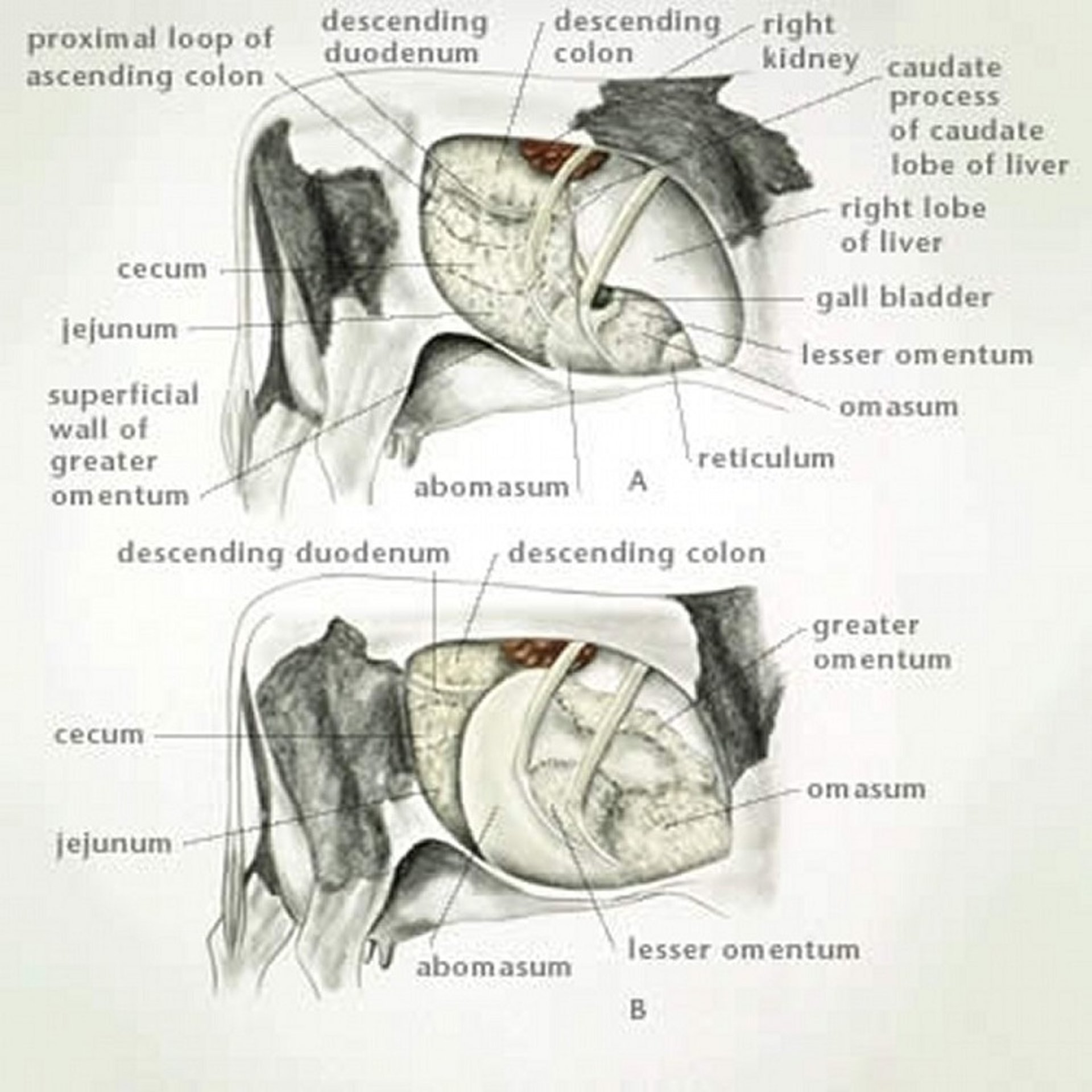
Illustration by Dr. Gheorghe Constantinescu. Adapted, with permission, from DeLahunta and Habel, Applied Veterinary Anatomy, W. B. Saunders, 1986.
Mild metabolic alkalosis with hypochloremia and hypokalemia is common. The hypochloremic metabolic alkalosis is due to abomasal hypomotility, continued secretion of hydrochloric acid into the abomasum, and the partial abomasal outflow obstruction, with sequestration of chloride in the abomasum and reflux into the rumen. Hypokalemia is due to decreased intake of feeds high in potassium, sequestration of potassium in the abomasum, and dehydration. Secondary ketosis is common and may be complicated by development of hepatic lipidosis (fatty liver disease).
In RDA, hypomotility, gas production, and displacement of the partially gas-filled abomasum occur as it does in LDA. Mild hypokalemic, hypochloremic, metabolic alkalosis develops as well. After this dilation phase, rotation of the abomasum leads to volvulus and local circulatory impairment and ischemia (hemorrhagic strangulating obstruction). The volvulus is usually in a counterclockwise direction when viewed from the rear and the right side of the animal. The omasum is displaced medially and can be involved in the volvulus with occlusion of its blood supply (called an omasal-abomasal volvulus) and displacement of the liver and reticulum. In rare cases, the reticulum can be involved in the volvulus (termed reticular-omasal-abomasal volvulus).
As much as 50L of chloride-rich fluid may accumulate in the abomasum, and hypochloremic, hypokalemic metabolic alkalosis develops. The blood supply to the abomasum, and often the omasum and proximal duodenum, is compromised, eventually resulting in ischemia of the abomasum and proximal duodenum as well as dehydration and circulatory failure. As circulatory failure progresses, a metabolic acidosis due to hyper-l-lactatemia and azotemia can develop along with the preexisting metabolic alkalosis.
Clinical Findings of Displaced Abomasum and Abomasal Volvulus in Cattle
The typical history of abomasal displacement includes anorexia (most commonly a lack of appetite for grain with a decreased or normal appetite for roughage) and decreased milk production (usually notable but not as extensive as with traumatic reticuloperitonitis or other causes of peritonitis). In abomasal volvulus, anorexia is complete, milk production is more markedly and progressively reduced, and clinical deterioration is rapid. In abomasal displacement, temperature, heart rate, and respiratory rate are usually normal. The caudal part of the rib cage on the side of the displacement may appear “sprung.” Hydration appears subjectively normal with LDA except in some chronic cases; in contrast, in abomasal volvulus, dehydration occurs very early. Rumen motility may be normal but often is reduced in frequency and strength of contraction. Feces are usually reduced in quantity and more fluid than normal; however, they may be shed with normal consistency.
The most important diagnostic physical finding is a ping on simultaneous auscultation and percussion of the abdomen, which should be performed in the area marked by a line from the tuber coxae to the point of the elbow, and from the elbow toward the stifle on both sides of the patient. The ping (detected during simultaneous percussion and auscultation) characteristic of an LDA is most commonly located in an area between ribs 9 and 13 in the middle to upper third of the left abdomen; however, the ping can be more ventral or more caudal, or both.
Pings associated with a rumen gas cap are usually more dorsal, less resonant, and extend more caudally through the left paralumbar fossa. Additionally, splashing sounds can be heard during the swinging auscultation of the displaced abomasum. Rectal examination can confirm a gas-filled rumen or an extremely empty rumen that correlates with the rumen ping in these cases.
Pings associated with pneumoperitoneum typically are less resonant, present on both sides of the abdomen, and inconsistent in location on repeated evaluation. Frequently, secondary ketosis develops, and ketones are present in blood, urine, or milk. Ketosis that develops in association with abomasal displacement responds only transiently to treatment and recurs (versus in primary ketosis, which develops early in lactation in high-producing cows and responds to therapy permanently if instituted early).
The ping associated with RDA also is most commonly located in the area between ribs 10 and 13 on the right abdomen, where the splashing sounds can be ausculted. Differentiation between various causes of a right-side ping can be difficult in some cases, although a ping cranial to rib 10 usually indicates the presence of abomasal volvulus because the liver is displaced medially by the distended viscus. The liver displacement can be confirmed via percussion or ultrasonographic examination.
A small, right-side ping underlying ribs 12 or 13 and extending as far forward as rib 10 is common in cows with functional ileus from a number of causes. This ping is most often associated with gas in the ascending colon and resolves with correction of the underlying condition. Cecal dilatation and rotation are characterized by a right-side ping. The ping extends through the dorsal paralumbar fossa in cecal dilatation and usually is located more caudally (well into the paralumbar fossa) in cecal rotation than the ping of RDA/abomasal volvulus. Another cause of a ping on the right side is the volvulus of the duodenal sigmoid flexure. Palpation per rectum is helpful in differentiating an RDA/abomasal volvulus from cecal dilatation or rotation. Other right-side pings are produced by pneumoperitoneum or gas in the rectum, descending colon, duodenum, or uterus.
Clinical signs associated with abomasal volvulus are more severe than with simple displacements because of the vascular compromise. However, an early abomasal volvulus can be difficult to distinguish from an RDA except by the presence of a right-side ping cranial to rib 10 (indicating medial displacement of the liver by the abomasal volvulus) and the anatomic position identified at surgery. In contrast to cases of displacement, an animal with abomasal volvulus has tachycardia proportional to the severity of the condition. The area of the ping is usually larger (extending as far forward as rib 8), and the amount of succussible fluid is greater. The animal is more depressed, and signs of weakness, toxemia, and dehydration develop as the disease progresses. The caudal extent of the abomasum is usually palpable per rectum. In addition, measurement of serum L-lactate concentration may help differentiate between the two conditions. Without therapy, the animal often becomes recumbent within 48–72 hours after developing volvulus. Death occurs from shock and dehydration and is sudden if the ischemic abomasum ruptures.
Diagnosis of Displaced Abomasum and Abomasal Volvulus in Cattle
Clinical evaluation (ie, percussion and auscultation of a ping)
Confirmed by results of laboratory testing as indicated
For abomasal displacement or volvulus, diagnosis is based on the presence of the characteristic ping on simultaneous auscultation and percussion and by exclusion of other causes of left- or right-side pings. Spontaneous fluid-splashing or gas-tinkling sounds may be heard on auscultation of the ping area or on simultaneous ballottement and auscultation of the abdomen (succussion). The characteristic rectal examination findings with LDA include a medially displaced rumen and left kidney. The abomasum is rarely palpable in LDA and only occasionally palpable in RDA. Because, in abomasal volvulus cases, the abomasum is severely dilated, it can be more frequently reached with the tips of the fingers during rectal examination. In LDA cases, abomasal displacement may only be present intermittently (pendulous LDA), which might be misleading during a physical examination. Ultrasonographic examination may help confirm a diagnosis of LDA, RDA, or abomasal volvulus, but it cannot reliably differentiate RDA from abomasal volvulus. Recent parturition, partial anorexia, and decreased milk production suggest displacement. A ketosis that is only temporarily responsive to treatment is consistent with abomasal displacement, which may be intermittent. The typical signs during a physical examination (in addition to the ping), rectal examination, and laboratory sample evaluation also support the diagnosis.
Melena or signs of peritonitis (eg, fever, tachycardia, localized abdominal pain, pneumoperitoneum) with an LDA may indicate a bleeding or perforated abomasal ulcer, respectively. In cattle with abomasal volvulus, blood l-lactate concentrations ≤2 mmol/L indicate a positive outcome with surgical correction, whereas cattle with blood l-lactate concentrations ≥6 mmol/L have a high probability of a negative outcome.
Treatment of Displaced Abomasum and Abomasal Volvulus in Cattle
For LDA, medical or surgical treatment can be successful
Abomasal volvulus and RDA require immediate surgical treatment
In patients with abomasal volvulus, antioxidative drugs are suggested prior to surgery.
The time from diagnosis to start of treatment, including supportive care, is highly relevant to outcome
Generally, both conservative (medical) and surgical therapy options are available for patients with LDA. However, surgery is the only possible treatment for those with abomasal volvulus. Because it cannot be reliably distinguished from abomasal volvulus, RDA is also corrected surgically.
A patient with LDA can be medically treated with administration of spasmolytic and analgesic drugs; in addition, rolling a cow through a 180° arc after casting it on its right side corrects most LDAs. Additionally, it has been reported that movement during transport of the patient with LDA may result in return of the abomasum to its normal position. A major disadvantage of all nonsurgical approaches is that recurrence is very likely.
Open (surgical) and closed (percutaneous) techniques can be used to correct abomasal displacements. LDA can be corrected surgically using right- or left-flank omentopexy, right-paramedian abomasopexy, combined left-flank and right-paramedian laparoscopy (a two-step procedure), or left-flank laparoscopy (a one-step procedure). Blind suture techniques (toggle-pin fixation or the “big needle” [blind-stitch] method), performed in the right-paramedian area, are percutaneous methods for correction of LDA.
The main disadvantage of these techniques is uncertainty of the exact location of the suture. Potentially fatal complications can develop after blind suture techniques, and the reported success rate is less than that of surgical correction by right-flank pyloric omentopexy.
With toggle-pin fixation, the typical smell of abomasal gas or pH can be checked to confirm that the pin is in the abomasum, which reduces the likelihood of attaching rumen, small intestine, or omentum to the body wall rather than the abomasum. More recently, endoscopic methods (one- or two-step procedures) have become routine procedures to correct LDAs. Both RDA and abomasal volvulus are corrected surgically using right-paralumbar fossa omentopexy. The time between onset of the disease and surgery is crucial for treatment success.
In all cases in which abomasal volvulus is on the list of differential diagnoses, limiting reperfusion injuries is essential to avoid postoperative abomasal atony. Antioxidative drugs (eg, vitamin C, vitamin E, and dexamethasone) can be administered in combination; however, any such medications should be administered before surgery. Right-paramedian abomasopexy should be performed only to correct RDA and abomasal volvulus in cattle unable to stand.
Ancillary treatment of patients with abomasal displacement includes treating any concurrent disease (eg, metritis, mastitis, ketosis). Administration of calcium borogluconate or calcium gluconate SC or calcium gels PO help restore normal abomasal motility in many cases. Administration of erythromycin (10 mg/kg, IM) at the time of surgery increases abomasal emptying rate and milk production in the immediate postoperative period. Because surgical correction of abomasal displacement or volvulus is frequently performed on the farm, the prokinetic effect of erythromycin suggests that it might be preferred if antimicrobials are administered to control intraoperative infection.
In simple displacement, fluid and electrolyte abnormalities correct spontaneously with access to water and a salt block. Providing electrolyte solutions (60 g sodium chloride and 30 g potassium chloride in 19 L of water) via nasogastric tube is helpful in cases of longer duration. Patients with notable dehydration and metabolic derangements require IV therapy, typically administered as hypertonic saline (7.2% NaCl, 5 mL/kg, IV over 5 minutes).
Occasionally, animals with abomasal displacement or volvulus have atrial fibrillation, thought to be of metabolic origin and primarily due to concurrent hypokalemia and metabolic alkalosis. Correction of the displacement or volvulus almost always results in correction of the atrial fibrillation within 5 days.
Aggressive treatment of ketosis plays an important role in successful treatment of abomasal displacement, because most of the patients that die after surgical correction of LDA and RDA do so from the metabolic consequences of prolonged anorexia.
The prognosis after correction of simple LDA or RDA is good, with survival rates of 95%. Abomasal volvulus has a variable and less favorable prognosis (average survival rate of 70%); a high heart rate, moderate to severe dehydration, a longer period of illness, a large quantity of fluid in the abomasum, increased blood or plasma l-lactate concentration, and the presence of omasal-abomasal or reticulo-omasal-abomasal volvulus are associated with a poorer prognosis.
Prevention of Displaced Abomasum and Abomasal Volvulus in Cattle
Maintaining optimal dry cow and calving management, avoiding rapid dietary changes, maintaining adequate roughage in the diet, avoiding postparturient hypocalcemia, avoiding endometritis/metritis, and minimizing and promptly treating concurrent disease and ketosis contribute to effective prophylaxis of LDA, RDA, and abomasal volvulus. The incidence of abomasal displacements can be decreased by ensuring a rapid increase in rumen volume after calving and by feeding a total mixed ration rather than feeding grain twice daily.
Key Points
Abomasal displacement is a common disease of high-producing dairy cows, with high variation in prevalence rates amongst herds.
Management and prophylactic measures can effectively decrease disease frequency.
Several treatment options are available that allow correction of the displacement and fixation of the abomasum to the anatomically correct position.


