


Aleutian Disease of Mink
Aleutian disease (AD), or mink plasmacytosis, is an important disease in mink resulting from infection with Aleutian mink disease virus (AMDV). An amdoparvovirus within the family Parvoviridae, AMDV is distinct from the parvovirus responsible for causing mink viral enteritis. Infection with AMDV is widespread among farmed and wild mink around the world.

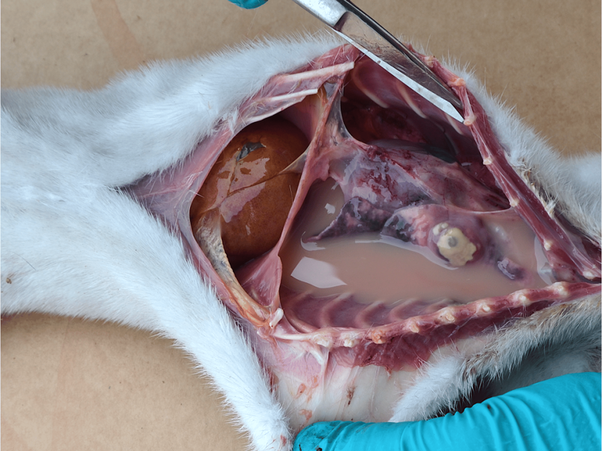
Pyothorax in a mink chronically infected with Aleutian mink disease virus.
Courtesy of Dr. John Easley.
Clinical signs depend on the age of the animals infected. In adult mink, AD is an immune-mediated disease that causes chronic wasting and unthriftiness, with poor reproductive performance and increased congenital malformations and abortions, poor pelt quality, oral and GI bleeding with tarry feces, renal failure and uremia, and increased mortality. In adult mink, viral infection results in increases in plasma cells that produce non-neutralizing antibodies. Massive increases in plasma cell production after infection result in lymphadenopathy, splenomegaly, and hypergammaglobulinemia. Viral-antibody complexes are taken up by macrophages, the target host cell, and immune complexes. Infected macrophages may be deposited in blood vessels and the kidneys, resulting in necrotizing arteritis and proliferative glomerulonephritis. In the acute neonatal form of AD, the virus targets pneumocytes, with subsequent interstitial pneumonia and respiratory disease, presenting as severe dyspnea with high litter mortality.
All coat colors (phases) of mink may be infected with AMDV; however, animals with dilute coat colors (eg, pastels) carry the Aleutian coat color gene and are genetically more susceptible to developing AD after AMDV infection. Virus transmission occurs both vertically, with viral replication within the placenta, and horizontally, by direct or indirect contact with infected mink; blood, saliva, and feces from infected mink; contaminated caging, feed, gloves, clothing, and equipment; via transfer by various biological vectors, such as flies, mosquitoes, and birds; and via various contaminated fomites, such as dust, bedding, or hair. Because the disease is chronic and may take a year or longer for clinical signs to appear, virus shedding can occur for months from inapparent infected carriers. Infected animals may be less tolerant of weather extremes and commonly develop secondary bacterial infections because of AMDV-induced immunocompromise. The extent of virulence varies among different strains of AMDV.
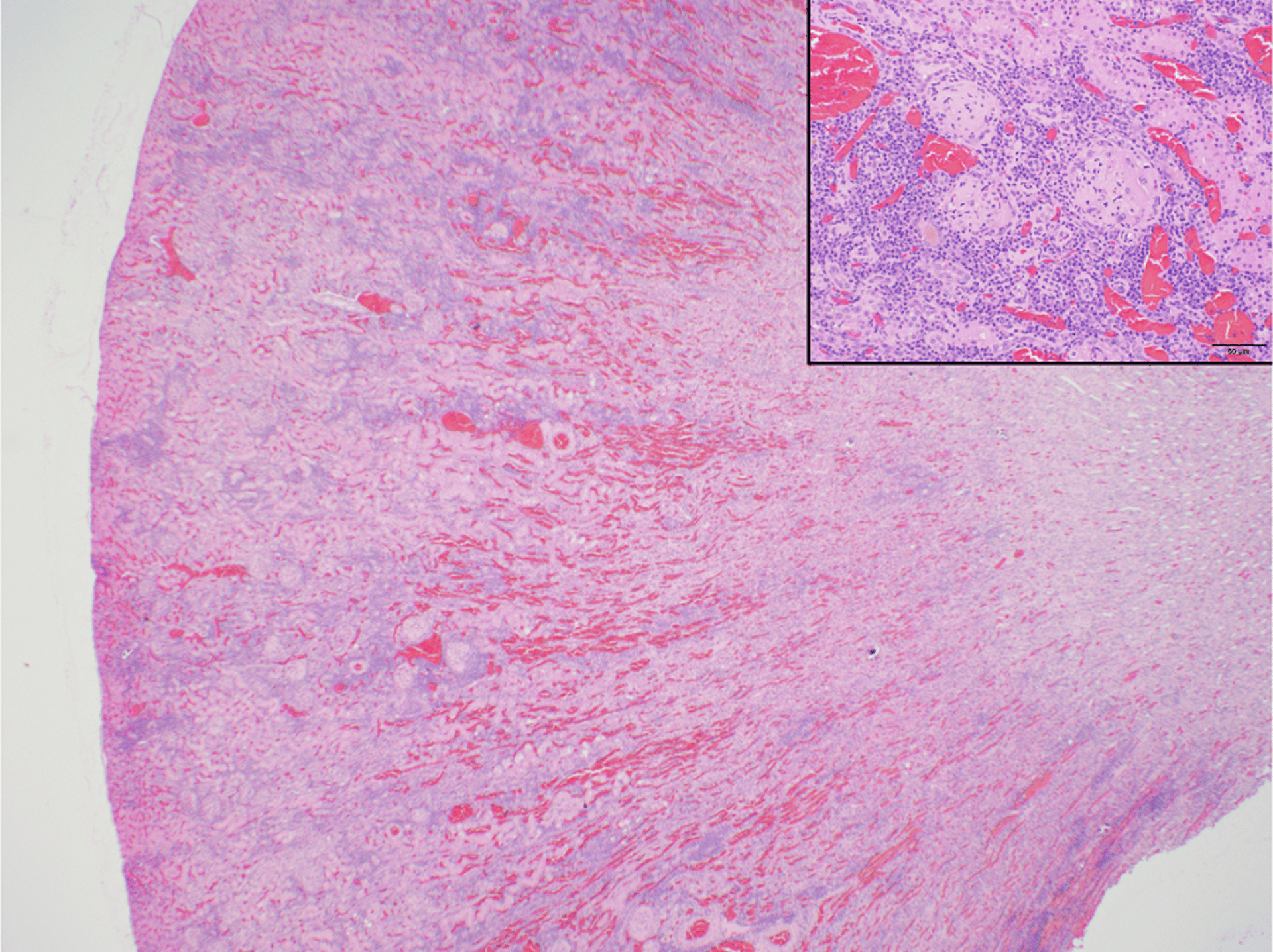
Subgross and histologic section (inset) showing chronic renal degeneration after immune complex deposition in an Aleutian mink disease virus-infected mink. Glomerular tufts are thickened (inset), and there is a moderate mononuclear leukocytic infiltrate within the surrounding stroma (H&E stain).
Courtesy of Dr. Emily Brouwer.
Aleutian disease is generally managed with a test and cull program. Positive mink are identified by testing blood samples for AMDV antibodies by counterimmunoelectrophoresis. A few drops of blood are collected by nail clip or lancet puncture of the footpad from live animals or from the heart blood of dead animals. The counterimmunoelectrophoresis assay is the most practical way to test large numbers of animals on a farm. Virus-specific antibody ELISA and dot immunoassays of blood samples are used for diagnosis in some places, and PCR assays for AMDV are also available at some veterinary diagnostic laboratories and may be used on rectal or saliva swabs or blood or tissue samples (eg, from the spleen). All AMDV-positive mink should be humanely killed on-farm. Mink to be kept for breeding should be tested in late fall before selection of breeding stock and pelting, as well as tested in January or February before breeding. New introductions to the herd also should be tested and only integrated into existing populations after receipt of negative test results.
There is no effective treatment or vaccine available for AD. Research is ongoing to select mink genetically resistant to AMDV infection. The virus is extremely resistant in the environment and can survive harsh temperatures and attempts at chemical inactivation. Pens and nest boxes housing known or suspect positive animals should be thoroughly cleaned and disinfected after they are emptied, and manure should be removed from sheds regularly. Hair adherent to caging and old bedding should be burned. The watering system should be cleaned periodically. Use of high-pressure sprayers in sheds is not recommended because this may aerosolize contaminated fomites. All equipment should be disinfected after handling, vaccinating, or testing mink. To help minimize transmission on a farm, AMDV-positive animals should be housed in separate sheds from AMDV-negative animals, if possible, with separate equipment and gloves used between these sheds. For all activities, AMDV-negative animals should be handled before AMDV-positive animals. On-farm integrated pest management programs are essential to help reduce spread of AMDV. Escape or release of farmed mink contribute to the spread of the virus within wild carnivore populations. Perimeter fences are recommended around sheds and manure holding areas to restrict domestic and wild animal contact with mink and their excreta.
Aleutian disease has zoonotic potential and has been rarely reported to cause clinical disease in humans.
Canine Distemper Infection of Mink
Mink of all ages are susceptible to infection with canine distemper virus (hard pad disease), a morbillivirus within the family Paramyxoviridae. The virus is highly infectious, and mortality can be very high, exceeding 80%.
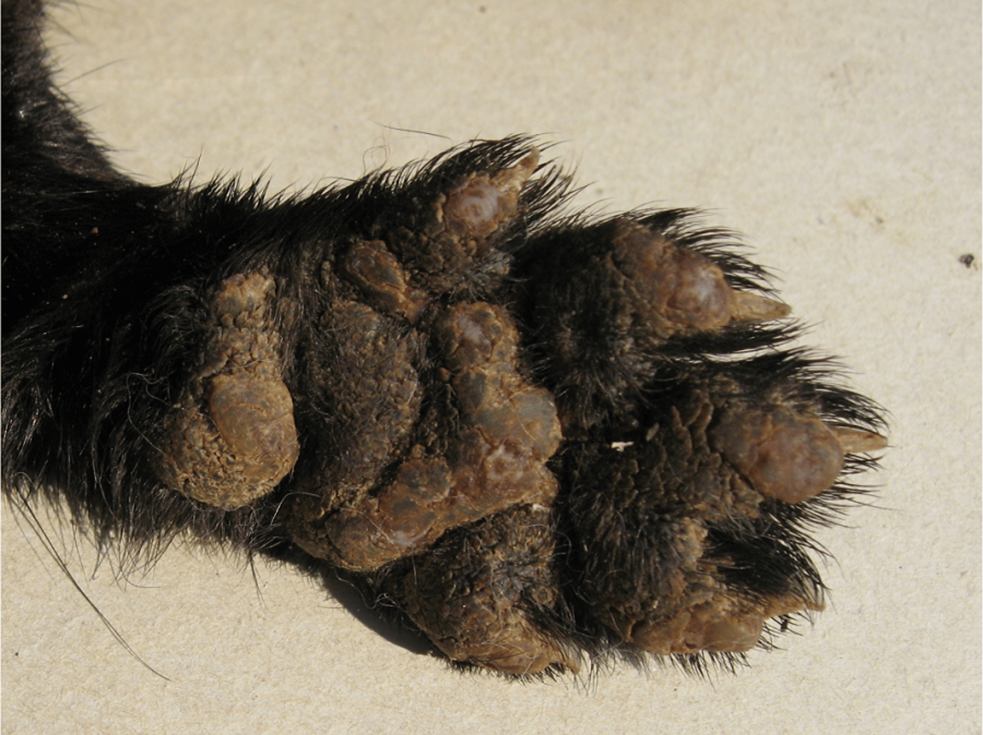
Thickening of the footpads is a common sign of canine distemper virus in mink.
Courtesy of Dr. John Easley.
Clinical signs are inconsistent and may include oculonasal discharge; conjunctivitis; photophobia; crusting of the skin around the eyes, on the muzzle, feet, and ventral abdominal wall; thickening (orthokeratotic hyperkeratosis) of the skin on the nose and footpads (hard pad); diarrhea; pneumonia; and neurologic signs (eg, seizures and vocalization) alone or in combination. Animals with viral pneumonia may die from secondary opportunistic bacterial infections.
The virus is typically transmitted via aerosol of respiratory droplets and other exudates between animals. Indirect transmission is less common because of virus fragility outside the host. The disease incubation period is variable, averaging 4–8 weeks, which makes retrospective epidemiologic investigation of outbreaks difficult. Recovered mink may continue to shed the virus for several weeks.
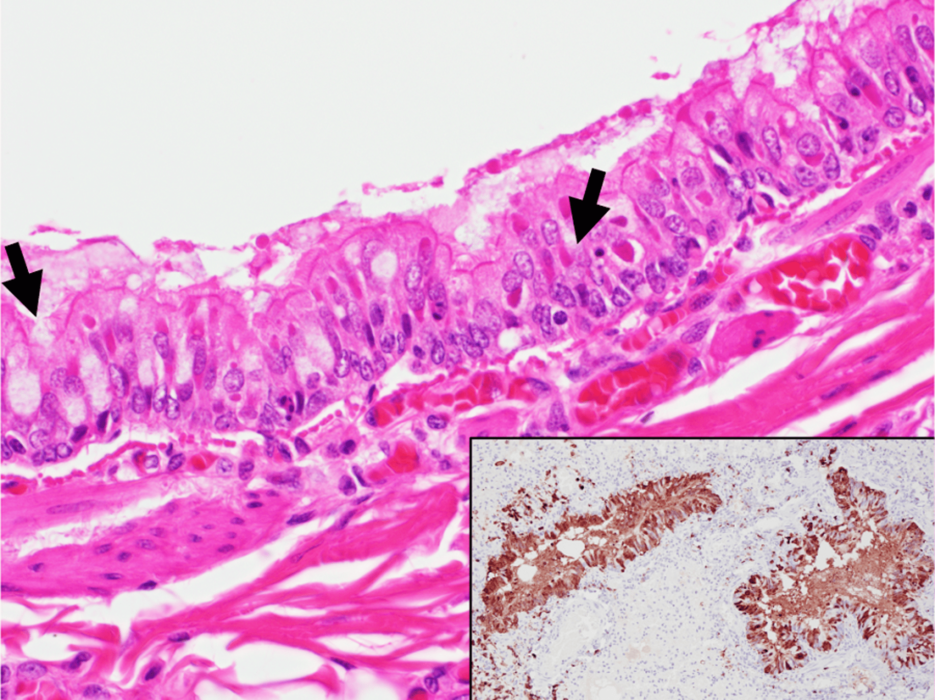
Photomicrograph of a histologic section of lung tissue from a mink infected with canine distemper virus. Eosinophilic intracytoplasmic inclusion bodies can be seen in the apical cytoplasm of some bronchial epithelial cells (black arrows). Inset: Section of lung tissue from an infected animal immunohistochemically labelled for canine distemper virus antigen, counterstained with Mayer’s hematoxylin. Note strong positive nuclear and cytoplasmic immunolabeling (brown staining) of airway epithelial cells.
Courtesy of Dr. Emily Brouwer and Dr. Marina Brash.
Diagnosis can be made by histologic evaluation, immunohistochemical evaluation, ELISA, or PCR assay. Characteristic intracytoplasmic and intranuclear inclusion bodies and syncytial cells are often present in the lungs, urinary bladder, kidneys, bile ducts, intestine, trachea, and occasionally brain of affected animals.
There is no specific treatment for canine distemper virus in mink. Affected mink should be humanely killed, and the balance of the herd vaccinated with a modified-live virus vaccine as soon as possible. Deaths from neurotropic distemper may occur until 12 weeks after vaccination. Kits should be vaccinated prophylactically when 10–12 weeks old with a modified-live virus vaccine. If vaccinated too early, before concentrations of maternal antibodies have waned, vaccination may be ineffective, resulting in no or only partial protection. Adults and kits on a farm are typically vaccinated at the same time. The virus does not persist for long periods outside the host and is susceptible to ultraviolet light, heat, and drying. Pens and nest boxes housing previously affected animals should be sanitized and disinfected between animals. Canine distemper virus can also be transmitted to farmed mink from domestic and wild carnivores; thus, it is important to exclude both from mink sheds with good on-farm biosecurity programs, including perimeter fences.
Mink Viral Enteritis
Mink viral enteritis is a highly contagious disease caused by infection with mink enteritis virus, a protoparvovirus related to that causing feline panleukopenia. Mink of all ages are susceptible, and morbidity is variable, with up to 10% mortality of affected animals (predominantly kits). Transmission occurs by the fecal-oral route, and the incubation period is approximately 4–10 days.
Clinical signs may include weight loss, poor pelage, anorexia, depression, watery to hemorrhagic diarrhea, dehydration, and death. Affected animals may die from secondary bacterial enteritis. Gross lesions include flaccid, dilated loops of small intestine with watery, sometimes hemorrhagic content. Some mink may die suddenly with no gross lesions. Microscopically, intestinal lesions are characterized by erosion or ulceration of the mucosa, with blunting and attenuation of villi and dilatation of crypts. Ballooned crypt epithelial cells may contain basophilic intranuclear inclusion bodies. To confirm the diagnosis, ELISA or fluorescent antibody procedures can be used. Splenic and lymph node lesions include lymphoid depletion and necrosis.
Early in an outbreak, all mink with clinical signs should be culled or isolated, and all clinically normal mink should be vaccinated immediately. Affected mink can be treated with a mixture of kaolin, pectin, and neomycin administered by mouth. Mink viral enteritis can be prevented by vaccination. All mink should be vaccinated when they reach at least 6–8 weeks of age with a combination 3-way vaccine containing mink enteritis virus, Clostridium botulinum toxoid, and Pseudomonas bacterin. Annual vaccination is recommended. The virus is highly resistant in the environment but is inactivated at high pH. Differential diagnoses include other viral and bacterial causes of enteritis.
Preweaning Diarrhea (Sticky Kit Syndrome) of Mink
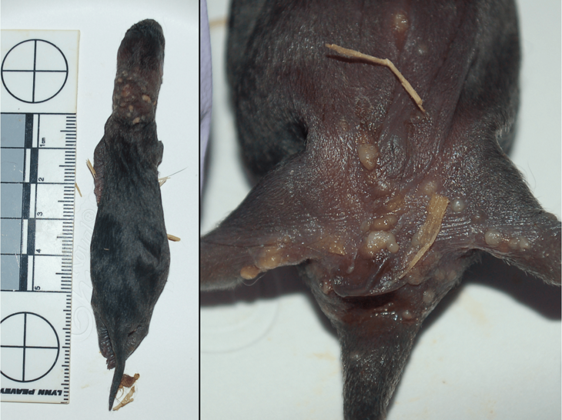
Superficial cervical and inguinal abscesses in mink kits that died due to preweaning diarrhea.
Courtesy of Dr. Patricia Turner.
Preweaning diarrhea, or sticky kit syndrome, is a multifactorial syndrome characterized by watery to catarrhal diarrhea, perianal irritation with or without rectal prolapse, and exudative or pustular dermatitis affecting the apocrine glands around the neck and perineal areas, conferring a greasy or sticky appearance. The syndrome affects kits between 1 and 4 weeks of age, and a few to large numbers of kits may be affected within a herd. Sometimes it presents as explosive outbreaks of diarrhea, with 30% or greater kit mortality. Affected kits may become dehydrated and die without supportive care. Even with treatment, animals may never fully recover in size, and pelts of affected animals may be of poor final quality. Infection with a GI variant of astrovirus is thought to be an important risk factor, as is infection with a mink-specific calicivirus. Various bacteria, including E coli, Staphylococcus delphini, and streptococci, also may be isolated from affected animals and may represent secondary infections.
Risk factors include increased farm size, increased litter sizes, and dam parity, with a higher proportion of affected kits coming from young (1-year-old) breeding females (both winter feed restriction and gestational feed restriction of females are associated with an increased incidence of the disease in their litters).
Virus may be shed from infected animals for up to 7 weeks after infection, emphasizing the need for good hygiene and disinfection of caging between animals. Sticky kit syndrome is an important differential diagnosis for mink viral enteritis. Treatment is nonspecific and supportive, and there is no preventive vaccine available. A mink-specific coronavirus and hepatitis E virus have also been isolated from mink; however, their role in inducing enteritis in mink is unknown.
Influenza of Mink
Mink of all ages are highly susceptible to influenza A strains of avian and mammalian origin. The virus is highly contagious, with variable, strain-dependent morbidity and mortality. Clinical signs include coughing and sneezing, and animals may be found dead suddenly in good body condition. Secondary bacterial infections may contribute to severity of the disease. Exposure to the influenza A virus can come from multiple sources. Contaminated raw pig offal or poultry products, incursions of infected wild birds, and sick humans all may be avenues for virus introduction. Treatment is nonspecific and supportive. Treating an entire farm may take 2–6 weeks. Immunity to the virus may be short-lived and is not cross-protective between strain variants. Humans with influenza should not enter mink sheds.
SARS-CoV-2 in Mink
The SARS-CoV-2 virus was first detected on two mink farms in the Netherlands in 2020; it has since been detected in many countries. Disease can range from mild to severe and resulting in death. Outbreaks are managed by culling of affected animals.
The virus can be transmitted to humans and other animals, and mink may be asymptomatic carriers.
Pseudorabies (Aujeszky Disease) of Mink
Pseudorabies occurs sporadically in mink fed pork offal contaminated with pseudorabies virus, a varicellovirus in the family Herpesviridae. On-farm mortality may be high, exceeding 80%, with a 100% case fatality rate. The incubation period is approximately 1–2 weeks, and clinical signs may include anorexia, abdominal and facial skin scratching, diarrhea, emesis, and lethargy, followed by tonic and clonic seizures, dyspnea, and death. Potentially contaminated feed should be removed immediately, with samples kept for testing. Diagnosis is confirmed by PCR assay of tissues (eg, spleen, lung, liver, or brain) from affected animals, virus isolation, or serology analysis. Because contaminated pork is the source of infection, all pork products should be thoroughly cooked before being fed to mink. Differential diagnoses include infection with mink enteritis virus or canine distemper virus or food poisoning (ie, bacterial- or toxin-contaminated feed). Pseudorabies is a reportable disease in most countries.
Astrovirus of Mink (Shaky Mink Syndrome)
A novel neurotropic astrovirus infection has been reported sporadically in mink kits. Morbidity is low, with only 1–2 kits affected per litter, and the case fatality rate is up to 25%. The disease is generally seen between late June and early August. Affected kits demonstrate tremors (shaking), ataxia, salivation, vocalization, inability to eat, and seizures. Some affected animals will recover if provided with food and water within easy reach. If clinical signs do not improve, euthanasia of affected mink is recommended for welfare concerns.
Microscopically, nonsuppurative encephalomyelitis with scattered neurodegeneration and lymphocytic and monocytic perivascular cuffing occurs in affected animals. Early in the course of the disease, astrovirus can be isolated; however, the virus is cleared by immunocompetent kits and may not be detected in chronically affected animals. This astrovirus is distinct from that associated with preweaning diarrhea of kits. Differential diagnoses include canine distemper virus infection, Aleutian disease, toxoplasmosis, and pseudorabies.
For Further Information
Koopmans M. SARS-CoV-2 and the human-animal interface: outbreaks on mink farms. Lancet Infect Dis. 2021;21(1):18-19.
Fenollar F, Mediannikov O, Maurin M, et al. Mink, SARS-CoV-2, and the Human-Animal Interface. Front Microbiol. 2021; April.


