

Miniature pet pigs (MPPs) are subject to the same infectious diseases that occur in commercial swine. When properly vaccinated and cared for, pet pigs only rarely die of infectious disease.
Gastrointestinal System

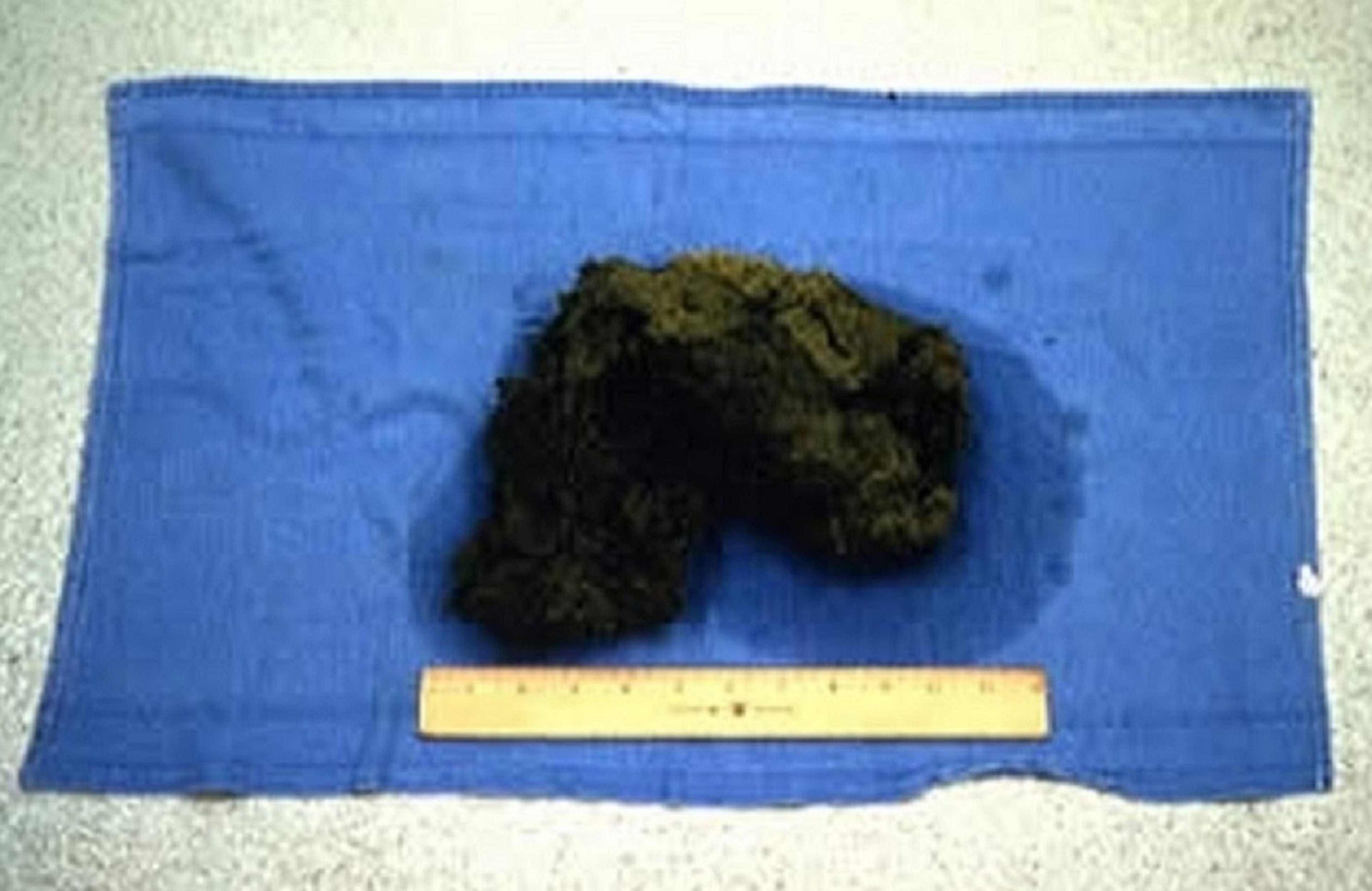
The foreign body (thought to be a piece of rug weighing ~2 pounds) in this photograph was removed from the stomach of a 45.5-kg (100-pound) miniature pig.
Courtesy of Dr. Bruce Lawhorn.
Gastritis and gastric foreign bodies are common in MPPs because they are omnivorous and prone to ingest many types of objects. Keeping MPPs indoors where they are unable to root and restricting calorie intake to prevent obesity probably contribute to this continual search for food. If an ingested foreign body is small or pliable enough, it may pass through the GI tract and cause mild gastritis that is self-limiting or requires only antimicrobial treatment. Larger objects may remain in the stomach or partially pass into the duodenum or a more distal part of the small intestine and cause obstruction.
Clinical signs may include anorexia, vomiting, and signs of abdominal pain. These can be acute, but they may be subtle and increase in intensity over several days or weeks. Fecal production may be decreased if a blockage develops.
Radiography may reveal obvious foreign material or delayed gastric emptying. CBCs may indicate infection, but they are usually not informative; serum enzyme and electrolyte panels may only reflect dehydration.
Treatment often requires surgical correction; however, if extensive necrosis of GI tissue is present surgery may not be successful. Fluid replacement and nutritional supplementation plus antimicrobial treatment, administration of appropriate analgesics, and tetanus prophylaxis are indicated after surgery in MPPs.
Lower GI obstruction due to bowel stricture occurs in geriatric MPPs. Anorexia, scant fecal production, and a bloated abdomen with massively distended intestines observed on radiographs are typical. Sedation and endoscopic examination of the oral cavity, esophagus, and stomach are indicated to exclude other problems. Exploratory laparotomy and anastomosis with or without bowel resection is usually remedial.
Colibacillosis or Escherichia coli diarrhea is generally an important disease in young MPPs. Mortality rate may be high in piglets that have not ingested adequate colostrum in their first 24 hours after birth. Older MPPs apparently develop resistance to colibacillosis. Diagnosis is based on signalment, history, and bacteriologic culture of feces. Sanitation to minimize infective doses of pathogenic coliforms in the environment of young, nursing MPPs is important for prevention. Commercial swine vaccines to prevent colibacillosis are available; however, they must be administered to sows before farrowing to stimulate immunity and secretion of IgA into the milk. Treatment is based on in vitro antimicrobial susceptibility testing.
Enterocolitis from Salmonella enterica serovar Typhimurium infection can affect MPPs of any age, but it usually occurs after weaning. Sources of salmonellae include waste food from overturned garbage cans, carrier swine (such as the dam), and fecal material from other animal species. Mild to severe diarrhea with mucus and blood can result. Diagnosis is based on signalment, history, and bacteriologic culture of feces or PCR assay.
Salmonella spp are characteristically resistant to many antimicrobials, so in vitro antimicrobial susceptibility testing is important. Untreated MPPs may die. Some recovered MPPs may develop rectal stricture after enterocolitis, resulting in megacolon and a distended abdomen. Subsequent straining to defecate can cause rectal prolapse. Surgical correction of the rectal prolapse will not correct the underlying problem. Owners should be advised that many Salmonella spp, including Salmonella Typhimurium, are zoonotic. Healthy MPPs may be tested via bacteriologic culture of feces or PCR assay to determine their Salmonella status. Multiple tests are more accurate predictors than single tests.
Bacteremia or septicemia after Salmonella enterica serotype Choleraesuis infection may also affect MPPs, usually after weaning. Sources of infection are similar to those for Salmonella Typhimurium. Mild to inapparent diarrhea may ensue, followed by fever, lethargy, anorexia, cyanosis of the extremities, recumbency, and death. Diagnosis, treatment, prevention, and zoonotic potential are similar to those of Salmonella Typhimurium infection; zoonosis is a threat mainly in immunocompromised people.
Constipationmay occur in MPPs; however, each normal bowel movement by a miniature pig is typically composed of one or more cylindrical formations made up of smaller, multiple fecal balls. Educating owners regarding the normal appearance and texture of the pet pig's feces is important. The pet owner can become familiar with the appearance of the fecal balls and fail to notice when they become very dry. True constipation can occur because of low water intake in sedentary MPPs or because of an actual disease. In these cases, the fecal balls are very dry, do not stick together at all, and can look more like single grapes than like clusters of grapes.
Careful evaluation is warranted before treatment is initiated. Enemas may be contraindicated in the instance of pathological changes such as colitis. In simple constipation, fecal softeners or mild laxatives, such as sodium sulfate or magnesium sulfate, may be used. These should be given with food, if possible, because forced oral administration can result in aspiration pneumonia and death, especially with mineral oil. Adding flavoring such as fruit juice or liquid gelatin may help to encourage increased water intake. Regular exercise is also beneficial to promote normal feces.

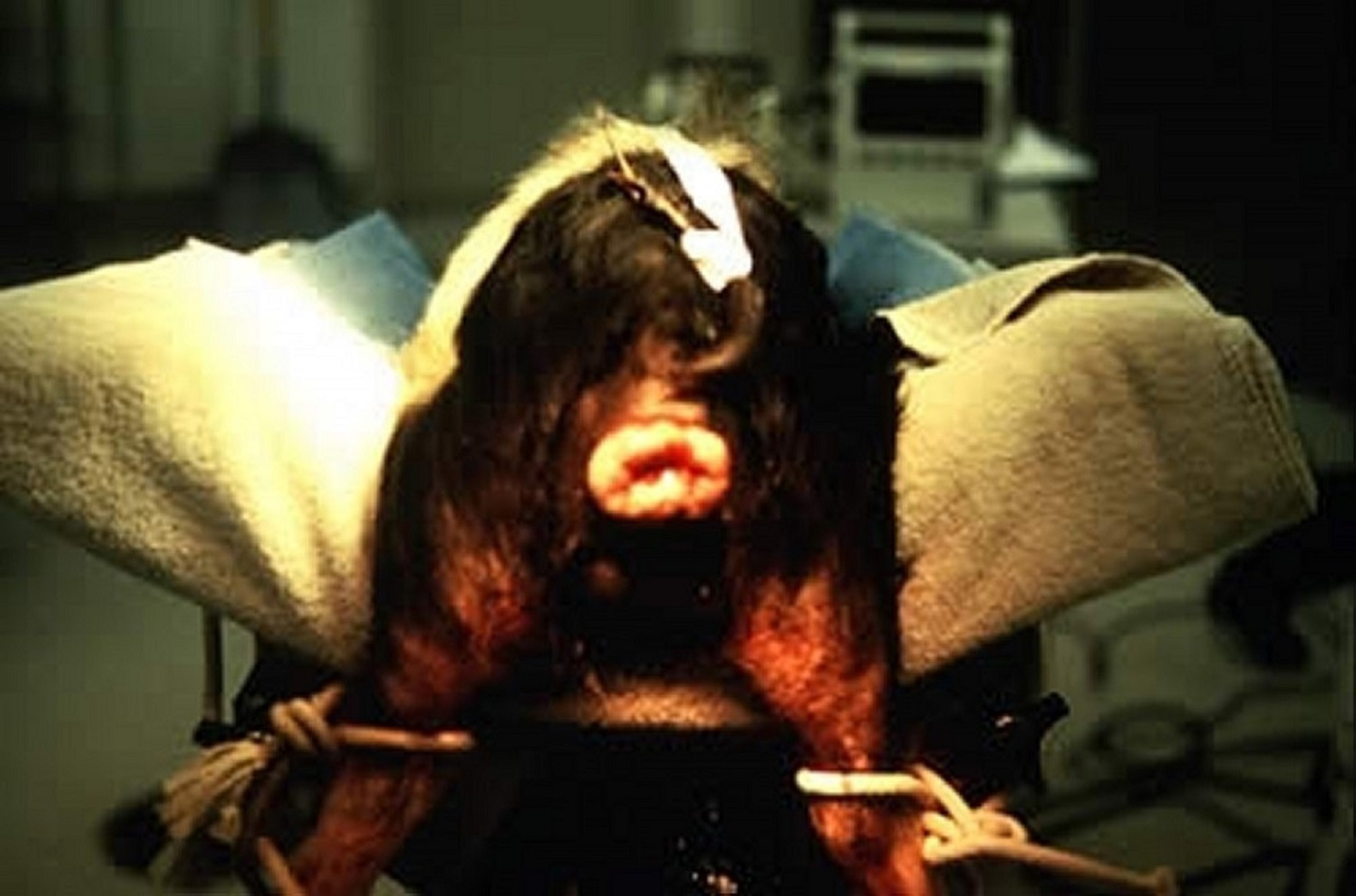
This photograph shows rectal prolapse in a miniature pig during surgery to repair a recurrence.
Courtesy of Dr. Bruce Lawhorn.
Rectal prolapse occurs as the result of straining due to bowel irritation from diarrheal disease, rectal stricture after Salmonella enterica serovar Typhimurium enterocolitis, or previous rectal prolapse repair; it may also be due to cystitis or urolithiasis, persistent coughing, dystocia, or possibly genetic predisposition. Small, uncomplicated, recent rectal prolapses may be repaired via anesthesia and purse-string closure of the rectum, which allows for minimal passage of feces. Larger, complicated prolapses require surgical excision. Recurrence is less likely after surgery; however, it is possible regardless of the method of repair.
Lymphosarcoma, lymphoma, and carcinoma of the intestines occur in aged MPPs. Clinical signs are vomiting, anorexia, melena, anemia, and chronic weight loss, followed by death (or euthanasia). Diagnosis is confirmed by postmortem histopathologic findings.
Integumentary System
Dry, flaky skin with minimal to severe pruritus occurs in virtually all MPPs. Wiping down the skin with wet towels each week will remove the flakes. Moisturizing lotions (eg, aloe vera) also temporarily alleviate this problem. Fatty acid supplementation can be used as a more longterm remedy; however, caution must be exercised not to promote obesity.
Sarcoptic mange is the most important ectoparasitic disease of MPPs. Intense pruritus and dermatitis are the basis for a presumptive diagnosis. In many cases, the owner has pruritic skin lesions on the arms or abdomen. Examination of skin scrapings (deep enough to contain some blood) from several sites usually confirms the diagnosis in advanced cases; however, false-negative results may occur in less advanced cases if very few mites are present. In young MPPs, the source of infestation is usually the dam; in older MPPs, the source is usually other infested pigs. Young MPPs isolated from other pigs and kept as pets may harbor mange mites as a subclinical problem until mite populations increase sufficiently to make the condition obvious. Recently acquired young MPPs should be administered ivermectin or doramectin for routine parasite prevention.
Melanoma is an important skin tumor in swine. Tumor removal and histologic evaluation of metastatic potential is important for prognosis. Spontaneous regression of melanomas, with subsequent depigmentation of the hair, skin, and iris, occasionally occurs in MPPs; affected swine usually have normal life spans.
Sunburn may develop in MPPs exposed to sudden, high-intensity sunlight. Skin lesions may or may not be obvious, but affected MPPs show signs of pain with vocalization and hind limb weakness or paresis. A thorough history is important for diagnosis. Exposure to further sunlight should be prevented. Supportive care is remedial.
Bleeding back syndrome (dippity pig syndrome) has an unknown etiology. Clinical signs are almost identical to those of sunburn (pigs dip their backs, vocalize, and show signs of extreme pain), but with no history of sun exposure. Serum-oozing lesions of various sizes and shapes occur on lumbar skin surfaces. Some pigs demonstrate the behavioral signs but have no visible lesions. Affected MPPs recover in several days with restricted activity with or without supportive care; however, pain medication should be administered in almost all cases. The condition may recur in some animals.
Erysipelas, due to Erysipelothrix rhusiopathiae, is a generalized bacterial infection that affects swine. For details on clinical signs, diagnosis, and treatment, see Swine Erysipelas.
Musculoskeletal System
Lameness due to lower back, hind limb, or forelimb weakness is common in MPPs. Their conformation makes MPPs susceptible to muscle pulls, ligament damage, and fractures of the back and limbs. Because MPPs usually struggle against manual restraint (predisposing to injury), sedation or anesthesia is recommended for most procedures (eg, prolonged examination, radiography, nail trimming, blood collection, and dental work).
Pain due to chronic arthritis, secondary to poor hoof care, poor conformation, or obesity (or a combination of the three), is a common reason for geriatric MPPs to be euthanized.
MPPs with injuries to the back or limbs are usually treated with anti-inflammatory drugs, such as buffered aspirin with antacid, flunixin meglumine, or glucocorticoids (eg, dexamethasone). Polysulfated glycosaminoglycan or glucosamine or chondroitin sulfate products may be tried in nonresponsive cases.

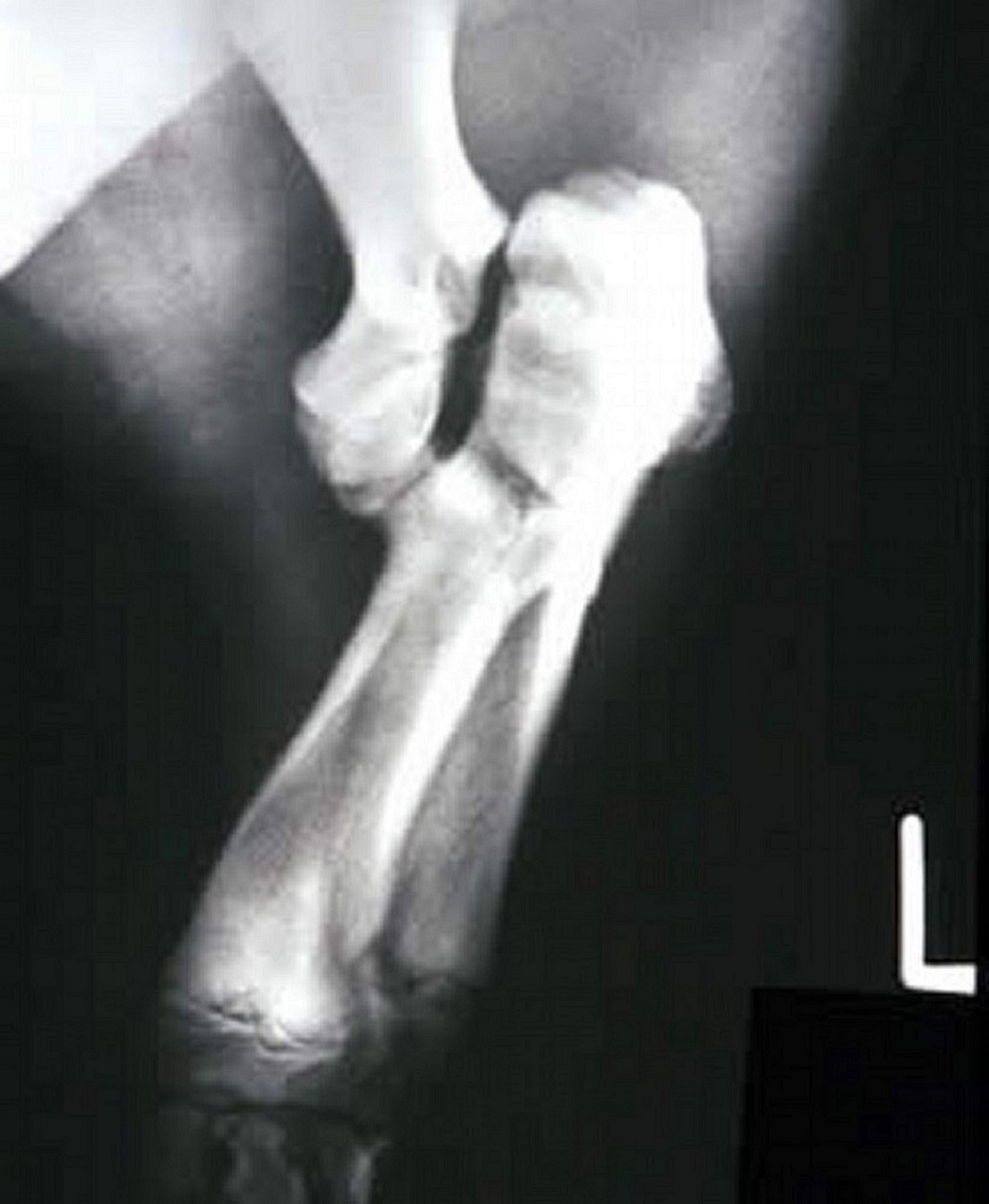
Lateral radiographic view of the left forelimb of a miniature pig with a fracture of the distal humeral condyle. This is the most common fracture in miniature pigs.
Courtesy of Dr. Bruce Lawhorn.

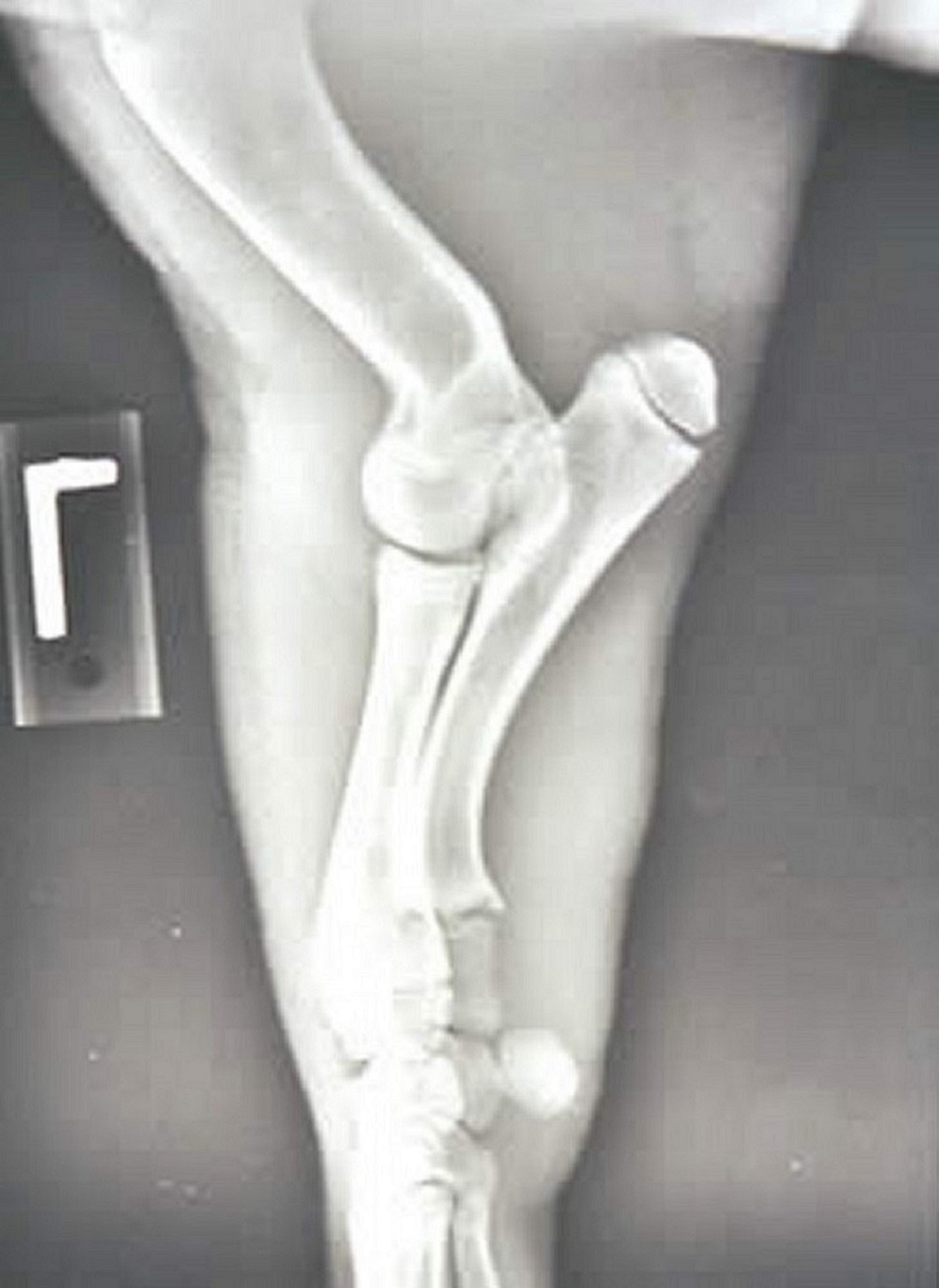
Lateral radiographic view of the left forelimb of a miniature pig. Note the normal radiographic appearance of the distal humerus and elbow, which is the most common area for fractures in miniature pigs.
Courtesy of Dr. Bruce Lawhorn.
Fractures of the distal humerus and elbow area and femur are common. Causes include jumps off furniture (distal humerus), dog bites (elbow area), restraint (elbow area and femur), equine kicks (femur), and other trauma. Repair via pins, screws, plates, and external devices successfully restores some range of motion if fractures are immobilized properly and any sepsis is controlled.
Infectious arthritis may affect MPPs of all ages and life stages. Lameness with or without joint swelling in one or more limbs is the usual clinical sign. Erysipelothrix rhusiopathiae, Streptococcus spp, Mycoplasma hyosynoviae, Mycoplasma hyorhinis, Staphylococcus spp, and Haemophilus parasuis are possible causes. Treatment early in the disease course with an appropriate antimicrobial may be effective. Treatment after chronic changes have occurred, antimicrobial ineffectiveness against the etiologic agent, and misdiagnosis are reasons for treatment failure and persistence of lameness. In chronic cases, pain management with anti-inflammatory drugs should be considered. Polyarthritis from neonatal infection of the navel may be due to various environmental bacteria, including Pseudomonas spp. If degenerative arthritis and joint fusion from chronic inflammation are present after polyarthritis, euthanasia may be warranted.
In shoulder, elbow, hip, and stifle lameness, osteochondrosis may also be considered; however, this condition is not common in slow-growing, light-muscled animals such as MPPs.
Overgrown or cracked hooves are a common cause of lameness. Regular exercise on abrasive surfaces (eg, concrete) will wear hoof ends and help keep them the appropriate length. Hoof cracks can be due to overgrown hooves. Miniature pet pigs with cracked hooves may also require antiseptic cleaning with tamed iodine and systemic antimicrobial treatment.
Mucormycosis from Mucor spp infection has been reported in the distal hind limb of a potbellied pig. The large growth that encompassed the entire foot was composed of infected and abscessed tissue that involved bone. Amputation was remedial.
Tetanus may occur after wound contamination from dog bites, skin abrasions, oral cavity abrasions, or surgical procedures. Tetanus toxoid should be part of the routine vaccination schedule of MPPs at high risk of exposure. If there is no current tetanus toxoid vaccination, tetanus antitoxin (500–1,500 U, depending on body weight) should be administered IM in the neck after recovery from any surgery or dental procedure (eg, trimming of canine teeth). The treatment for tetanus is to administer high doses of tetanus antitoxin and penicillin early in the disease, along with tranquilizers, to isolate the pig to minimize external stimuli, and to provide supportive care.
Nervous System
Systemic bacterial infection can be due to (in approximate decreasing order of importance) Streptococcus suis type 2, other Streptococcus spp, Salmonella Choleraesuis, Haemophilus parasuis, Escherichia coli, other gram-negative bacteria, and Listeria monocytogenes. CNS signs may include fever, depression, incoordination, staggering, postural abnormality, head tilt, circling, nystagmus, seizures, and death. Miniature pet pigs are most commonly affected from birth through 4–6 months old. Treatment with the appropriate antimicrobial (eg, extralabel administration of florfenicol, which penetrates the blood-brain barrier) in the early stages of infection is most effective; however, death may be the first clinical sign. Because Streptococcus suis type 2 is a zoonotic disease agent, care should be taken to prevent infections in humans when performing necropsies on pigs dying from suspected CNS disease.
Overheated MPPs may be depressed, inactive, and recumbent and show open-mouth breathing or panting with an initial fever followed by a subnormal and decreasing temperature. The prognosis is grave; however, some overheated MPPs may respond to resting on a cool surface and cooling only the head with water for 10–15 minutes, followed by packing ice bags around the head. If the temperature is still not controlled, cold-water enemas can be used while additional areas of the skin surface are packed in ice. Supportive care is continued as indicated.
Salt toxicosis occurs after water deprivation for ≥ 36 hours followed by sudden rehydration, or less commonly, after prolonged consumption of high-salt foods. Affected MPPs may have seizures, walk aimlessly, or show other CNS signs, such as blindness or postural abnormalities. Diagnosis in the affected live animal is confirmed by high concentrations of serum sodium, usually 160–183 mEq/dL (normal range, 142–153 mEq/dL). Gradual rehydration and other supportive care to counteract cerebral edema is indicated; however, severely affected MPPs may be stabilized to only a vegetative and blind status. The histopathologic finding of eosinophilic infiltration into brain tissue is also diagnostic.
Seizures due to unknown causes occur in MPPs. Pigs < 1 year old seem the most susceptible. The frequency may range from 1–2 seizures per month to several per day. Infrequent seizures may require no preventive medication. Diazepam is used to control more frequent episodes. Phenobarbital in addition to diazepam may be required to control the most severe cases. Seizures may cease as the affected MPP ages.
Respiratory System
Atrophic rhinitis is an infectious disease of swine that initially causes sneezing, nasal discharge, tearing, and growth delay. Younger MPPs are more commonly affected. For details about clinical signs, diagnosis, and treatment, see Respiratory Diseases of Pigs.
Pneumonia can be a very serious disease in MPPs because of their relatively small lung capacity. The most common cause of pneumonia is initial Mycoplasma hyopneumoniae infection (see Mycoplasmal Pneumonia), which immunocompromises the lungs, followed by Pasteurella multocida infection. Young pigs contract these infectious agents from their dams or from mixing with infected pigs after weaning. Antimicrobial treatment may be more effective if directed against P multocida, because this bacterium becomes the most important pathogen once coughing has been present for several days. Vaccines available for M hyopneumoniae in domestic commercial swine have been used in young MPPs to prevent mycoplasmal pneumonia and subsequent Pasteurella pneumonia. Vaccination in adult MPPs is probably unnecessary, unless risk of exposure warrants continued use.
Actinobacillus pleuropneumoniae (see Pleuropneumonia) causes a life-threatening pneumonia that may occur after infection from the sow or exposure to carrier animals. Clinical signs range from coughing, fever, and lethargy to sudden death, depending on the serotype of A pleuropneumoniae. Prompt antimicrobial treatment with penicillin or ceftiofur is indicated. Recovered MPPs usually have permanent tissue loss in affected lung areas and may have recurrent respiratory problems. Vaccines available for domestic commercial swine may be used in MPPs if there is risk of exposure.
Swine influenza is an important viral pneumonia in MPPs that are taken to fairs, exhibitions, and petting zoos and exposed to other pig populations. It is usually self-limiting after 7–10 days, but it can be fatal. H1N1, H3N2, H1N2, and H2N3 are the most common strains in domestic swine. Multivalent vaccines available for domestic swine could be used in MPPs if indicated. Swine influenza is a zoonotic disease.
Urinary System
Cystitis and urolithiasis are common in MPPs. Clinical signs include frequent urination or straining to urinate. Urinalysis, bacteriologic culture of urine, CBC, serum biochemical analysis, radiography, and ultrasonography are important diagnostic aids. A sterile urine sample for culture can be obtained via cystocentesis. Cystitis without triple phosphate crystalluria should respond to extended antimicrobial treatment based on in vitro antimicrobial susceptibility testing. Acidification of the urine may minimize recurrence of infection. Nephritis can occur after cystitis as an ascending infection. Leptospirosis may be a primary cause of nephritis. Increased BUN and creatinine concentration may aid in the diagnosis of nephritis and kidney failure. Vaccination is recommended because it may decrease renal shedding of leptospires if MPPs become chronically infected and, therefore, minimize transmission of this zoonotic disease.
In an MPP that is straining and unable to urinate, the bladder size should be decreased immediately by cystocentesis after sedation and radiography (plain or contrast) or ultrasonography to evaluate the location of urethral and bladder stones. If the blockage is in the urethra, cystotomy is recommended (both sexes) to identify and remove calculi in all possible locations.
Calculi in the urethra of males may be removed by cutting through the sheath to expose the distal penis, catheterizing the urethra, and backflushing into the bladder. Calculi that cannot be removed by this method must be surgically removed by incision of the urethra at the location of the blockage. However, scar tissue at the healed incision may also cause urethral obstruction. Suturing of the urethra is followed by cystotomy and bladder flushing to minimize recurrence, and then by inspection for more calculi. The bladder is then closed, and a Foley catheter is inserted into the bladder, tunneled through the abdominal muscles, and sutured to the skin. Several days later, the Foley catheter is occluded, and the urethra is inspected to determine patency and urine flow; if the urethra is not patent, the Foley catheter is opened again, and the process is repeated several days later. When the urethra becomes patent, the Foley catheter is removed.
Although the urethra in females is short, blockage can still occur. Because urethral catheterization is difficult without endoscopy, a Foley catheter is inserted into the vagina and inflated, and a purse-string suture is placed at the vulva. Retrograde flushing through the urethral opening in the vaginal floor is attempted. A cystotomy is then performed to remove all possible calculi, followed by routine closure of the bladder. Foley catheter placement into the bladder may not be necessary. Further treatment includes administration of antimicrobials and acidification of the urine.
Despite these efforts, some affected MPPs do not recover, and they require euthanasia. Perineal urethrostomies are usually only temporarily successful, because the surgical site becomes occluded by amorphous material or urethral polyps, and patency cannot be reestablished. However, surgical methods have been described to correct failed perineal urethrostomies in MPPs. Rupture of the bladder is a grave complication because normal bladder tone may not return, even after stones have been removed and the bladder has been surgically repaired. Laser lithotripsy has been used to fracture urethral calculi that cannot be removed by flushing.
Routine urinalysis as part of an annual examination may enable early diagnosis and prevention of serious urinary tract disease in MPPs.
Psychogenic water consumption should be considered in MPPs (especially young MPPs) with polydipsia and polyuria. MPPs may develop a habit of drinking water and urinating frequently, especially if the amount of food they are being given is highly restricted. Pigs are unusual in that they will drink to fill their stomach—not only in response to osmotic cues. Owners should be questioned regarding diet and feeding practices. Cystitis and crystalluria should be eliminated as differential diagnoses.
Measuring urine specific gravity before and after a 12-hour water fast will demonstrate whether the affected pig is able to concentrate urine. The ability to concentrate urine indicates normal kidney function and helps exclude diabetes insipidus. Estimating the daily water intake and urine output will further aid the diagnosis of psychogenic water intake or establish that water consumption and urination are, in fact, normal. Ensuring proper diet, using foraging devices for feeding, and providing adequate foraging opportunities and other opportunities for species-typical behaviors will help control this behavior. Affected young MPPs, appropriately managed, typically outgrow this condition. Water should never be restricted.
Chronic kidney disease is a common cause of death in geriatric MPPs. Lethargy, anorexia, dehydration, azotemia, halitosis (ammonia breath odor), and low body temperature are possible clinical signs. Supportive care (eg, rehydration) and administration of antimicrobials may be at least temporarily helpful in less severe cases.


