

Leptospirosis is a zoonotic disease with a worldwide distribution caused by infection with any of several pathogenic serovars of Leptospira. The disease affects virtually all mammals and has a broad range of clinical effects, from mild, subclinical infection to multiple-organ failure and death. Kidney, liver, and respiratory disease are key findings. Diagnosis is best made by a combination of serologic testing and PCR assay. Antimicrobial therapy is usually effective; however, organ damage can be permanent.
Leptospirosis is a zoonotic disease caused by infection with any of several pathogenic serovars of Leptospira. The disease affects virtually all mammals and has a broad range of clinical effects, from mild, subclinical infection to multiple-organ failure and death. Leptospira are maintained in nature through chronic renal infection of carrier animals—commonly rats, dogs, cattle, horses, sheep, goats, and pigs. These animals can shed leptospires in their urine for years. Dogs and rats are probably common sources of human infection.
Leptospirosis in cats remains poorly described, but there is evidence from serologic testing and PCR assay studies that cats can be infected with leptospires and can shed the organisms, with outdoor cats likely at higher risk. Cats could contribute to environmental contamination and potentially transmit the infection. There is much work to be done in this area; however, it is likely that cats are susceptible to leptospirosis and could potentially have clinical signs similar to those of dogs, although most infections are suspected to be mild or subclinical.
Etiology of Leptospirosis in Animals

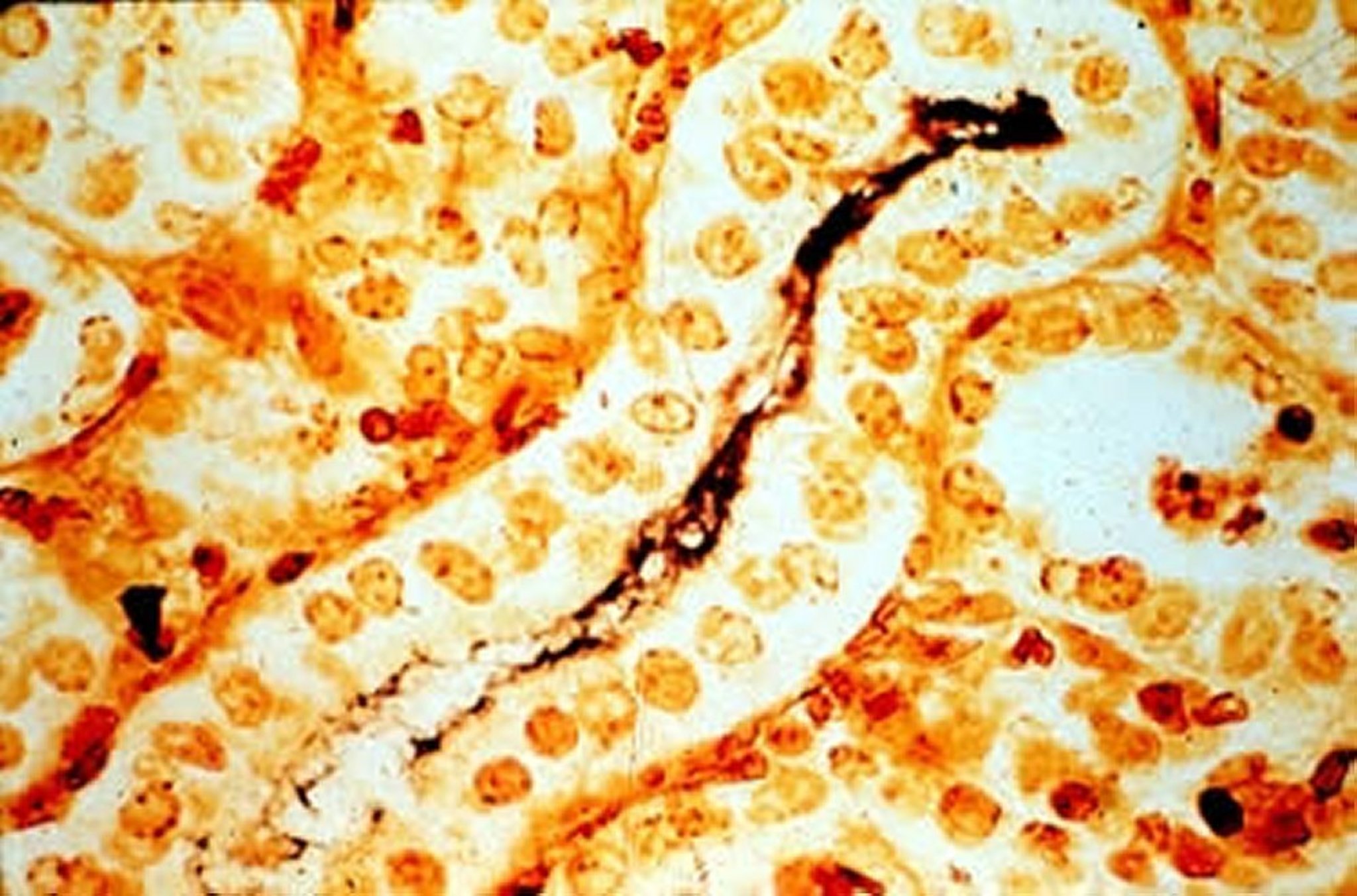
Photomicrograph of a histologic section from the kidney of a pig demonstrating Leptospira interrogans. Note the large number of threadlike black bacteria within the tubule lumen of the proximal convoluted tubules. Warthin-Starry silver stain; original magnification, medium power (10X).
Courtesy of the Department of Pathobiology, University of Guelph.

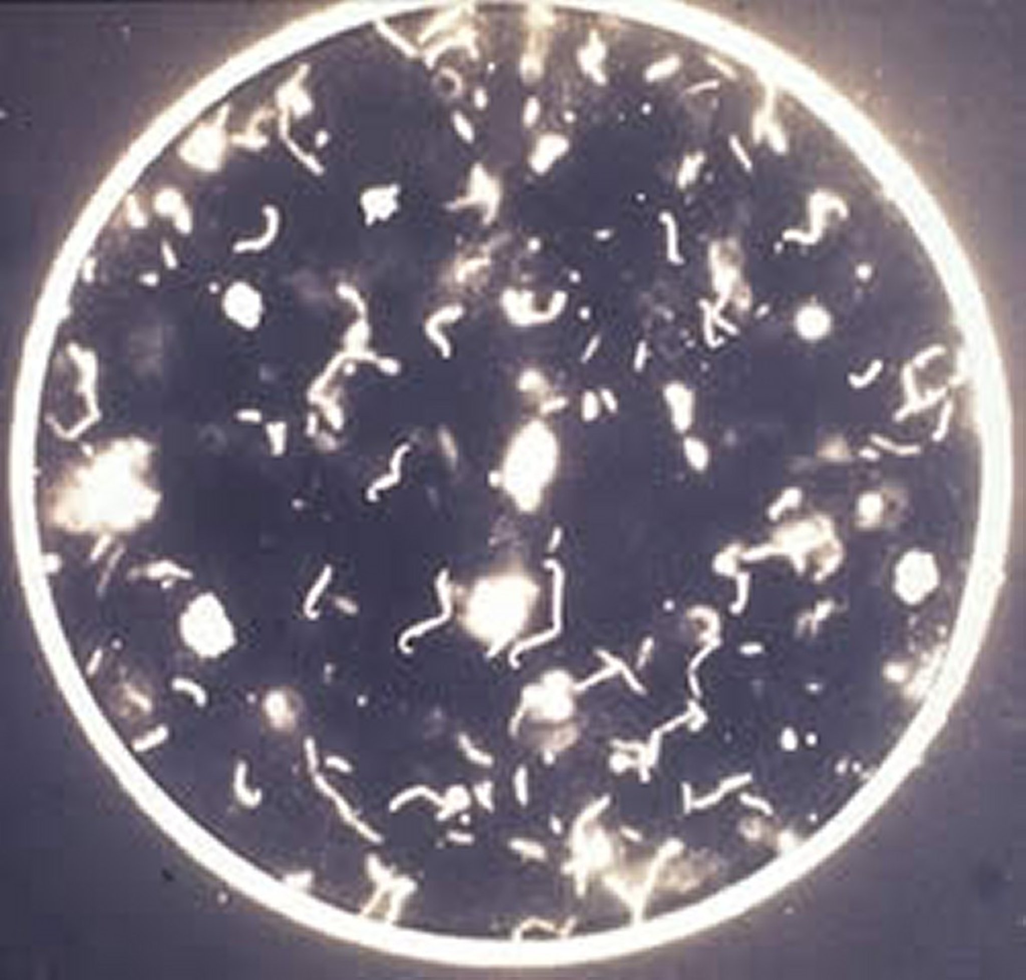
Photomicrograph of L interrogans in a microscopic agglutination test. The characteristic hooked ends (described as resembling "question marks" or "interrogation points," which give the species its name) are visible in nonagglutinated bacteria. Dark-field microscopy; original magnification, low power (4X).
Courtesy of the Department of Pathobiology, University of Guelph.
Leptospira are aerobic, gram-negative spirochetes that are fastidious, slow growing, and have characteristic corkscrew-like motility. The taxonomy of Leptospira is complex and can be confusing. Traditionally, Leptospira were divided into two groups; the pathogenic L interrogans and the saprophytic L biflexa. Within each of these species, leptospiral serovars were often grouped into antigenically related serogroups. With the increased use of genomic information for the classification of bacteria, the genus Leptospira was reorganized into genomospecies. The revised nomenclature is now reflected in the scientific literature, but not on labels for vaccines and pharmaceutical products. Fortunately for clinicians, the serovar and serogroup names remain in common use and are useful when discussing the epidemiology, serologic testing, clinical features, treatment, and prevention of leptospirosis.
Host Susceptibility, Epidemiology, and Transmission of Leptospirosis in Animals
All mammals are potentially susceptible to infection with pathogenic Leptospira. Among common companion animals and production animals, leptospirosis is most frequently recognized in cattle, swine, dogs, and horses. Leptospirosis in wildlife is common, although the disease is most often noticed only when the wildlife serve as a source of infection for domestic animals or humans.
Leptospirosis is found throughout the world and is regarded as a reemerging infectious disease. The infection (and disease) is more prevalent in warm, moist climates and is endemic in much of the tropics. In temperate climates, the disease is more seasonal, with the highest incidence after periods of rainfall.
Although >250 serovars of pathogenic Leptospira are recognized, leptospiral serovars in any given subset are prevalent within a particular region or ecosystem and are associated with one or more maintenance hosts, which serve as reservoirs of infection. Maintenance hosts are often wildlife species and, sometimes, domestic animals and production animals. In maintenance hosts, leptospirosis is generally characterized by a high prevalence of infection, low antibody responses, few organisms in tissues, relatively mild acute clinical signs, and persistent infection in the kidneys and sometimes the genital tract.
Common Maintenance Hosts of the Pathogenic Leptospires Associated with Disease in Domestic Animals in the US and Canada
Leptospiral Serovar | Maintenance Hosts |
|---|---|
Canicola | Dogs |
Pomona | Pigs, cattle, opossums, skunks |
Grippotyphosa | Raccoons, muskrats, skunks, voles |
Hardjo | Cattle |
Icterohaemorrhagiae | Rats |
Bratislava | Pigs, mice (suspected), horses (suspected) |
In incidental hosts, leptospirosis is characterized by a low prevalence of infection, severe clinical signs, and a short renal phase of infection. Incidental host infections produce a marked antibody response, and there are larger numbers of organisms in tissues of infected animals. Examples of this type of infection are serovar Grippotyphosa infection in dogs or serovar Icterohaemorrhagiae infection in cattle and swine.
Transmission among maintenance hosts is often direct and involves contact with infected urine, placental fluids, or milk. In addition, the infection can be transmitted venereally or transplacentally. Infection of incidental hosts is more commonly indirect, by contact with areas contaminated with urine of subclinically affected maintenance hosts that shed leptospires in their urine. Survival of leptospires is favored by moisture and moderately warm temperatures; survival is brief in dry soil or at temperatures < 10°C or >34°C. The organisms are killed by freezing, dehydration, or direct sunlight.
Pathogenesis of Leptospirosis in Animals
Leptospires invade the body after penetrating exposed mucous membranes or damaged skin. After a variable incubation period (4–20 days), leptospires circulate in the blood and replicate in many tissues including the liver, kidneys, lungs, genital tract, and CNS for 7–10 days. The phase of bacteremia and tissue colonization leads to the clinical signs of acute leptospirosis. Agglutinating antibodies can be detected in serum soon after leptospiremia occurs and coincide with clearance of the leptospires from blood and most organs. As the organisms are cleared, the clinical signs of acute leptospirosis begin to resolve, although damaged organs may take some time to return to normal function or, in severe cases, may not recover.
Leptospires remain in the tubules of the kidneys of incidental hosts for a short period of time and are shed in the urine for a few days to several weeks. In maintenance hosts, however, leptospires often remain in the renal tubules, genital tract, and less commonly, the eyes, despite the presence of high levels of serum antibody. Leptospires are shed in the urine and genital secretions of persistently infected animals for months to years, and these animals become an important reservoir of infection, with the potential to transmit infection to other reservoir hosts or to incidental hosts at risk of developing disease.
Clinical Findings of Leptospirosis in Animals
The clinical signs of leptospirosis depend on the host species, the pathogenicity of the strain and serovar of Leptospira, and the age and physiologic state of the animal. Subclinical infections are common, particularly in the maintenance host. In incidental hosts, leptospirosis is an acute, systemic, often febrile illness characterized by renal or hepatic damage. In addition, there may be effects on other body systems resulting in clinical problems such as uveitis, pancreatitis, bleeding, hemolytic anemia, muscle pain, or respiratory disease.
In both incidental and maintenance hosts that are pregnant at the time of infection, localization and persistence of the organism in the uterus may result in fetal infection, with subsequent abortion, stillbirth, birth of weak neonates, or birth of healthy but infected offspring. In general, incidental hosts abort acutely, whereas in maintenance hosts, abortions or other reproductive sequelae may be delayed by several weeks or months.
Diagnosis of Leptospirosis in Animals
Clinical signs
Combination of serologic testing to detect antibodies and PCR assay to detect organisms
Clinicians must be aware of the range of clinical signs for leptospirosis or the diagnosis will be missed. The best approach is a combination of serologic testing and PCR assay to detect, respectively, antibodies and organisms.
Available Antibody Tests
The microscopic agglutination test (MAT) is the most frequently used serologic test to diagnose leptospirosis, although patient-side rapid tests are increasingly used in small animals (see Leptospirosis in Dogs). The MAT involves mixing appropriate dilutions of serum with live leptospires of serovars prevalent within the region. The presence of antibodies is indicated by the agglutination of the leptospires, with the reported titer being the highest dilution of serum that results in 50% agglutination. The MAT is a complex test to perform and interpret, and it requires the maintenance of live leptospiral cultures. An ELISA to diagnose canine leptospirosis is offered by a commercial laboratory in the US. This test detects antibodies to LipL32, a membrane protein found on pathogenic leptospires. The currently available assay provides a qualitative negative or positive result and will also detect antibodies induced by vaccination. In general, the numerical titers provided by the MAT reveal more diagnostically useful information than a qualitative ELISA does, particularly when acute and convalescent MAT titers are performed.
Interpretation of MAT Results
Antibodies produced in an animal in response to infection with a given serovar of Leptospira often cross-react with other serovars in the MAT. Paradoxical reactions may occur also with the MAT early in the course of an acute infection, with a marked agglutinating antibody response to a serovar other than the infecting serovar. In addition, there is evidence of lack of consistency between diagnostic laboratories. For these reasons, the infecting serovar in an individual animal cannot be reliably identified from MAT results. The real value of the MAT is in providing a numerical titer to allow comparison of acute and convalescent values.
How to Use MAT Results
With a compatible clinical history and vaccination >3 months earlier, a single titer of 1:800 to 1:1,600 is good presumptive evidence of acute leptospirosis; however, a low initial antibody titer does not exclude a diagnosis of leptospirosis, because titers are often low in acute disease and in maintenance host infections. Therefore, the use of paired acute and convalescent titers is strongly recommended whenever possible, with infection confirmed usually by a 4-fold rise in antibody titer in paired serum samples collected 7–10 days apart. Antibody titers can persist for several months after infection and recovery, but they gradually decline with time. Vaccination of dogs and production animals with leptospiral vaccines also complicates the interpretation of leptospiral serologic testing. In general, vaccinated animals develop relatively low agglutinating antibody titers (1:100 to 1:400) in response to vaccination, and these titers persist for 1–4 months after vaccination. However, some animals develop high titers after vaccination that persist for ≥6 months. Again, paired acute and convalescent titers will discriminate between vaccinal titers and natural infection.
Other Tests
Immunohistochemistry is useful to identify leptospires in formalin-fixed tissue; however, because small numbers of organisms may be present in some tissues, the sensitivity of this technique varies. A number of PCR procedures are available, and each laboratory may select a slightly different procedure. Unfortunately, few publications have confirmed the validity of all the commercially available PCR assays, which likely vary considerably in their performance. Use of PCR techniques allow detection of pathogenic leptospires in blood, urine, or tissue samples, but they do not determine the infecting serovar. Culture of blood, urine, or tissue specimens, followed by molecular typing, is the only method to definitively identify the infecting serovar. Blood may be cultured early in the clinical course; urine is more likely to be positive 7–10 days after clinical signs appear. Culture rarely yields growth after antimicrobial therapy has begun. Culture of leptospires requires specialized culture medium, the organisms are fastidious and slow-growing, and diagnostic laboratories rarely culture specimens for the presence of leptospires. Thus, culture is of little value to clinicians.
Prevention of Leptospirosis in Animals
Avoidance of exposure to free-ranging wildlife and domestic animals that may be maintenance hosts for Leptospira is difficult because rodents, raccoons, opossums, and skunks are frequently found in rural and urban environments. The cornerstone of leptospirosis prevention is vaccination with polyvalent inactivated vaccines. Immunity to leptospirosis is assumed to be serovar specific; however, this assumption has been questioned.
Zoonotic Risk of Leptospirosis in Animals
Humans are susceptible to infection with most of the pathogenic serovars of Leptospira; however, they are incidental hosts and, therefore, not important reservoirs of infection. Occupational exposure is a risk factor, and veterinarians, veterinary staff, people who raise production animals, and dairy workers are at increased risk. In addition, recreational exposure to waters contaminated with urine of domestic animals or wildlife presents a risk.
The principal route of infection with Leptospira is contact with infectious body fluids (blood in acute cases or urine) via mucous membranes.
In humans, leptospirosis varies from subclinical to severe and can be fatal as a result of renal or hepatic failure or leptospiral pulmonary hemorrhage syndrome. The most common clinical signs are fever, headaches, rash, ocular pain, myalgia, and malaise. Transplacental infection, abortion, and infection of infants via breast feeding have been described, making exposure of pregnant women of particular concern. Laboratory techniques are necessary for a definitive diagnosis.
Because diagnosis of leptospirosis in animals is difficult based on clinical signs, veterinarians may wish to implement an infection control program in which animal body fluids are handled only with gloved hands and hand washing is routine. It is also essential for staff to take precautions when handling or nursing animals suspected or confirmed to have leptospirosis. Appropriate precautions include wearing gowns, shoe covers, and gloves to avoid contaminating exposed skin or transmitting organisms. Face shields should be worn when handling wet bedding or cleaning cages, stalls, or runs to avoid contact of aerosolized organisms with mucous membranes.
Key Points
Leptospirosis is maintained in the environment by a variety of carrier animals that typically are not clinically affected but shed organisms in the urine.
Disease occurs when incidental hosts are exposed to environments contaminated by urine from carrier animals.
Leptospirosis is a zoonotic disease. Clinicians should handle leptospirosis suspects with barrier nursing precautions.
Individuals should seek medical attention if they are exposed to leptospirosis or have clinical signs that could be compatible with the disease.
For More Information
Leptospirosis information for pet owners from the American Veterinary Medical Association.
Leptospirosis in Humans - CDC.
Also see pet health content regarding leptospirosis in horses, dogs, and cats.


