Chronic wasting disease is a transmissible spongiform encephalopathy that affects deer and other cervids, primarily in North America. It is a fatal progressive neurodegenerative disorder that affects both wild and farmed animals. The primary clinical signs are marked weight loss, behavioral changes, ataxia, and hypersalivation. Diagnosis is made by ELISA and Western blot, with confirmation by immunohistochemistry in brain (obex) and lymphatic tissue. There are no treatments or vaccines, so control relies on surveillance and depopulation of affected herds in farmed settings.
Chronic wasting disease (CWD) is a contagious and fatal neurodegenerative disease of captive and free-ranging cervids, including deer, elk, moose, and reindeer (1). It is a member of the transmissible spongiform encephalopathy (TSE) family of diseases, or prion diseases, that includes bovine spongiform encephalopathy; scrapie of sheep and goats; transmissible mink encephalopathy; camel prion disease; and, in humans, kuru, Creutzfeldt-Jakob disease (CJD), and variant CJD.
TSEs are caused by prions, infectious agents consisting solely of protein that do not use encoding nucleic acid for replication. They consist of PrPSc, a misfolded and aggregation-prone isoform of the host-encoded cellular prion protein (PrPc). Upon direct binding of PrPc to PrPSc, PrPc adopts the disease-associated conformation that is the basis for prion replication. In contrast to PrPc, PrPSc is partially resistant to protease digestion and accumulates within neurons, which eventually leads to neuronal death.
The main clinical signs of CWD, which occur after a long incubation period of up to several years, are marked weight loss, behavioral changes (including loss of fear of humans), loss of awareness, separation from the group, low head carriage, drooping ears, ataxia, hypersalivation, polydipsia, and polyuria. There is no curative or prophylactic treatment available for CWD.
CWD was first identified as a clinical syndrome in the late 1960s among captive mule deer in Colorado; a decade later, it was recognized to be a spongiform encephalopathy with characteristics similar to those of scrapie (2). It is found in farmed or free-ranging populations of mule deer, white-tailed deer, red deer, and elk (wapiti) in 36 states (US) and 5 Canadian provinces. Cases of CWD in free-ranging moose have been diagnosed in Colorado, US, and in Alberta and Saskatchewan, Canada. (Compare and images.)

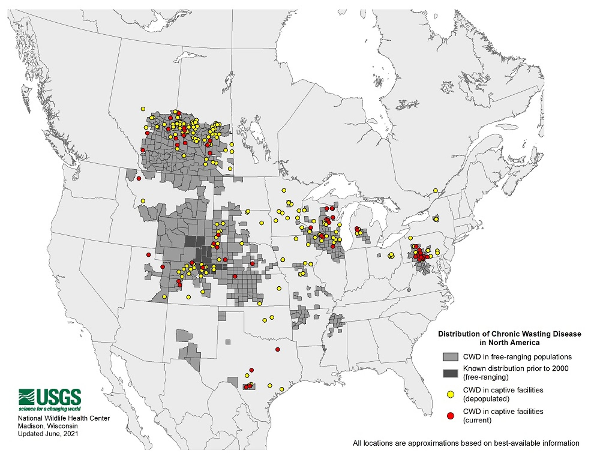
Distribution of chronic wasting disease in North America, June 2021.
Courtesy of USGS and Bryan Richards, USGS National Wildlife Health Center. Public domain.


Distribution of chronic wasting disease in North America, July 2025.
Courtesy of USGS and Bryan Richards.
Many states and provinces have developed regulations for control and management of CWD in farmed populations, and federal regulations are in place in Canada and the US. It is a reportable disease in most jurisdictions.
CWD has been identified outside of North America. In South Korea, elk imported from Canada in 1997 were CWD infected and caused outbreaks of CWD nationwide (3, 4). In 2016, CWD was diagnosed in wild reindeer in Norway, which marked the first finding of the disease in Europe and the first case in wild reindeer (5). Since then, CWD has been discovered in several Norwegian red deer and moose, and in 2018 and 2019, single cases in moose were documented in Finland and Sweden (6).
Etiology of Chronic Wasting Disease
Chronic wasting disease is mostly acquired by infection with CWD prions, except for rare cases in aged red deer and moose in Scandinavia, which are considered sporadic/atypical cases, similar to sporadic CJD in humans, or atypical bovine spongiform encephalopathy and scrapie. It is known to naturally affect mule deer, white-tailed deer, elk, red deer, and moose.
Experimentally, CWD can be transmitted by intracerebral inoculation to cattle, sheep, goats, domestic ferrets, mink, mice, hamsters, and squirrel monkeys (7). Transmission efficiency to cattle varies depending on the cervid species from which the CWD-infected brain homogenate used for infection is derived (8, 9).
Investigations of cases of CWD in moose and red deer in Scandinavia suggest an additional form of atypical/sporadic CWD. In these animals, PrPSc distribution was restricted to the brain, and affected animals were approximately 15 years old; therefore, a sporadic origin of the disease is possible (6).
Transmission, Epidemiology, and Pathogenesis of Chronic Wasting Disease
Chronic wasting disease is transmitted horizontally and vertically. Because prions are highly resistant to environmental and chemical inactivation, they can accumulate in the environment and thus be available to infect susceptible cervids. Consequently, close confinement of farm-raised cervids will likely potentiate the spread of CWD.
Winter feeding of deer and elk will concentrate cervid populations and likely potentiate horizontal transmission of CWD in wild populations. Foraging on feeding grounds contaminated by urine or feces of infected animals or contact with either infected animals or decomposing carcasses of animals with CWD results in transmission of disease to other susceptible cervids (10).
The CWD agent probably enters a susceptible host via ingestion and is taken up by lymphoid tissues associated with the GI tract. The agent can be detected in lymphoid tissue, nervous tissue, muscle tissue, antler velvet, blood, saliva, urine, and feces. Prions can be detected in the blood and saliva of infected animals as soon as 3 months after infection. In urine and feces, detection of prions is possible at a preclinical stage of the disease (7).
The CWD agent most likely arrives in the brain by retrograde movement up the vagus nerve to the dorsal motor nucleus of the vagus at the obex region of the medulla oblongata. Spongiform lesions in the brain develop first in the vagal nucleus at approximately the time of onset of clinical disease. This occurs naturally, with an incubation period of approximately 1.5–3 years; however, incubation periods are influenced by PrP polymorphisms.
One of the highest prevalences of CWD in wild deer has been reported in Saskatchewan, reaching > 90% in male mule deer (11).
The movement of CWD in populations of free-ranging deer and elk follows natural migration routes, often along waterways and natural corridors. In the past, movement of CWD in farmed deer and elk in commerce was through human-facilitated transportation of animals incubating CWD. Now that programs and regulations are in place in most jurisdictions, movement of CWD in live animals should be curtailed. Surveillance, however, should continue, and submission of samples from hunter-harvested animals might be mandatory in areas with a high prevalence of CWD.
Clinical Findings of Chronic Wasting Disease
Animals with clinical chronic wasting disease are > 16 months old and show a spectrum of clinical signs. The earliest and most difficult to appreciate are subtle changes in behavior and weight loss. These changes are often detectable only by animal caretakers familiar with the individual animal.
As CWD progresses, behavioral changes can include alterations in how the animal interacts with herdmates and caretakers, loss of wariness, somnolence, persistent walking, polydipsia and polyuria, and hyperexcitability when handled. Affected animals can show variable locomotor signs, including ataxia (especially hindlimb ataxia) and head tremors.
Animals with late-stage CWD can have a low head carriage, drooped ears, and a fixed, staring gaze (see ); they might hypersalivate and grind their teeth. Death after routine chemical immobilization has been noted. Aspiration pneumonia can be the only presenting clinical sign and is often the cause of death. Therefore, CWD should be suspected in any adult cervid with aspiration pneumonia.
Pearls & Pitfalls
|


A bull elk in Wind Cave National Park with late-stage chronic wasting disease (CWD).
Photograph by National Park Service. Public Domain.
Weight loss is progressive throughout the course of disease, even when adequate feed is present; however, it is important to recognize that CWD can be present in cervids that are not emaciated. Death of CWD-affected animals can be precipitated by cold weather or other acute stressors.
Affected cervids are more susceptible to hunting, predation, vehicle collisions, and other forms of death by misadventure. Carcasses and offal should be disposed of in a manner that limits the exposure of farm-raised and free-ranging cervids to any potentially infectious material.
Lesions
Lesions from chronic wasting disease are present in the gray matter of the CNS. Lesions are bilaterally symmetrical and anatomically constant among animals. Spongiform appearance is obvious; vacuolization occurs in neuronal perikarya and neuronal processes.
Along with neuronal degeneration, astrocytic hyperplasia and hypertrophy can appear. H&E staining of brains of affected animals reveals amyloid plaques that appear as pale, fibrillar, eosinophilic areas of neuropil that are sometimes surrounded by vacuoles (florid plaques).
Detection of PrPSc in brain sections by immunohistochemistry (IHC)provides a very good way to visualize CWD pathology while maintaining the structural context. PrPSc is detected by IHC or immunoblot in a variety of tissues in cervids showing clinical signs. Although PrPSc can be found in regions of the brain not exhibiting spongiform change, typically there is correlation between PrPSc deposition and spongiform appearance.
Diagnosis of Chronic Wasting Disease
Detection of PrPSc by ELISA or Western blot
Confirmation by immunohistochemistry
Diagnosis of chronic wasting disease by clinical signs is not reliable, because they are unspecific and mild at the beginning of disease. Therefore, diagnosis relies on the detection of protease-resistant PrPSc by ELISA or Western blot and confirmed by immunohistochemical detection of PrPSc in brain (obex region) or lymphatic tissue.
In mule deer and white-tailed deer, the CWD PrP accumulates in the retropharyngeal lymph node before arriving in the brain; thus, it is considered to be the most important tissue to collect for testing. Both brain and lymph node samples should be collected from elk. The correct portion of the brain (ie, the obex, at the caudal end of the fourth ventricle below the cerebellum) must be collected for a meaningful test.
The laboratory should be consulted to determine whether samples must be fixed in 10% buffered formalin, chilled or frozen, or sent both fixed and frozen. Samples should be, and in many jurisdictions are required to be, submitted to certified laboratories to be tested for evidence of CWD. It is good practice to either send the carcass to the diagnostic laboratory or collect a wide variety of samples, so if diseases other than CWD are present, they will be identified. At a minimum, samples for CWD testing should include brain and retropharyngeal lymph nodes. Many laboratories accept whole heads from cervids for testing.
Pearls & Pitfalls
|
Surveillance programs for free-ranging cervids vary depending on the jurisdiction and are usually conducted by the local wildlife management agency, which should be consulted if CWD is suspected in a free-ranging deer or elk. Surveillance depends mainly on submission of heads from hunter-harvested animals.
In some areas, active CWD surveillance is done by taking biopsies from tonsils, retropharyngeal lymph nodes, and rectoanal mucosa–associated lymphoid tissue (RAMALT). Diagnostic tests include detection of PrPSc by IHC, ELISA, or Western blot in brain and/or lymphoid tissues. In the US, these tests are run only at USDA-certified laboratories. ELISA is used as a screening test, and IHC, which is considered the preferred test, is used to confirm positive ELISA.
In vitro conversion methods for amplification and detection of minute amounts of PrPSc include protein misfolding cyclic amplification or real-time quaking-induced conversion assay. Using these tests, CWD prions are detectable at a preclinical stage in specimens that can be obtained antemortem by noninvasive methods, such as blood, urine, feces, saliva, or nasal brushings. These newly developed assays are experimental, undergoing validation, and have not been approved for diagnostic use.
Differential diagnoses for animals suspected of CWD include:
brain abscess
traumatic injury
pneumonia
arthritis
starvation and nutritional deficiency
dental attrition
anesthetic death
Treatment and Control of Chronic Wasting Disease
Depopulation
Surveillance
There is no treatment, prophylaxis, or supportive care available for any transmissible spongiform encephalopathy. In the US and Canada, control in farmed cervids is by depopulation with indemnity and development of chronic wasting disease herd certification programs. These are voluntary cooperative programs between industry and federal or state/provincial governments. They are administered by the USDA Animal and Plant Health Inspection Service and the Canadian Food Inspection Agency.
Herd certification programs typically require 5 years of monitoring to achieve the highest status. The bases for CWD control programs in the farmed cervid industry are individual animal identification, CWD testing in all animals in the herd that die over a certain age, and limiting additions to animals from herds of comparable or higher CWD status.
Control of CWD in free-ranging populations is extremely difficult. All jurisdictions have banned movement of live cervids from endemic areas for translocations, and many have regulations on movement of parts of hunted deer and elk. In areas where CWD occurs, attempts at control have included population reduction, test and removal, and intensified surveillance; however, these have had limited success.
Only a few disinfectants and methods of disposal inactivate prions. Fresh household bleach at 50% concentration for 30–60 minutes or sodium hydroxide (1 M) for 60 minutes will inactivate the agent (12). This is inexpensive and readily available, however, it can be corrosive to some surfaces and instruments. Additional disinfectants are being considered for general use but are not yet approved. Incineration in a medical incinerator, alkaline digestion in specially designed equipment, and disposal in certified municipal landfills are used to dispose of tissues and carcasses of animals with CWD.
Zoonotic Risk of Chronic Wasting Disease
Although chronic wasting disease has been present in hunted populations of deer and elk for > 30 years, no case of human CWD has been identified. The risk to humans appears to be minimal. Experimental studies to gain insight into the zoonotic potential of CWD, using infection of humanized transgenic mice or cynomolgus macaques as models, provided conflicting results. While studies using these models did not indicate transmissibility, other studies demonstrated transmission and atypical disease presentation and features (13, 14, 15, 16). Therefore, zoonotic potential cannot be excluded; however, the transmission barrier is considered high.
The CWD agent has been detected in muscle tissue of infected cervids. Public health authorities and wildlife management agencies suggest the following precautions for hunters and people handling cervids in areas where CWD is found, to further decrease the risk of human exposure:
do not harvest deer or elk that appear to be sick or abnormal
wear rubber, plastic, or latex gloves when dressing the carcass
avoid contact with brain, spinal cord, and lymphoid tissues
debone the meat when processing
disinfect knives, saws, and tables with 50% bleach
have the animal tested for CWD
All public health authorities state that animals positive for any transmissible spongiform encephalopathy should not be consumed by humans or other animals.
Key Points
Chronic wasting disease is a fatal and transmissible neurodegenerative disease of wild and captive cervids caused by prions.
CWD has been reported in 36 US states, 5 Canadian provinces, and 3 European countries.
Clinical signs include progressive weight loss, ataxia, hypersalivation, and behavioral changes.
CWD is typically diagnosed postmortem by detection of disease-associated PrPSc in brain or lymphatic tissue.
No therapy or prophylaxis for CWD is available.
For More Information
References
Bartz JC, Benavente R, Caughey B, et al. Chronic Wasting Disease: State of the Science. Pathogens. 2024;13(2):138. doi:10.3390/pathogens13020138
Williams ES, Young S. Chronic wasting disease of captive mule deer: a spongiform encephalopathy. J Wildl Dis. 1980;16(1):89-98. doi:10.7589/0090-3558-16.1.89
Sohn H-J, Kim J-H, Choi K-S, et al. A case of chronic wasting disease in an elk imported to Korea from Canada. J Vet Med Sci. 2002;64(9):855-858. doi:10.1292/jvms.64.855
Choi YP, Lee Y-R, Park HC, et al. Chronic wasting disease in farmed cervids, South Korea, 2001–2024. Emerg Infect Dis. 2026;32(4):614-618. doi:10.3201/eid3204.251046
Benestad SL, Mitchell G, Simmons M, Ytrehus B, Vikøren T. First case of chronic wasting disease in Europe in a Norwegian free-ranging reindeer. Vet Res. 2016;47(1):88. doi:10.1186/s13567-016-0375-4
Tranulis MA, Gavier-Widén D, Våge J, et al. Chronic wasting disease in Europe: new strains on the horizon. Acta Vet Scand. 2021;63(1):48. doi:10.1186/s13028-021-00606-x.
Arifin MI, Hannaoui S, Chang SC, Thapa S, Schatzl HM, Gilch S. Cervid prion protein polymorphisms: role in chronic wasting disease pathogenesis. Int J Mol Sci. 2021;22(5):2271. doi:10.3390/ijms22052271
Hamir AN, Kunkle RA, Miller JM, Greenlee JJ, Richt JA. Experimental second passage of chronic wasting disease (CWD (mule deer)) agent to cattle. J Comp Pathol. 2006;134(1):63-69. doi:10.1016/j.jcpa.2005.07.001
Greenlee JJ, Nicholson EM, Smith JD, Kunkle RA, Hamir AN. Susceptibility of cattle to the agent of chronic wasting disease from elk after intracranial inoculation. J Vet Diagn Invest. 2012;24(6):1087-1093. doi:10.1177/1040638712461249
Sorensen A, van Beest FM, Brook RK. Impacts of wildlife baiting and supplemental feeding on infectious disease transmission risk: a synthesis of knowledge. Prev Vet Med. 2014;113(4):356-363. doi:10.1016/j.prevetmed.2013.11.010
Government of Saskatchewan. 2024-25 Chronic Wasting Disease Surveillance Program Results. Accessed April 15, 2026. https://pubsaskdev.blob.core.windows.net/pubsask-prod/149701/CWD%252BSurveillance%252BProgram%252BResults%252B-%252B2024-25.pdf
Williams K, Hughson AG, Chesebro B, Race B. Inactivation of chronic wasting disease prions using sodium hypochlorite. PLoS One. 2019;14(10):e0223659. doi:10.1371/journal.pone.0223659
Race B, Meade-White KD, Miller MW, et al. Susceptibilities of nonhuman primates to chronic wasting disease. Emerg Infect Dis. 2009;15(9):1366-1376. doi:10.3201/eid1509.090253
Race B, Williams K, Orrú CD, Hughson AG, Lubke L, Chesebro B. Lack of transmission of chronic wasting disease to cynomolgus macaques. J Virol. 2018;92(14):e00550-18. doi:10.1128/JVI.00550-18
Hannaoui S, Zemlyankina I, Chang SC, et al. Transmission of cervid prions to humanized mice demonstrates the zoonotic potential of CWD. Acta Neuropathol. 2022;144(4):767-784. doi:10.1007/s00401-022-02482-9
Hannaoui S, Pritzkow S, Jürgens-Wemheuer WM, et al. Limited transmission of cervid prions to nonhuman primates provides insights into the zoonotic potential of chronic wasting disease. Sci Adv. 2026;12(22):eaeb7613. doi:10.1126/sciadv.aeb7613