


Also see Diseases of the Peripheral Nerves and Neuromuscular Junction.
Large Animals
Bovine spastic paresis andbovine spastic syndrome are seen in many breeds of cattle and has been referred to as “contraction of the Achilles tendon,” “straight hock,” "barn cramp," and “Elso heel.” (Also see Lameness in Cattle.) It can be divided into two syndromes, one that affects calves (spastic paresis) and one that affects adults (spastic syndrome). Affected muscles include the gastrocnemius, quadriceps, or mixed muscles.
In calves, the condition appears to be familial and can be seen in many breeds, with signs beginning between 1 week and 1 year of age. It is characterized by progressive hyperextension of the stifle and tarsus and by spastic contracture of the muscles of one or both pelvic limbs. The leg is usually held in extension behind the calf and does not touch the ground during walking. The disease is progressive, but the gastrocnemius and quadriceps forms usually respond to partial neurectomy of the tibial nerve. Only a partial response to neurectomy is seen with mixed muscle involvement. The cause is unknown despite much research, and genetic studies have not yet identified the responsible mutation(s). No lesions are seen in peripheral nerves, and the condition is thought to involve excessive activity of the neuromuscular spindle reflex arc.
Adult cattle are affected at 3–7 years of age. Extensor muscles of one or both pelvic limbs are affected, progressing to the spinal muscles, causing kyphosis and caudal extension of the limbs. This condition is also thought to be familial and is usually progressive. Mephenesin (30–40 mg/kg, PO, every 24 hours for 2–3 days) may produce variable control of signs. Quadriceps muscle hypoplasia as a cause of congenital lameness has been described in Holstein calves. Reduced numbers of spinal cord motor neurons suggest that there is failure to innervate the muscle on the affected side.
Hyperkalemic periodic paralysis is seen in Quarter horses 2–3 years old and is due to an inherited mutation of the sodium channel (SCN4A). It causes episodes of muscle tremor and sometimes recumbency, both of which may be precipitated by exercise. Hyperkalemia is usually present during an attack, and electromyography can also be helpful for diagnosis. Acetazolamide (2–4 mg/kg, PO, every 8–12 hours) may lessen the frequency and severity of attacks.
Myotonia congenita is an inherited/familial disorder in goats and Shropshire lambs and is occasionally seen in horses. It causes muscle rigidity; marked dimpling on percussion of the muscle belly; and a stiff, stilted gait. Electromyography is a useful aid to diagnosis. This disease results from a mutation in a chloride channel (CLCN1).
Muscular dystrophy is an inherited disease in Merino sheep and has also been reported in some horses, including Thoroughbred, Quarterhorse, and Swedish half-bred horses. It results in a slowly progressive stiffness that affects the limbs and neck from 3–4 weeks of age onward, although some are noticeably younger. Clinically affected sheep have high resting and postexercise concentrations of serum CK and lactate dehydrogenase. On histopathology, there is selective atrophy of Type I muscle fibers.
Porcine stress syndrome or malignant hyperthermia is a hypermetabolic and hypercontractile syndrome that, when triggered by anesthesia or stress, produces a sustained increase of intracellular calcium levels within skeletal muscle fibers. This in turn causes muscle stiffness, hyperventilation, hyperthermia, and pale exudative pork. It results from a mutation in a calcium-channel gene that is inherited as an autosomal dominant trait, usually in Landrace pigs. It may occur rarely in horses. Dantrolene sodium may be used to reduce incidence or for treating signs. Swine (3.5 mg/kg, IV) or horses (4–6 mg/kg after fasting for at least 4 hours, intragastric, 1–1.5 hours before induction of anesthesia, with additional doses of 2.5 mg/kg every hour) may be used. IV dosing in horses is rapidly cleared and not as effective, but dosages of 4 mg/kg, IV, should maintain therapeutic levels for 2 hours.
Mitochondrial myopathy has been reported in horses and may occur alone or in combination with other myopathies, including polysaccharide storage myopathy. Affected horses show exercise intolerance and muscle stiffness. Diagnosis is by altered oxidative phosphorylation activity and profound lactic acidosis, or by histopathologic assessment of muscle biopsy. Ragged red fibers and increased staining for mitochondria may be identified. There is no treatment, and activity should be limited in affected horses.
Startle disorders include congenital myoclonus in Poll Hereford calves (GLRA1 mutation) and Peruvian Peso horses andcongenital muscular dystonia in Belgian Blue Cattle (SLC6A5 mutation). Clinical signs are stimulus-induced and include muscle rigidity and tremor in response to tactile, acoustic, or visual stimulation. Standing and feeding are often impaired, and affected animals often die within days to weeks.
Small Animals
Hypertrophic neuropathy of Tibetan Mastiffs is an autosomal recessive disease that has been recognized in the USA, Switzerland, and Australia. Onset is 7.5–10 weeks of age, and clinical signs include rapidly progressive paresis and muscle hypotonia. Hyporeflexia is marked, but sensory function is preserved. Demyelination and remyelination are seen on nerve biopsy. Prognosis is guarded. The disease can stabilize, and some puppies may regain the ability to walk but remain weak. There is no treatment.
Alaskan Malamute polyneuropathy affects Alaskan Malamutes 10–18 months old and is caused by a mutation in the NDRG1 gene. Clinical signs include laryngeal paralysis, exercise intolerance, decreased postural reactions, paraparesis progressing to tetraparesis, hyporeflexia, and muscle atrophy. Electromyography shows diffuse fibrillation potentials and positive sharp waves. On nerve biopsy, there is axonal necrosis with demyelination. There is no effective treatment, although clinical signs stabilize in some dogs. In most affected dogs, however, progressive disability leads to euthanasia.
Congenital laryngeal paralysis is seen in Bouvier des Flandres (autosomal dominant) and Siberian Huskies, Rottweilers, and Bull Terriers < 1 year old. It results in exercise intolerance and inspiratory dyspnea. Diagnosis is confirmed by visualization on laryngoscopy. Congenital laryngeal paralysis with diffuse peripheral neuropathy is seen in several breeds, including Dalmatians, Rottweilers, and Pyrenean Mountain Dogs (Great Pyrenees). An autosomal recessive mode of inheritance is suspected. Prognosis is guarded to poor without treatment. Some dogs with unilateral cricoarytenoid lateralization have been able to resume activity.
Primary hyperoxaluria (l-glyceric aciduria) is a rare, inherited (autosomal recessive) neurofilament disorder of domestic shorthaired cats and Coton de Tulear and Tibetan Spaniel dogs. Neurologic signs have not been reported in dogs, whereas clinical signs in cats include renal disease and weakness due to a peripheral neuropathy. Signs develop at 5–9 months of age in cats and 3–4 weeks in dogs. A plantigrade stance is the most prominent sign, and spinal reflexes are sometimes reduced. Urine contains increased oxalate and l-glycerate levels. Genetic mutations in AGXT and GRHPR genes have been identified in the Coton de Tulear. There is no treatment.
Neuropathy of hereditary hyperchylomicronemia (hyperlipidemia) is a suspected autosomal recessive disorder that causes a generalized peripheral neuropathy in cats. Affected cats show reduced lipoprotein lipase activity. Serum cholesterol and triglyceride concentrations are typically increased. Clinical signs do not develop until at least 8 months of age. The hyperlipidemia results in deposition of lipid granules within nerves. There is evidence that the clinical signs can be controlled by a low-fat diet. Blood samples from affected cats have the appearance of “cream of tomato soup.”
Sensory neuropathy of longhaired Dachshunds (probably autosomal recessive) causes pelvic limb ataxia at 8–12 weeks of age. Urinary and GI function may also be disturbed. Paw position sense, spinal reflexes, and pain sensation are depressed, and self-mutilation can occur. There is a loss of myelinated fibers in sensory nerves and in selected areas of the spinal cord. There is no treatment, but affected dogs may have a relatively normal quality of life, provided self-mutilation does not occur.
Sensory neuropathy of Border Collies is an autosomal recessive disease that manifests as ataxia, lack of proprioception, and abnormal sensory testing. Onset is usually at 5–7 months of age and progresses relentlessly. Euthanasia is the common endpoint. Disruption of the FAM134B gene was identified in association with this disease.
Sensory neuropathy in Pointers is seen in English Pointers (autosomal recessive) in the USA and Shorthaired Pointers in Europe. Self-mutilation of the digits is the main clinical sign, and disease onset is before 6 months of age. Pain perception is absent in the pelvic limbs and depressed in the thoracic limbs. There is neuronal loss in dorsal root ganglia. Prognosis is poor. There is no treatment.
Inherited polyneuropathy of Leonberger dogs is a distal neuropathy, with an age of onset of 1–9 years in this breed. Clinical signs include weakness, exercise intolerance, change in bark, and dyspnea. A high-stepping gait, representative of sensory dysfunction, is often present. Pedigree analysis suggests an X-linked inheritance.

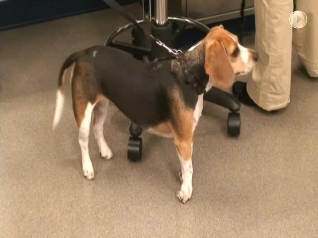
Musladin-Lueke syndrome is an autosomal recessive connective tissue disorder recognized in Beagles that affects muscles, bone, heart, and skin. It is caused by a mutation in the ADAMTSL2 gene. Clinical signs most prominently reflect muscle fibrosis and contractures. The resulting posture is that of the animal walking on its “tip-toes.” This is an autosomal recessive disorder. The abnormal posture is present shortly after birth. Other clinical signs include thickened cartilage of the ear and wide-set eyes. Seizures may occur concurrently. It is not yet known whether the seizures are related to the primary disorder or are a concurrent disorder. There is no treatment. The human counterpart to this disease is progressive and frequently fatal; however, the disease appears to stabilize in dogs.
Congenital myasthenia gravis (autosomal recessive) has been described in a number of dog and cat breeds, including Parson Russell Terrier, Smooth-haired Fox Terrier, Smooth-haired Miniature Dachshund, Springer Spaniel, Labrador Retriever, Golden Retriever, Sphinx cat, and Devon Rex cat. It is due to either a deficiency or dysfunction of the acetylcholine receptor, and there is none of the circulating antireceptor antibody seen in the more common acquired form of the disease. Several mutations have been associated with congenital myasthenic syndromes: CHRNE (Jack Russell Terrier), CHAT (Old Danish Pointing dogs), and several mutations in COLQ (Labrador Retriever, Golden Retriever, Sphinx cat, and Devon Rex cat). Clinical signs usually start at 5–10 weeks of age (dogs) and 10–16 weeks of age (cats). The characteristic finding is an exercise-induced weakness, often associated with megaesophagus. The prognosis is more guarded than in acquired myasthenia gravis. The congenital disease has also been described in cats, in which weakness, ventroflexion of the head and neck, and difficulty swallowing also occur. A presynaptic form is seen in 12- to 16-week-old Gammel Dansk Hønsehund dogs (autosomal recessive). Treatment with anticholinesterase medications may be effective (pyridostigmine at 0.2–2 mg/kg, PO, every 8–12 hours) for some breeds, although anticholinergic medications were shown to worsen clinical signs in the Labrador variant.
Scotty cramp (episodic muscle hypertonicity) is an inherited disease most commonly seen in Scottish Terrier puppies. The mode of inheritance is not yet known, but may be associated with CFA X. These episodes are exacerbated by excitement, exercise, stress, and poor health and are characterized by a hypermetric gait and arching of the spine, which can cause the dog to somersault when it runs. Clinical signs are usually apparent by 2–6 months of age, or up to 4 years for other breeds. The disorder seems to be related to faulty serotonin metabolism. Diazepam (0.5–1 mg/kg, IV; 0.25 mg/kg, PO, 3 times daily)) and promazines (acepromazine 0.5–2.2 mg/kg, PO, 3 times daily) help to relieve signs. Fluoxetine (1.2 mg/kg, PO, once daily, or 0.8 mg/kg, PO, twice daily) may also be effective.
Congenital myoclonus of Labrador Retrievers (familial reflex myoclonus) causes stimulus-induced muscle spasms/hypertonicity from an early age. Puppies may be unable to walk or even maintain a sternal position due to extensor rigidity that is triggered with movement. At rest, muscles are relaxed. Treatment with diazepam and clonazepam was not effective. The neurologic examination is normal, but stimulus from performing parts of the examination (eg, reflexes) results in generalized stiffness. The prognosis is very poor.
Hypokalemic myopathy of Burmese cats (autosomal recessive) causes periodic paralysis or weakness with ventral flexion of the neck. In some cases, weakness is persistent. Cats are affected at 3–4 months of age. Serum CK is markedly increased. Dietary supplementation of oral potassium usually produces a favorable response (eg, potassium gluconate solution at 2–4 mEq or mmol/cat/day, PO, until serum potassium levels are stable). In some cases, the condition may be outgrown.
Myotonia congenita is seen in Chow Chows, Staffordshire Terriers, Great Danes, and Miniature Schnauzers (autosomal recessive), as well as Domestic Short-haired cats, and causes signs similar to those seen in myotonic goats. There is often a degree of muscle hypertrophy, and marked stiffness is seen when dogs first rise. Dimpling is seen on percussion of several muscles, including the tongue. Diagnosis can usually be confirmed by electromyography (characteristic “dive bomber” sound); muscle biopsy changes are mild and nonspecific. Prognosis is guarded, although membrane-stabilizing drugs (procainamide at 40 mg/kg, PO, every 6–12 hours; mexiletine at 4–8 mg/kg, PO, every 8 hours) result in significant improvement. Signs do not resolve but may stabilize by 6–12 months.
X-linked muscular dystrophies have been described in Irish Terriers, Golden Retrievers, Miniature Schnauzers, Rottweilers, Samoyeds, German Shorthaired Pointers, Groenendaeler Belgian Shepherds, Brittany Spaniels, Rat Terriers, Labrador Retrievers, Japanese Spitz, Border Terriers, and also in cats. All are due to mutations in the dystrophin gene. Males show muscle stiffness, dysphagia, and weakness at an early age, along with a plantigrade stance and muscle atrophy as the animal gets older. Initial muscle hypertrophy may be marked, particularly in cats. Diagnosis is facilitated by the initial massive increases in serum levels of CK and by demonstration of hyalinized and mineralized muscle fibers on biopsy. Prognosis is guarded to poor. Currently, there is no treatment. A novel congenital muscular dystrophy has been reported in cats associated with deficiency of merosin (laminin alpha-2), in which degenerative changes occur in both muscles and peripheral nerves.
Centronuclear myopathy (including Labrador Retriever myopathy) is an autosomal recessive disease caused by a mutation in the PTPLA gene and is seen in Labrador Retrievers. Age of onset is 6 weeks to 7 months, and clinical signs are progressive but variable in severity. Some cases may stabilize. Weakness, exercise intolerance, ventroflexion of the neck, and marked muscle atrophy are the predominant clinical signs. Signs may worsen with cold, stress, or exercise, and affected dogs may be unable to keep their heads elevated in a normal position from as early as 3 months of age. Tendon reflexes are usually absent. In some cases, signs may stabilize. Muscle biopsy shows loss of Type II fibers, and myofiber nuclei are centralized. Within the family of centronuclear myopathies, an X-linked myotubular myopathy also occurs in dogs and exhibits a similar onset and phenotype. However, it is more rapidly and severely progressive and eventually causes fatal respiratory failure, with affected dogs unlikely to survive past 6 months.
Dermatomyositis of Collies and Shetland Sheepdogs (inherited as a dominant trait with variable expressivity) causes atrophy and weakness of the masticatory and distal limb muscles from a few months of age, sometimes associated with trismus and megaesophagus. These signs are combined with a dermatitis over the face and extremities. The clinical signs may wax and wane and, in general, do not become severely debilitating. Polymyositis and dermatitis are evident on histopathologic examination. This disorder has also been seen in Beauceron Shepherds, Pembroke Welsh Corgis, Australian Cattle Dogs, Lakeland Terriers, Chow Chows, German Shepherds, Kelpies, and Kuvasz dogs.
Mitochondrial myopathy has been described in Clumber and Sussex Spaniels and in Old English Sheepdogs. Mitochondrial myopathies result in exercise intolerance and collapse. Blood lactate and pyruvate levels are often increased after exercise. Ragged red fibers, indicating increased numbers of mitochondria, may be seen on muscle biopsy. Inherited disorders of carnitine metabolism cause a lipid storage myopathy and are another type of mitochondrial myopathy, which may cause accumulation of lipid vacuoles within muscle fibers.
Nemaline rod myopathy (probable autosomal recessive) in cats causes weakness and later a hypermetric gait at 6–18 months of age. Patellar reflexes are depressed, and muscle atrophy develops progressively. Large numbers of nemaline rods are found in skeletal muscle fibers. The prognosis is poor. A similar disorder is seen sporadically in young dogs.
Inherited myopathy of Great Danes (Central core myopathy) has been described as a cause of progressive weakness, muscle atrophy, and exercise intolerance/collapse in young Great Danes in the UK. Signs begin at approximately 6 months to 3 years. Prognosis is poor, and euthanasia often occurs within months of the diagnosis.
Congenital megaesophagus is inherited in Wirehaired Fox Terriers and Miniature Schnauzers and possibly also in German Shepherds, Great Danes, Irish Setters, Newfoundlands, Chinese Shar-Pei, Greyhounds, and Siamese cats. Clinical signs include regurgitation and aspiration pneumonia. Prognosis is guarded. Treatment with sildenafil oral suspension (1 mg/kg, PO, twice daily) may reduce the number of regurgitation episodes and increase weight gain.
Devon Rex cat hereditary myopathy (autosomal recessive) is seen in kittens about 4–7 weeks old and is characterized by exercise intolerance and passive ventroflexion of the head and neck, which is especially noticeable during locomotion, urination, or defecation. Some cats assume a “dog-begging” position. Megaesophagus is present. Prognosis is guarded.
Polyneuropathy of Bengal cats is a chronic, relapsing polyneuropathy that results in weakness from demyelination. Age of onset ranges from 3 to 44 months of age. Clinical signs include paresis, plantigrade stance, exercise intolerance, and stiff or stilted gait. Signs predominantly affect the pelvic limbs but may progress to tetraparesis. Muscle atrophy, weight loss, and stunted growth may occur. There is no specific treatment. Remyelination and recovery can occur, but residual deficits often remain, and relapses are possible.
Startle disease is an inherited disease in Irish Wolfhounds and causes episodes of stimulus-induced hyperreflexia, muscle stiffness, and apnea. A mutation in SLC6A5 is thought to be responsible. Sudden death may occur in affected dogs, and often-affected dogs are euthanized due to poor quality of life.
Exercise-induced collapse is an autosomal recessive disease described in Labrador Retrievers, Chesapeake Bay Retrievers, Curly-coated Retrievers, German Wirehaired Pointers, and Welsh Corgis. It is associated with a mutation in dynamin 1 (DNM1). A genetic test is available for some breeds. The onset of clinical signs is 6 months to 3 years. Affected dogs show an exercise-induced ataxia, weakness, and collapse during mild-moderate activity. After 5 to 25 minutes of rest, they recover without residual signs. No treatment is available, and control is by exercise moderation. Swimming should be avoided due to risk of drowning if an episode occurs in water.


