

The protozoon Histomonas meleagridis infects a wide range of gallinaceous birds and causes histomoniasis (blackhead disease). Chickens are typically subclinical carriers, but mortality rate in turkeys is commonly 80%–100%. Clinical signs include drooping head and wings, prolonged standing, closed eyes, ruffled feathers, emaciation, and sulfur-colored feces. Diagnosis is based on pathognomonic ulceration of the ceca and necrotic lesions in the liver. There are no approved treatments or vaccines.
Etiology of Histomoniasis in Poultry
Histomonas meleagridis, an anaerobic protozoal parasite of the order Trichomonadida, is the causative agent of histomoniasis (blackhead disease). It can exist in flagellated (8–15 mcm in diameter) and amoeboid (8–30 mcm in diameter) forms.
H meleagridis is primarily transmitted in the egg of the cecal nematode Heterakis gallinarum. Chickens and other gallinaceous birds act as a reservoir for H gallinarum. Nematode eggs infected with H meleagridis remain viable in the environment for years.
Three species of earthworms can act as paratenic hosts for H gallinarum larvae containing H meleagridis. Chickens and turkeys that consume these earthworms can become infected with both H gallinarum and H meleagridis.
In turkeys, transmission by direct cloacal contact with infected birds or via fresh feces results in H meleagridis quickly spreading throughout the flock.
Historically, histomoniasis has been thought of as affecting turkeys while doing little damage to chickens. However, outbreaks in chickens may cause morbidity and moderate mortality rates. Liver lesions tend to be absent or less severe in chickens but can involve secondary bacterial infections. In most cases, chickens recover from clinical signs but remain carriers, whereas turkeys die from the infection.
Clinical Findings of Histomoniasis in Poultry
Clinical signs of histomoniasis (see ) are apparent in turkeys 7–12 days after infection and include the following:
listlessness
decreased appetite
drooping wings
unkempt feathers
yellow feces in the later stages of the disease

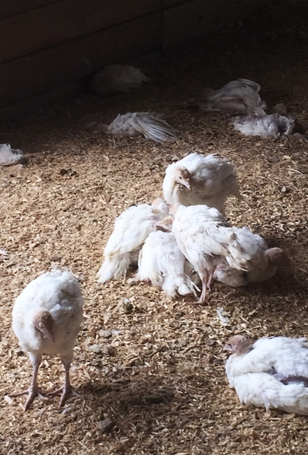
Flock of turkeys showing clinical signs of histomoniasis, including drooping wings, unkempt feathers, huddling, and death.
Courtesy of Dr. Robert B. Beckstead and Elle Chadwick.
The colloquial name for the disease, “blackhead,” is misleading; birds do not display a cyanotic head.
Pearls & Pitfalls
|
Young birds have a more acute disease course and die within a few days after clinical signs appear. Older birds may be sick for some time and become emaciated before death.
Lesions
The primary lesions of histomoniasis are in the ceca, where the parasite migrates into the submucosa and muscularis mucosae (see ). This leads to inflammation and development of a yellowish green caseous exudate or, in later stages, a dry, caseous core. Occasionally, these ulcers erode the cecal wall, leading to peritonitis and involvement of other organs.

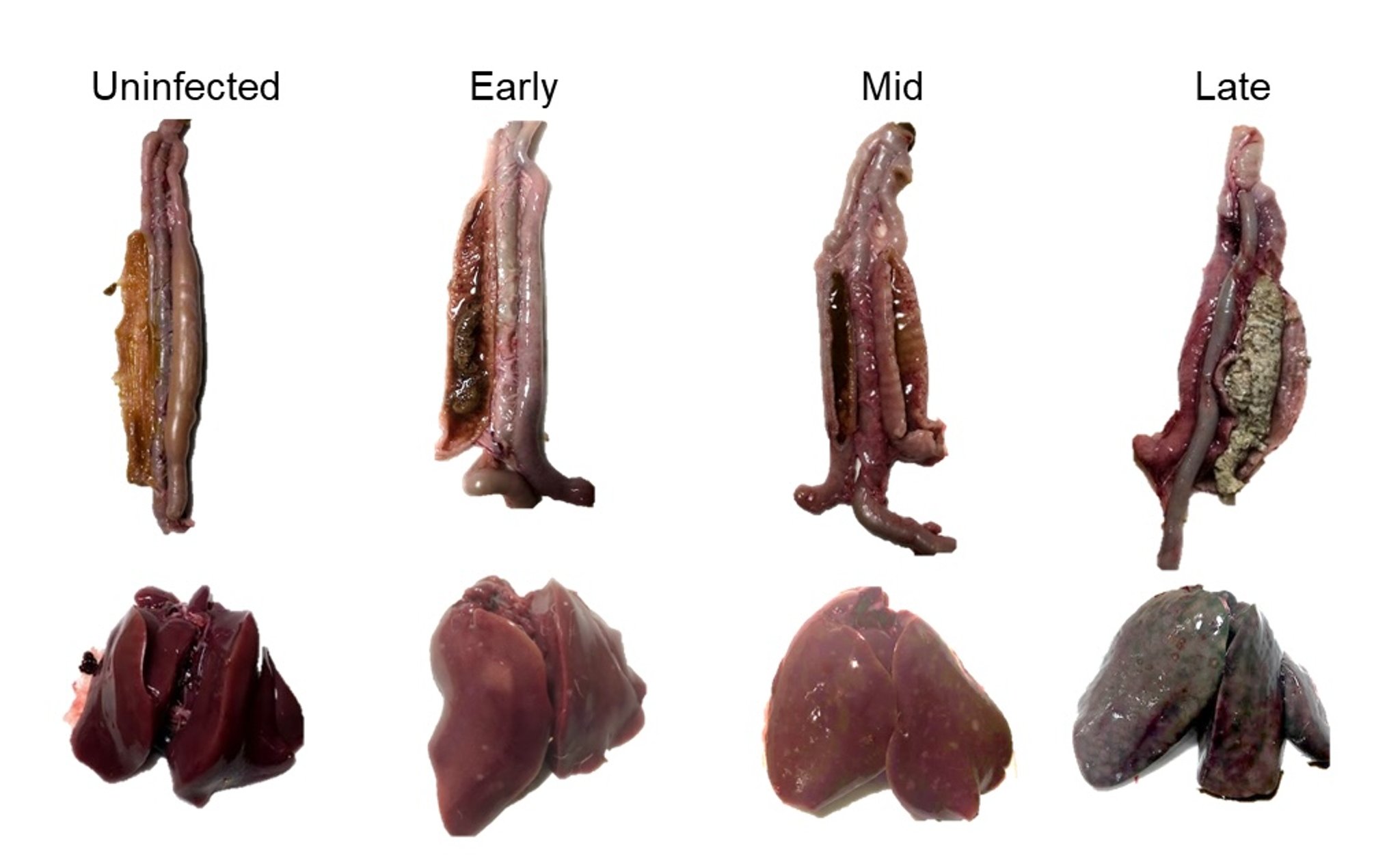
Progression of histomoniasis in livers and ceca (attached to small intestine) from turkeys aged 14–24 days.
Courtesy of Dr. Robert Beckstead and Dr. Elle Chadwick.
Clinical signs in the ceca are apparent 3–4 days after H meleagridis invasion. Histomonads can reach the liver via either the vascular system or the peritoneal cavity (see ).

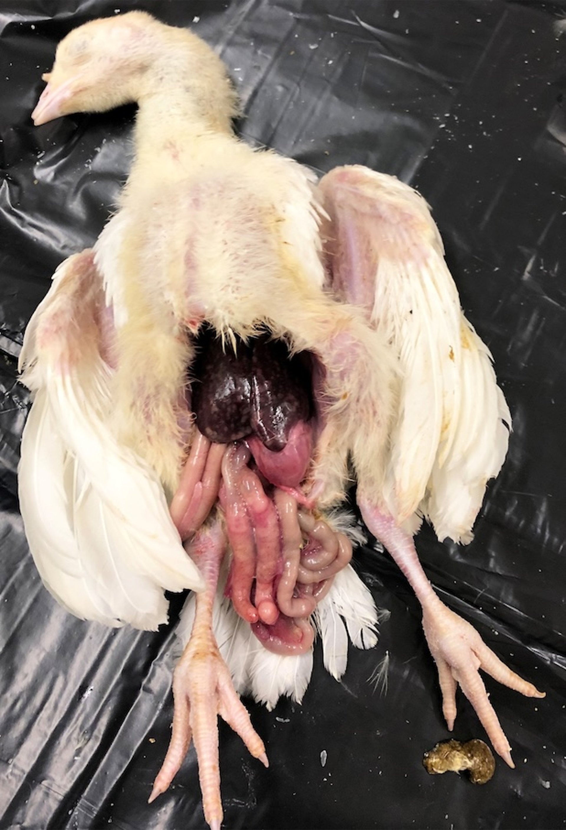
Postmortem photograph of a 24-day-old turkey with histomoniasis showing liver lesions and enlarged ceca.
Courtesy of Dr. Robert B. Beckstead and Elle Chadwick.
Liver lesions are highly variable in appearance; however, large, umbilicated lesions (often described as "target" or "bullseye" lesions) are typical and are strongly suggestive of histomoniasis. In turkeys, liver lesions appear 6–8 days after infection and may be up to 4 cm in diameter and involve the entire organ. In some cases, the liver will appear green or tan. Lesions are also present in other organs, such as the kidneys, bursa of Fabricius, spleen, and pancreas.
Diagnosis of Histomoniasis in Poultry
PCR assay
Microscopic examination of cecal content, or cecal or liver scrapings (see Histomonas meleagridis)
Histological examination of liver or cecal tissue
The liver and cecal lesions together are pathognomonic. H&E stain is sufficient for histological examination.

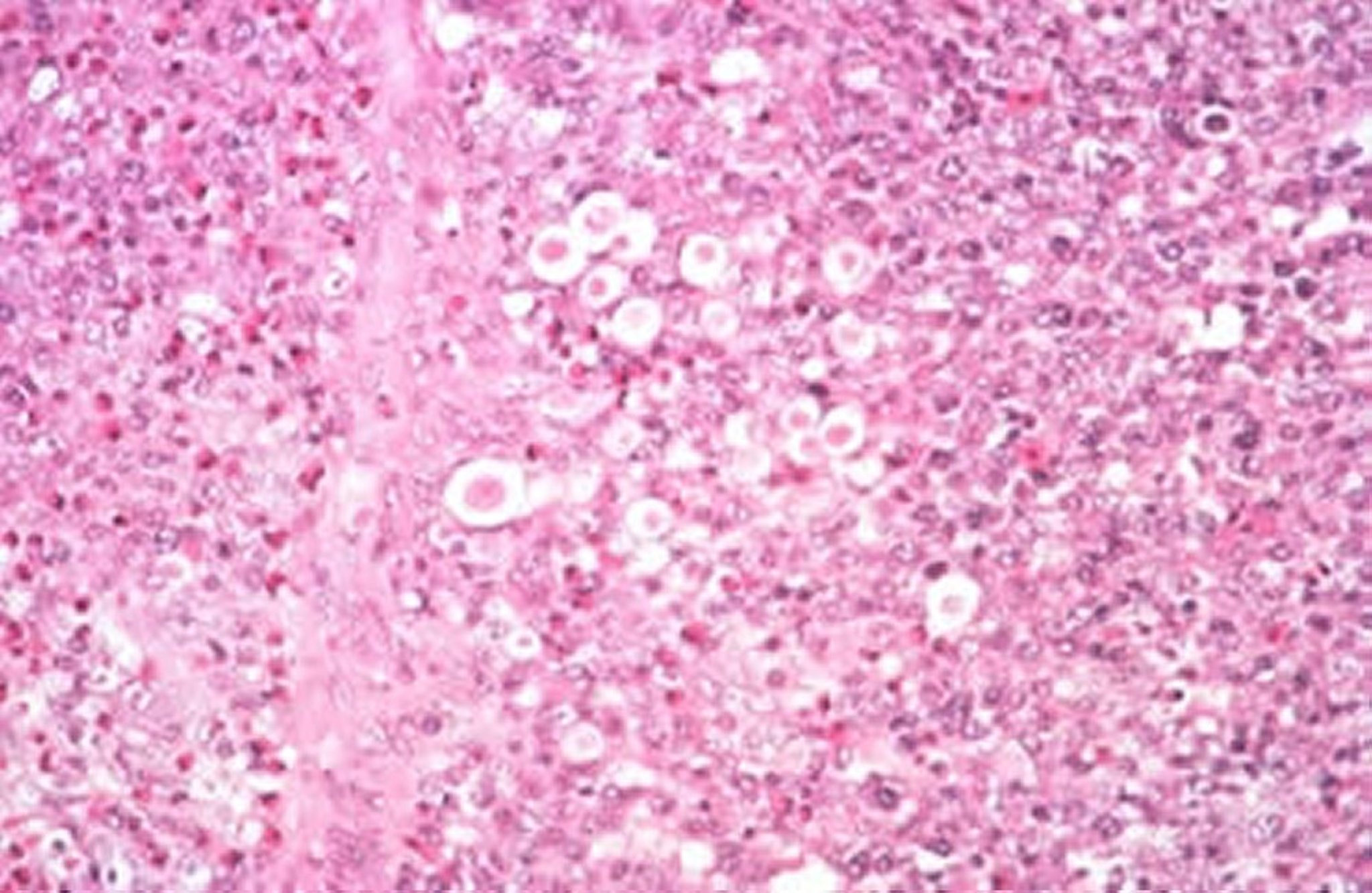
Histomonas meleagridis in the liver of a turkey. H&E stain; original magnification, 40X.
Courtesy of Dr. Jean Sander.
To diagnose histomoniasis, the liver lesions must be differentiated from those of the following diseases:
Histomonads are extracellular, although they may be so closely packed as to appear intracellular. The nuclei are much smaller than those of the host cells, and the cytoplasm is less vacuolated. Histomonas spp must be differentiated from other cecal flagellates.
Prevention and Treatment of Histomoniasis in Poultry
Raise turkeys apart from other galliformes
Divide turkey facility with barriers
Because healthy chickens and game birds often carry the cecal nematode vector, any contact between turkeys and other galliformes should be avoided, and care should be taken to decrease the H gallinarum population to prevent histomoniasis.
H gallinarum eggs from contaminated soil can be tracked inside by workers, causing infection.
Arthropods such as flies may also serve as mechanical vectors.
Because H gallinarum ova can survive in soil for many months or years, turkeys should not be put on ground contaminated by chickens.
Histomonads that are shed directly into the environment die quickly. Thus, in a turkey facility where H gallinarum is unable to complete its life cycle, decontamination is not required.
Immunization has been only partially successful in controlling histomoniasis, and reports differ on its effectiveness. The immune response of turkeys to live, attenuated H meleagridis requires 4 weeks to develop. Vaccination of 18-week-old pullets 5 weeks before experimental infection has been shown to prevent a decrease in egg production. Killed histomonads stimulate some immunity when administered SC or IP but do not offer protection.
No drugs are currently approved for use as treatments for histomoniasis. Historically, nitroimidazoles such as ronidazole, ipronidazole, and dimetridazole were used for prevention and treatment and were highly effective, while arsenicals such as nitarsone were used for prevention. Some of these products can be used by veterinary prescription in non–food-producing birds; however, this class of drugs is strictly prohibited from extra-label drug use in food-producing animals in the US.
Frequent worming of chickens with benzimidazole anthelmintics helps decrease exposure to H gallinarum worms that carry H meleagridis.
Key Points
Chickens are a reservoir for Histomonas meleagridis and its vector Heterakis gallinarum.
H gallinarum eggs maintain H meleagridis in the environment.
Currently, there are no approved vaccines or treatments for histomoniasis.
For More Information
Blackhead disease in poultry. FDA Center for Veterinary Medicine. Updated December 13, 2019. Accessed November 30, 2023.


