

Tumors are abnormal growths of cells. Tumors affecting the skin or the tissue just under the skin are the most commonly seen tumors in dogs. Skin tumors are diagnosed more frequently than other tumors in animals in part because they are the most easily seen tumors and in part because the skin is constantly exposed to many tumor-causing factors in the environment. Chemicals, solar radiation, and viruses are just some of the things that can cause skin tumors. Hormonal abnormalities and genetic factors may also play a role in the development of skin tumors.
All of the various layers and components of skin have the potential for developing distinctive tumors. Distinguishing a tumor from an inflammatory disease can sometimes be difficult. Tumors are usually small lumps or bumps, but they also can occur as hairless, discolored patches, rashes, or nonhealing ulcers. Because skin tumors are so diverse, identifying them should be left to a veterinarian.
Tumors may be benign or malignant (cancerous). Benign tumors are not invasive, do not spread to other areas of the body, and are easy to remove surgically. Malignant tumors can spread and cause harm to the animal. Malignant tumors can invade surrounding tissue and spread to distant organs. Distinguishing a benign tumor from a cancerous tumor requires specialized knowledge and laboratory equipment. A veterinarian can perform a fine needle aspiration of cells or a biopsy (which removes a small amount of tissue from a tumor) for evaluation.
Treatment for a particular tumor depends largely on the type of tumor, its location and size, and the overall physical condition of the dog. For benign tumors that are not ulcerated and do not impair the dog’s normal routine, treatment may not be necessary. This may be the most prudent option, especially in aged dogs.
There are several treatment options for cancerous tumors and benign tumors that inhibit normal activities or are cosmetically unpleasant. For most tumors, surgical removal is the most effective option. It is also probably the least costly option and the one with the fewest side effects. If malignancy is suspected, tissue surrounding the tumor will also be removed to increase the chance that none of the tumor cells are left behind. For tumors that cannot be completely removed, partial removal may prolong the life of the dog. Radiation treatment or chemotherapy may also be used to provide your pet with a better outcome.
After surgical removal, tumors should be evaluated under a microscope (called a histopathology test) to confirm the type of tumor and whether all of the tumor was likely removed. The latter is done by microscopically evaluating the edge of the resected tissue (the "margins") to see whether tumor cells are present. "Dirty" margins describe tumor cells that extend to the edge of the removed tissue, indicating that tumor cells still remain in the dog's body. "Narrow" margins describe tumor cells close to—but not at—the edge, indicating that tumor cells could possibly be left behind at the surgical site. "Wide" or "clean" margins describe tumors cells far from the edge of the removed tissue, indicating it is unlikely that tumor cells remain at the surgical site.
In addition to skin and hair follicle tumors, there are also tumors that affect the ceruminous glands. These are discussed in the section on ear diseases.
Apocrine Gland Tumors of the Anal Sac
These tumors most commonly appear as deep, firm, masses near the anal sacs. Older English Cocker Spaniels, Springer Spaniels, Dachshunds, Alaskan Malamutes, German Shepherds, and mixed-breed dogs are most at risk. As the tumors grow, they may compress the rectum and induce constipation. Some of these tumors are associated with a syndrome that is characterized by abnormally high calcium in the blood. Elevated calcium causes poor appetite, weight loss, kidney disease, and increased water intake and urine output. The tumors often spread to local lymph nodes and other organs. In most cases, surgery requires removal of the mass and tissues surrounding it, including involved lymph nodes. If the entire tumor cannot be removed, reducing the size of the tumor can help improve signs. Chemotherapy and radiation treatment may also be provided. Few dogs live more than a year after this type of tumor has been diagnosed.
Basal Cell Tumors and Carcinomas
Basal cells lie at the base of the top layer of the skin (the epidermis). A benign growth of these cells is a basal cell tumor. A malignant growth is a basal cell carcinoma.
Basal cell tumors are common in dogs and most are benign. Canine basal cell tumors most commonly develop in middle-aged to older dogs. Many breeds are predisposed, especially Wirehaired Pointing Griffons and Kerry Blue and Wheaten Terriers. These tumors are found most commonly on the head (especially the ears), the neck, and forelimbs. These tumors generally appear as firm, solitary, dome-shaped elevated masses, which are often hairless or ulcerated. The lumps may stick out like stalks from the skin surface. They vary in size from less than 0.4 inches (1 centimeter) to more than 4 inches (10 centimeters) in diameter. These tumors are sometimes dark in color. Cysts may also form. Although basal cell tumors are benign, they can be large and may cause extensive ulceration and secondary inflammation. These tumors can break the skin, cause the death of skin tissue, and drain fluid or pus. The dog is often uncomfortable. Surgical removal is effective treatment and reduces the chance of secondary infection and inflammation.
Basal cell carcinomas are less common in dogs than in cats. They are also often called basosquamous cell carcinomas in dogs. They can appear almost anywhere on the body. These carcinomas may be flattened or raised above the skin surface. However, they spread, forming new ulcers. Consequently, surgical removal is the treatment of choice. These tumors generally occur in older dogs. Saint Bernards, Scottish Terriers, and Norwegian Elkhounds are most at risk. Unlike basal cell tumors, basal cell carcinomas can be found almost anywhere on the body. These tumors spread to neighboring skin but seldom spread to other organs. Surgical removal is the treatment usually recommended. When performing this surgery, the veterinarian will remove a sufficient amount of normal skin around the tumor to make certain that the entire tumor has been removed.
Benign Fibroblastic Tumors
Collagenous nevi are benign collections of fibrous proteins known as collagen. They are common in dogs. Generally collagenous nevi are found in middle-aged or older animals, most frequently on the legs, head, neck, and areas prone to trauma. They are flat to raised lumps that develop in the skin or fat beneath the skin. Surgical removal of both forms is generally effective. Infrequently, some may grow too large to be surgically removed.
A disorder called generalized nodular dermatofibrosis (dermatofibromas) is rarely seen in German Shepherds. Affected dogs have multiple collagenous nevi that are associated with kidney and uterine tumors. The skin tumors are recognized first, and kidney disease develops 3 to 5 years later. There is no known treatment to prevent the formation of the kidney tumors.
Skin tags are distinctive, benign, skin lumps on older dogs. These are common, may be single or multiple, and can develop in any breed, although large breeds may be at increased risk. Most commonly, skin tags look like extended stalk-like growths, often covered by a wart-like surface. Surgical removal is optional, but a biopsy is recommended to confirm the diagnosis. Dogs that develop one are likely to develop others.
Fibromas resemble collagenous nevi or skin tags. Fibromas occur in all breeds but are primarily a tumor of aged dogs. Doberman Pinschers, Boxers, and Golden Retrievers are most at risk. The head and legs are the most likely sites. Fibromas appear as isolated, generally raised, often hairless lumps originating under the skin surface. They feel firm and rubbery (fibroma durum) or soft and mushy (fibroma molle) These tumors are benign and treatment is optional. However, complete surgical removal is recommended if they change appearance or grow large.
Blood Vessel Tumors
Blood vessel (vascular) tumors of the skin and soft tissues are growths that closely resemble blood vessels. Some forms are benign while others are highly malignant.
Hemangiomas are benign tumors of adult dogs. Many breeds (including Gordon Setters; Boxers; and Airedale, Scottish, and Kerry Blue Terriers) are considered to be at risk. These tumors are most common on the legs and trunk. Hemangiomas are single to multiple, circular, often compressible, red to black lumps and can look like a “blood blister.” Although they are benign, they tend to develop ulcers and some grow quite large. Because of this, and because it is important to identify whether the tumor is cancerous, they should be removed.
Hemangiopericytomas develop most frequently on the lower legs and chest of older dogs. They occur more often in females than in males. Siberian Huskies, Irish Setters, German Shepherds, and mixed-breed dogs are most at risk. These sarcomas are typically firm, solitary tumors with irregular looping borders. They occur most commonly in the fat under the skin. They can invade surrounding tissues but rarely spread to other sites. Complete surgical removal is the treatment of choice. Because these tumors are locally invasive, tumor cells may remain after surgery unless a wide area around the tumor is also removed. Regrowth is common within 1 year. If the first surgical removal of any sarcoma is not adequate, followup surgery to completely remove the tumor is normally prescribed. Followup radiation treatment may also be necessary if surgical removal is incomplete. If available, chemotherapy and/or radiation treatments may also be performed on the area during surgery to reduce the risk of regrowth.
Cutaneous (skin) angiosarcomas (also known as angioendotheliomas) start out looking like benign hemangiomas but then progress to become malignant blood vessel tumors. They occur most often in dogs with short, often white coats, with high amounts of sun exposure. The breeds prone to sun-caused angiosarcomas are Whippets, Italian Greyhounds, white Boxers, and Pit Bull Terriers. Irish Wolfhounds, Vizslas, Golden Retrievers, and German Shepherds are also prone to develop these tumors, but not in response to sun exposure. In dogs, they most frequently develop on the underside of the trunk, hip, thigh, and lower legs. Small surface tumors are easily controlled with freezing (cryosurgery) or laser surgery as needed. Avoidance of further sun exposure may reduce the development of new tumors; however, more tumors can appear over several years.
Angiosarcomas are highly malignant and can vary greatly in appearance. Most commonly, they appear as one or more red lumps in the skin or underlying soft tissues. Less frequently, they appear as a poorly defined bruise. All grow rapidly and often cause death of nearby normal tissue. These tumors spread, especially to the lungs and liver. A biopsy is needed to confirm the diagnosis. Wide surgical removal is the treatment of choice for angiosarcomas below the skin surface. During surgery, chemotherapy drugs may be placed in the area to treat any remaining tumor cells.
Cornifying Epitheliomas
Other names for these benign tumors of dogs include keratoacanthoma and infundibular keratinizing acanthoma. These growths are nests of tough, layered lumps that stick up from the skin surface. They can look a little like a horn, which is why they are described as cornifying. In other cases, the epitheliomas may appear solely as cornified cysts. They most likely arise from a hair follicle. These tumors can develop anywhere on the body, but they occur most frequently on the back, tail, and legs. Middle-aged dogs are most at risk. Norwegian Elkhounds, Belgian Sheepdogs, Lhasa Apsos, and Bearded Collies are most likely to develop these tumors. Norwegian Elkhounds and Lhasa Apsos are at risk for developing widespread tumors. The condition is diagnosed by finding the tumors on the animal. Treatment is optional, provided there is no self-trauma, ulceration, or secondary infection. Some dogs find the tumors annoying and attempt to scratch, rub, or bite them off. This leads to skin trauma that can easily become infected. Surgical removal is the treatment of choice. However, dogs are prone to develop additional tumors. For animals with a generalized form of the disease, oral retinoid medications may help.
Hair Follicle Tumors
Trichilemmomas are rare, benign, hair follicle tumors of dogs, most commonly found on the head. Poodles may be predisposed. They appear as firm, oval masses, 0.4 to 2.75 inches (1 to 7 centimeters) in diameter that are compact but gradually grow.
Trichoepitheliomas are multiple small lumps in which an entire hair follicle is filled with condensed, yellow, granular, “cheesy” material. They occur mostly on the skin of the face. They can be either benign or malignant.
In dogs, they can occur at any age but are found most commonly during late middle age. Many breeds are predisposed, including Basset Hounds, Bull Mastiffs, Irish Setters, Standard Poodles, English Springer Spaniels, and Golden Retrievers. Tumors can develop anywhere on the body but most commonly on the trunk in dogs. Benign forms appear as cysts in or under the skin. Growth of the cysts or self-trauma may cause skin ulcers. Treatment is by surgical removal. However, dogs that develop one such tumor are prone to develop more at other sites. This is especially true for Basset Hounds and English Springer Spaniels.
Malignant trichoepitheliomas are much less common than benign trichoepitheliomas. They invade surrounding tissues, spread to the skin surface, and cause extensive inflammation, tissue death, and fibrosis. It is uncommon for these tumors to spread to other organs. Surgery is the usual treatment. During the surgery, your veterinarian will remove tissue around the tumor to reduce the chances of it recurring.
Pilomatricomas are hair follicle tumors that appear similar to trichoepitheliomas, but their cystic contents are often gritty. They can be benign or malignant. Benign tumors are most common on the trunk of middle-aged dogs. Kerry Blue and Wheaten Terriers, Bouvier des Flandres, Bichons Frises, and Standard Poodles are most at risk. Surgical removal is the treatment of choice, but additional tumors can develop in other locations.
Malignant pilomatricomas are rare tumors of old dogs. They are cystic tumors that firmly attach to surrounding tissues, making them difficult to remove surgically. Recurrence is common after surgery, and they often metastasize to lymph nodes, the lungs, and other organs.
Histiocytic Cell Tumors
These tumors form a group of poorly defined skin diseases all characterized by a proliferation of cells called histiocytes (tissue macrophages). The cause for these diseases is unknown.
Histiocytomas are common skin tumors typically seen in younger dogs (less than 3½ years old). They can occur in dogs of any age, however. English Bulldogs, Scottish Terriers, Greyhounds, Boxers, and Boston Terriers are most at risk. The head, ears, and limbs are the most common sites. The tumors appear as solitary, raised, generally hairless, and sometimes ulcerated lumps that are freely movable. Diagnosis is through microscopic examination of samples of the tumor cells from fine needle aspiration or biopsy. Canine histiocytomas are normally considered benign tumors; most resolve spontaneously and without treatment within 2 to 3 months. Surgical removal is optional and normally performed only if the tumors cause severe problems for the dog.
Cutaneous histiocytosis is associated with development of numerous raised or flat bumps involving the deep layer of the skin or fat under the skin. It is rare in dogs and can develop at any age but is most common in young adults. Chinese Shar Peis, Collies, Border Collies, Shetland Sheepdogs, Briards, Bernese Mountain Dogs, Golden Retrievers, and German Shepherds may have a higher risk of disease. The skin bumps may come and go and do not typically cause itching. The legs and trunk are most commonly affected. The disease can also affect the face and can cause trouble breathing if present on the nostrils. This form of histiocytosis does not typically affect any internal organs but can cause dogs to appear unsightly. A number of therapies have been tried to treat this condition, but the response is variable. Some dogs will respond rapidly and permanently, whereas others will only improve temporarily, if at all.
Two forms of histiocytosis affect Bernese Mountain Dogs. Systemic histiocytosis of Bernese Mountain dogs is an aggressive skin disease that causes multiple skin lesions that wax and wane. The disease tends to become more severe with each new wave of eruptions. Skin bumps develop across the skin (especially on the scrotum of males), inside the nose, and on the eyelids. Although the skin masses may resolve, they usually recur several months later. Masses may also develop in internal organs, such as the lymph nodes, spleen, and bone marrow. The disease eventually becomes progressive and results in death. Chemotherapy and other drugs may be used to treat systemic histiocytosis.
Malignant histiocytosis is the other form of disease that affects Bernese Mountain dogs. Although uncommon, the disease can also affect other breeds. The disease is more likely to affect male dogs, with an average age of onset of 7 years. This disease first appears in the internal organs, such as the liver, lymph nodes and lungs and usually does not affect the skin. The disorder progresses rapidly, causing illness, pain, and eventually death. Few dogs survive longer than 6 months after diagnosis. Chemotherapy and other drugs may be used to treat malignant histiocytosis.
Keratinized Skin Cysts
Some dogs develop cysts that are filled with keratin, a skin protein. Such cysts have a hard or solid core. The most common type of cyst contains a gray, brown, or yellowish, granular, “cheesy” material. Most are malformations of hair follicles. There are several kinds of keratinized skin cysts, each of which affect a different part of the hair follicle. The ones found in dogs include infundibular follicular cysts, isthmus catagen cysts, matrix cysts, hybrid cysts (panfollicular cysts), and dermoid cysts. Dermoid cysts are congenital (the animal is born with them). Among dogs, they are most commonly found in Boxers, Kerry Blue Terriers, and Rhodesian Ridgebacks. Most dermoid cysts are multiple and contain fully formed hair shafts. Diagnosis is by finding the cysts on the dog. They can be solitary or multiple and are benign. Surgical removal is the best treatment. You should not to attempt to remove the cysts by squeezing them because this can spread the cyst contents into the surrounding tissues. Your dog’s body will react to the cyst contents as a foreign substance, which can cause severe inflammation.
Lipomas and Liposarcomas
Lipomas are benign tumors of fat (adipose tissue) and are common in dogs. Lipomas generally occur in older, obese females, most commonly on the trunk and near the tops of the legs. The breeds most at risk are Doberman Pinschers, Labrador Retrievers, Miniature Schnauzers, and mixed-breed dogs. Lipomas typically appear as soft, occasionally thin, discrete lumpy masses; most move freely when touched. A rare variant of this tumor, diffuse lipomatosis, has been identified in Dachshunds, in which virtually the entire skin is affected, resulting in prominent folds in the skin on the neck and trunk.
Many lipomas merge with healthy fat tissue next to them, making it difficult to determine the edges of the tumors. A fine needle aspiration is necessary in order to exclude other types of tumors that can mimic lipomas, such as mast cell tumors (see below).
Despite their benign nature, lipomas should not be ignored. Some tend to grow, and they may be indistinguishable from infiltrative lipomas or liposarcomas. Surgical removal is the cure. In dogs, dietary restriction (weight loss diet) starting several weeks before surgery may make it easier for the surgeon to identify the edges of the tumor and remove all of it.
Infiltrative lipomas are rare in dogs. They are most common in middle-aged females, usually on the chest and legs. The breeds most at risk are Doberman Pinschers, Labrador Retrievers, Miniature Schnauzers, and mixed-breed dogs. These tumors are soft, lumpy swellings in the fat layer under the skin. They can spread to underlying muscle and connective tissue. Infiltrative lipomas are considered sarcomas of partial malignancy. They rarely spread to other sites. The treatment of choice for infiltrative lipomas is surgery to remove the tumor and a margin of normal tissue surrounding it. In some cases, this may mean amputation of a limb.
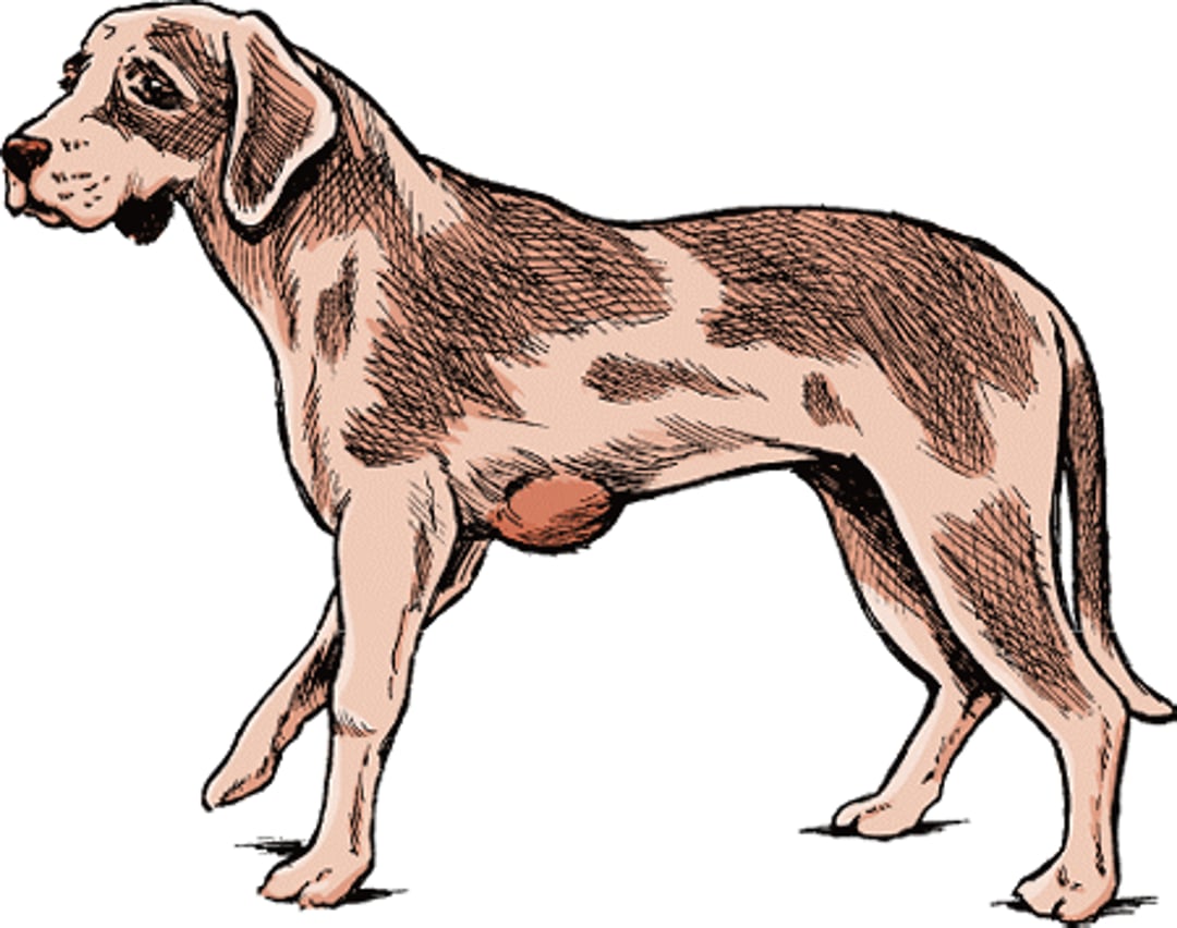
Lipoma, dog

Liposarcomas are rare tumors in all domestic animals. Most are recognized in older male dogs in which they usually develop on the chest and legs. Shetland Sheepdogs and Beagles are most at risk for liposarcomas. Liposarcomas are lumpy and can be soft or firm. They are malignant tumors with a low potential to spread to other sites. Wide surgical removal (removing both tumor and some surrounding tissue) is most often recommended. Recurrence is common, so follow up radiation treatment may be required.
Lymphoid Tumors of the Skin
Canine extramedullary plasmacytomas are relatively common skin tumors in dogs. They are most frequently identified on the head, ears, lips, mouth, and legs of mature to aged animals. Cocker Spaniels, Airedales, Scottish Terriers, and Standard Poodles are most at risk. The tumors are generally small (less than 2 inches [5 centimeters]) in diameter and sometimes narrow. Diagnosis is by finding the tumors on the animal and confirming the type of tumor with a fine needle aspiration or a biopsy. Most of these tumors do not spread and surgical removal is the usual treatment. When these tumors develop in the mouth, they may multiply. Treatment for the multiple form is more difficult, because the tumors are more likely to return following surgery. In such cases, tissue around the tumors may have to be removed. When tumors are multiple, or surgical removal is not feasible, radiation treatment is considered. Chemotherapy is commonly recommended for patients if radiation treatment is declined or if the tumor is resistant to radiation treatment.
Cutaneous (skin) lymphosarcoma is a rare form of skin cancer that may occur in a form in which the skin is the first and primary site of lymphoid tumor involvement. However, this disease may also be secondary to whole-body, internal diseases, such as canine malignant lymphoma. This uncommon tumor occurs in 2 distinct forms—epitheliotropic cutaneous lymphosarcoma and nonepitheliotropic cutaneous lymphosarcoma.
Epitheliotropic lymphosarcoma is the most frequently recognized form of skin lymphosarcoma in dogs. It is primarily a disease of middle-aged and older dogs, most often found in Poodles and Cocker Spaniels. The disease progresses slowly or moderately. Signs vary widely and may include flaky skin, red patches on the skin surface, raised and ulcerated areas, or lumps deep within the skin. These changes may also appear in the mouth or on the lips, eyelids, or footpads. Because of the variable appearance, diagnosis can be very difficult. The early stages can be confused with allergies, immune-mediated disease, or infections. Thus, your veterinarian may suggest a tissue biopsy of any tumor or tumor-like growth found on your pet. The presence of tumors with simultaneous leukemia is known as Sézary syndrome.
Many treatments for skin lymphosarcoma have been tried, though no treatment has been shown to be completely successful. Thus far, all the tested treatment procedures improved the signs of the disease but did not lengthen an affected dog’s life. Your veterinarian or a veterinary cancer specialist will have access to the latest treatment information for skin lymphosarcoma and will recommend the treatment program that is best suited for your pet and its overall health.
Nonepitheliotropic cutaneous (skin) lymphosarcoma is most common in middle-aged or older animals. The tumors are lumps or plaques that often develop on the trunk. Generally, these are multiple tumors. In many cases, nonepitheliotropic skin lymphosarcoma is, by appearance, indistinguishable from epitheliotropic skin lymphosarcoma. A definitive diagnosis is important because the nonepitheliotropic form in dogs is generally more serious than the epitheliotropic form. These tumors frequently spread to other organs and do so early in the course of the disease. Thus, an early, accurate diagnosis is extremely important in treating this disease.
Various treatments, including surgical removal, chemotherapy, and, less frequently, radiation treatment have been used both singly and in combination. Surgical removal is usually the first choice when the disease is limited to a single tumor. Removing the tumor can potentially completely cure the dog. For diffuse or multiple forms, surgical removal or freezing have been less successful. Chemotherapy can relieve signs but this form of cancer often recurs. The average remission time is 8 months.
Mast Cell Tumors
Mast cell tumors are named for the type of cell from which they grow. Mast cells are involved in allergic reactions. They release histamine, which causes irritation and itching, and other chemicals that may cause shock. Mast cell tumors are the most common malignant tumor seen in dogs. They may be seen in dogs of any age but occur most commonly in dogs 8 to 10 years old. They may develop anywhere on the body surface as well as in internal organs, but the limbs (especially the back of the upper thigh), lower abdomen, and chest are the most common sites. In about 10% of cases, tumors are found in multiple locations. Tumor size at the time of surgery often predicts the outcome; tumors larger than 1¼ inches (3 cm) are associated with decreased survival time. Tumors located near mucous membranes, feet, prepuce, or on the lower surface of the body are more likely to spread than mast cell tumors in other areas. Tumors that grow rapidly or that are not removed completely during surgery are also more likely to spread. Many breeds appear to be prone to the disease, especially Boxers and Pugs (in which tumors are often multiple), Rhodesian Ridgebacks, and Boston Terriers.
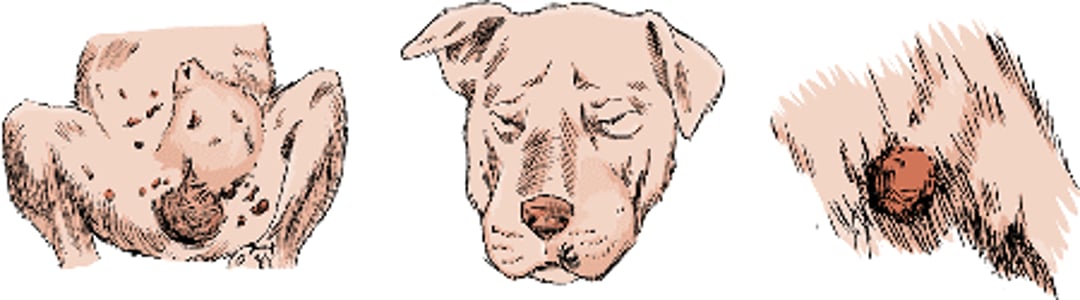
Mast cell tumors

These tumors vary greatly in size and rate of growth. They can mimic lipomas; therefore, visual signs alone cannot establish a diagnosis. Most commonly, a mast cell tumor appears as a raised lump or mass that may be soft to solid to the touch. Mast cell tumors are tricky and difficult to deal with because they appear as a large central tumor but are in fact surrounded by a halo of smaller, microscopic nests of mast cells that infiltrate normal-looking skin. Dogs can also develop signs associated with the release of toxins from the malignant mast cells. For example, up to a quarter of dogs with mast cell tumors also have stomach ulcers due to histamine release. Diagnosis is by microscopic examination of tumor samples obtained by fine needle aspirations, impression smears, or biopsy samples.
Small mast cell tumors may remain quiet and seem inactive for long periods before spreading. Other tumors can be aggressive and spread rapidly. Thus, all mast cell tumors are normally treated as at least potential malignancies. Treatment depends on the clinical stage of the disease and the predicted spread of an individual tumor. For a small solitary tumor confined to the skin (Stage I), the preferred treatment is complete surgical removal. Most veterinarians will remove at least 1¼ inches (3 centimeters) of healthy tissue surrounding all borders of these tumors to remove both the lump and any surrounding nests of tumor cells. If, during surgery, biopsy of a sample of the removed tissue suggests that the tumor extends beyond the initial edge of the surgery, additional surrounding tissue will be removed. Alternatively, because mast cells are sensitive to radiation, radiation treatment may be effective if there is remaining tumor. In some cases, a combination of radiation treatment, chemotherapy at the tumor location, and hyperthermia of the site (carefully increasing the body temperature of the affected area while under anesthesia) may be more effective than radiation alone. If the margins are not clean and radiation treatment is not elected, chemotherapy is also an effective followup treatment. Some dogs grow new tumors periodically. When surgery is not performed for residual disease or for small recurrent tumors, injections of steroids and antihistamines into the tumor, which may be followed by freezing (cryotherapy), can be considered as an option.
At present, there is no agreed upon treatment for Stage II to IV mast cell tumors. Options include surgical removal of the mass and affected regional lymph nodes (if feasible), radiation, and chemotherapy. Your veterinarian may consult with a veterinary oncologist for assistance in the treatment of these tumors. Veterinary cancer specialists may recommend new types of treatments that may help manage difficult cases.
Melanomas
A melanoma is a dark-pigmented skin tumor that may be either benign (not cancerous) or malignant (cancerous). Benign melanomas (also called melanocytomas) are diagnosed much more frequently in dogs than malignant melanomas. They most commonly develop on the head and forelimbs in middle-aged or older dogs. Miniature and Standard Schnauzers, Doberman Pinschers, Golden Retrievers, Irish Setters, and Vizslas are the breeds in which these tumors are most commonly found. They can appear as spots or patches, or raised or flat masses. Most have a dark surface. Although generally solitary, melanomas may be multiple, especially in the breeds at risk. When these tumors are benign, surgical removal cures the condition.
Malignant melanomas most commonly develop in older animals. Among dogs, Miniature and Standard Schnauzers and Scottish Terriers are most at risk. The lips, mouth, and nail beds are the most common sites of development. Malignant melanomas on haired skin are rare, and most arise on the lower abdomen and the scrotum. Males are affected more often than females.
Most malignant melanomas appear as raised, generally ulcerated lumps that may or may not be darkened. When present on the lips or in the mouth, the tumors appear as dark to light gray or pink raised lumps. When present in the nail bed, they appear as swellings of the toe, often with loss of the nail and destruction of underlying bone.
For this reason, a veterinarian who finds a festering toe in an older dog will often order x-rays and remove a tissue sample from deep in the toe (including bone) for a biopsy. These steps will help the veterinarian treat this form of cancer as early as possible. Malignant melanomas grow quickly and have great potential to spread to other organs. Early treatment is critical for a positive outcome for a dog with a malignant melanoma.
Treatment consists of complete surgical removal. However, the spreading nature of the tumor may make this difficult. When present on a toe, amputation of the involved toe is the standard treatment. When present on the mouth, surgical removal of part of the jaw may allow for complete tumor removal and an acceptable postsurgical cosmetic appearance and survival. Melanomas are generally considered resistant to radiation treatment, and there is no established chemotherapy known to be highly effective. Typical survival times for dogs with malignant melanomas range from 1 to 36 months. A vaccine is available that helps shrink the size of malignant melanomas in dogs and may prolong survival.
Perianal (Hepatoid) Gland Tumors
Perianal gland tumors are a type of tumor found near the anus in dogs. They occur mostly in oil glands found around the anus. They may also occur in similar glands along the abdomen from the anus to the base of the skull, the tail, and in the skin of the back and areas near the tail. The tumors occur in male dogs three times more often than in females. They are most common in aged dogs. Siberian Huskies, Samoyeds, Pekingese, and Cocker Spaniels are the breeds most commonly affected.
The tumors appear as one or (more commonly) multiple lumps 0.2 to 4 inches (0.5 to 10 centimeters) in diameter. Larger tumors commonly form ulcers and bleed. In addition, large tumors can compress the anal canal and make defecation difficult.
Up to 95% of male dogs with these tumors are cured by castration. Surgical removal of the tumors may be used to remove extremely large or ulcerated tumors that have become secondarily infected. Surgery is the treatment of choice for females but may need to be repeated because recurrence is common in females. Radiation treatment is also an option and may be prescribed either alone or in combination with surgery. Laser surgery and cryosurgery (freezing) are other options, but because fecal incontinence is very common following extensive surgery involving the sphincter, this option is used only when tumors cannot be removed using regular surgical techniques.
Perianal gland adenocarcinomas are uncommon in dogs. These tumors are found in male dogs 10 times more commonly than in females. Siberian Huskies, Alaskan Malamutes, and Bulldogs are most likely to develop this tumor. Biopsy evaluation (laboratory study of a tissue sample taken from the tumor) is the best means of diagnosis. These tumors often spread to draining lymph nodes that are located along the lower back. Therefore, during surgery the tumor itself and a wide margin of tissue around it will be removed. If any of the lymph nodes are involved, they may also be surgically removed. Radiation treatment is often added following the surgery. Newer drugs and chemotherapy may also help. These tumors are generally not responsive to castration or to estrogen treatment. The outlook is guarded.
Peripheral Nerve Sheath Tumors
There are several tumors that arise from the connective tissue that provides a covering for nerves. These include amputation neuromas, neurofibromas, and neurofibrosarcomas.
Amputation neuromas are disorganized growths that form after amputation or traumatic injury. They most commonly happen after tail docking in dogs. They are most commonly found in young dogs that continuously bother a docked tail. Surgical removal is the cure.
Neurofibromas and neurofibrosarcomas are other tumors that grow in the connective tissue around a nerve. In dogs, they are found in older animals. These tumors appear as white, firm, lumps. There are both benign and malignant forms. In dogs, most are locally invasive but do not spread to other sites. Complete surgical removal is the treatment of choice. Followup radiation treatment or chemotherapy may slow regrowth.
Sebaceous Gland Tumors
The sebaceous glands secrete the oil known as sebum into the hair follicles and onto the skin. Tumors of sebaceous glands are common in dogs.
Sebaceous gland hamartomas are solitary tumors of dogs. These are elongated or circular, roughly 2 inches (5 centimeters) in length or diameter. They are usually identified shortly after birth.
Sebaceous gland overgrowth (hyperplasia) occurs in old dogs and cats. It appears as lumps seldom more than 0.4 inches (1 centimeter) in diameter, often with a shiny, horn-like surface. Among dogs, Manchester, Wheaten, and Welsh Terriers are at greatest risk. The head and abdomen are affected most often.
Sebaceous gland adenomas are common in older dogs. Coonhounds, English Cocker Spaniels, Cocker Spaniels, Huskies, Samoyeds, and Alaskan Malamutes are the breeds most likely to develop these tumors. They are often indistinguishable from sebaceous gland overgrowth, but they tend to be larger (typically over 0.4 inches [1 centimeter] wide). They are often multiple and may occur anywhere on the body but are commonly found on the head. Sebaceous adenomas may be covered with a crust and may become inflamed or infected. Sebaceous gland epitheliomas are a type of sebaceous adenoma that may extend deep into the skin. They often appear as ulcerated lumps.
Sebaceous gland adenocarcinomas are a rare malignant form of sebaceous gland tumor. They occur in middle-aged or older dogs. Cavalier King Charles Spaniels, Cocker Spaniels, and Scottish, Cairn, and West Highland White Terriers are the breeds most at risk. Male dogs may be predisposed. These may be indistinguishable from sebaceous epitheliomas or other skin carcinomas. They spread within the skin and may spread to regional lymph nodes late in the disease.
Once a diagnosis is established, surgery is optional for benign sebaceous gland tumors unless they are inflamed and infected. For malignant adenocarcinomas, surgery is the treatment of choice. Your veterinarian will remove not only the tumor but also tissue around the tumor, including involved lymph nodes. In addition, chemotherapy and radiation treatment may also be prescribed. Dogs that develop a sebaceous gland overgrowth or adenoma often develop new tumors at other sites. Your veterinarian may consult with a veterinary oncologist or veterinary dermatologist for assistance in the treatment of these tumors.
Smooth Muscle Tumors of the Skin
These tumors (called leiomyomas or leiomyosarcomas) occur rarely in the skin and little is known about them. Those that have been reported have been malignant and found in dogs and cats. In most cases, these are firm masses that can be felt through the skin. Treatment is surgery to remove the tumor. Chemotherapy may also be recommended.
Soft Tissue Giant Cell Tumors (Fibrous Histiocytomas)
These tumors are not well understood. They appear more like an inflammatory reaction than a tumor and are generally treated with steroids. An abnormality called canine fibrous histiocytoma has been found in the eye of some dogs, most commonly Collies 2 to 4 years old.
Some soft tissue giant cell tumors are malignant (cancerous). Malignant fibrous histiocytomas are rare in dogs. They seldom spread to other sites but tend to return after surgical removal. Surgery to remove these tumors is the treatment of choice. Because these tumors can grow into surrounding tissues, most veterinarians will also remove a wide margin of tissue surrounding the tumor, to be as sure as possible that the entire tumor has been taken out.
Soft Tissue Sarcomas
Sarcomas are a group of connective tissue tumors that often rapidly invade surrounding tissues but are less likely to spread to other sites in the body. Sarcomas on the surface of the skin tend to be benign. Deep sarcomas tend to be malignant. The larger the tumor, the more likely it is to be malignant. A rapidly growing tumor is more likely to be malignant than one that develops slowly. Benign tumors have few blood vessels, whereas most malignant tumors have many blood vessels.
Diagnosis is made by biopsy of the sarcoma. Many sarcomas are shaped like an octopus, with tentacles that extend deeply into the tumor bed. They are often called “spindle-cell” sarcomas.
Wide surgical removal is the treatment of choice for soft-tissue sarcomas. The veterinarian will remove a 2- to 3‑centimeter margin of tissue surrounding the sarcoma. This is necessary because there is a strong possibility that the sarcoma has spread beyond the mass into surrounding tissues that appear normal. Occasionally, depending on location, amputation may be required to remove the entire tumor. It is important to remove all of the tumor during the first surgery, because sarcomas that recur have a greater potential to invade local tissue, and they may also spread to other parts of the body. Each future attempt at surgical removal can increase the rate of spread. Freezing (cryosurgery) is usually not used for sarcomas because some tumor types are resistant to freezing. Spindle-cell sarcomas generally do not respond well to conventional doses of radiation. Higher doses have been somewhat successful. Surgical removal followed by radiation is an option, as well as radiation prior to surgical removal. Chemotherapy for sarcomas has become a more common method of treatment. Although chemotherapy may improve the quality and prolong the life of an affected dog, it is seldom a complete cure. There is only a guarded outlook for dogs with soft-tissue sarcomas.
Fibromatosis is a thickening and invasive growth in tendon sheaths. They are generally seen on the heads of dogs, especially Doberman Pinschers and Golden Retrievers, where they are commonly called nodular fasciitis. Fibromatoses are generally indistinguishable from infiltrative fibrosarcomas (see below) except by microscopic examination. Small lymphoid lumps are scattered throughout the tissues. Fibromatosis can spread to nearby tissue but does not spread through the bloodstream to distant organs. If feasible, surgical removal is the treatment of choice. Recurrence is common, and radiation treatment may be helpful in reducing regrowth.
Fibrosarcomas are fast-growing malignant tumors. They are common in dogs, with most located on the trunk and legs. Gordon Setters, Irish Wolfhounds, Brittany Spaniels, Golden Retrievers, and Doberman Pinschers are most likely to develop these tumors. Fibrosarcomas vary greatly in appearance and size. Tumors arising under the skin surface may look lumpy. Those arising in the fat or nearby soft tissues may require hands-on examination to detect. They are firm and fleshy and appear deep in the skin and the fat underneath. They often invade underlying muscles. When multiple tumors are present, they usually occur within the same area of the body. In dogs, fibrosarcomas are invasive tumors that spread into surrounding tissues; only about 10% of these tumors spread to other parts of the body (metastasize). Wide and deep surgical removal is the best treatment, although complete removal is difficult. Because it is hard to determine the tumor’s edges during surgery, recurrence is common (more than 70% return within 1 year of the initial surgery). Even when surgical removal is complete, recurrence is still the rule. Followup radiation treatment and chemotherapy has been recommended after surgery, for tumors that are inoperable, and for tumors that cannot be removed completely. Your veterinarian may refer you to a veterinary cancer specialist for the treatment of these tumors.
Squamous Cell Carcinomas
Squamous cell carcinomas are malignant tumors. They can be found in all domestic animals. In dogs, these are the most frequently diagnosed carcinomas of the skin. Two forms occur in dogs—skin and subungual. Skin squamous cell carcinomas are tumors of older dogs. Bloodhounds, Basset Hounds, and Standard Poodles are at greatest risk. They appear on the head, lower legs, abdomen, and rear. Most appear as firm, raised, frequently ulcerated patches and lumps. Sometimes they can grow outward with a surface like a wart. Some are caused by prolonged sun exposure. These usually develop on the lower abdomen, especially on or near the pubic area in white-skinned, shorthaired breeds such as Dalmatians, Pit Bull Terriers, Bull Terriers, and Beagles. They develop on the underside of dogs because the poorly haired skin offers minimal shielding from ultraviolet radiation. Many animals sun themselves lying on their backs. They also get some solar radiation that reflects from the ground. Before a malignant tumor develops, dogs develop solar keratosis. Solar keratosis is thickened and discolored skin. Thus, finding areas on your dog where the skin is thick and discolored is cause for a veterinary checkup. Early diagnosis is important for successful treatment.
Subungual squamous cell carcinomas are tumors that originate under a nail (claw). They are most commonly found in Giant and Standard Schnauzers, Gordon Setters, Briards, Kerry Blue Terriers, Scottish Terriers, and Standard Poodles. Generally, these are dark-haired breeds, and a dark coat color has been associated with the development of subungual squamous cell carcinomas arising on multiple toes, often on different legs. Females are slightly more likely to develop these tumors than males, and both fore and hind legs are equally likely to have tumors. They may spread to other organs, including lymph nodes and the lungs.
Most squamous cell carcinomas are solitary tumors. However, multiple tumors may develop, especially in areas with prolonged sun exposure. They appear as raised, irregular masses with either ulcers or pimples. Dogs with subungual squamous cell carcinomas first show lameness or malformation, infection, or loss of a claw. These tumors usually invade into surrounding tissues. Some tumors grow slowly, whereas others are more likely to spread or return within 20 weeks of surgical removal. In general, when treatment fails it is due to late diagnosis and lack of control of the original tumor rather than spread of new tumors. For this reason, you need to be alert to any problems your dog may be having with its skin, toes, or claws and have these problems checked by your veterinarian promptly after discovery.
For dogs, surgical removal, such as the amputation of the involved toe or ear, is the proper treatment. A margin of skin at least ¾ of an inch (2 centimeters) around the tumor needs to be removed. Surgical removal may be combined with radiation treatment or chemotherapy. Survival rates depend on the malignancy of the tumor and its size before treatment.
Limiting exposure to excessive sunlight may help prevent squamous cell carcinomas. This may be accomplished by using ultraviolet window screens, sunscreen, and keeping the animals indoors during hours of peak sunlight.
Sweat Gland Tumors
There are 2 types of sweat glands in dogs, called apocrine and eccrine. In domestic animals, all hair follicles have apocrine glands, as do anal glands and ceruminous glands in the ears.
Apocrine gland cysts are found in middle-aged or older dogs. They can occur either in or outside of hair follicles. They appear most commonly on the head and neck. One or more cysts develop in the middle to upper skin layer with a loose association with hair follicles. Another form is more diffuse and involves cysts within the glands associated with multiple hair follicles in uninjured skin. Both forms of apocrine gland cysts are benign (not cancerous). Treatment is by surgical removal, though this may be difficult if the cysts are diffuse.
Apocrine gland adenomas include apocrine adenomas and apocrine ductular adenomas. Apocrine adenomas appear as firm to soft cysts, seldom larger than 1.6 inches (4 centimeters) in diameter. They contain varying amounts of clear to brownish fluid. They are found in older dogs, cats, and, in rare cases, horses. Among dogs, Great Pyrenees, Chow Chows, and Alaskan Malamutes are the most commonly affected breeds. The head, neck, and legs are the most frequent sites.
Apocrine ductular adenomas are less common than apocrine adenomas. They are found in older dogs and cats and appear closer to the surface of the skin. In dogs, these tumors are most commonly recognized in Peekapoos, Old English Sheepdogs, and English Springer Spaniels. They are often smaller, firmer, and less cystic than apocrine adenomas.
Apocrine adenomas and apocrine ductular adenomas are benign, and complete surgical removal cures the condition.
Apocrine gland adenocarcinomas are malignant tumors of sweat glands. They are rare in all domestic animals but occur most often in older dogs and cats. In dogs, Treeing Walker Coonhounds, Norwegian Elkhounds, German Shepherds, and mixed-breed dogs are most at risk. This tumor most commonly occurs where the front legs meet the trunk and near the groin. The appearance varies from thick lumps to ulcers. They are likely to spread into surrounding tissue and lymph nodes. Complete surgical removal is the treatment of choice. Little is known about the effectiveness of chemotherapy in treatment of these tumors.
Eccrine sweat glands are found in the footpads of dogs. Eccrine gland tumors are extremely rare. When they do occur, most are severely malignant and have a high potential to spread to the lymph nodes. The treatment of choice involves removal of not only the malignant gland but also surrounding tissue and any involved lymph nodes. In addition, chemotherapy and radiation treatment may also be provided.
Tumors Originating Outside the Skin (Metastatic Tumors)
All malignant tumors, wherever they originate, are capable of spreading to the skin. However, the spread of a primary tumor from inside the body to the skin is unusual. Although the appearance is variable, the tumors that spread to the skin are usually multiple, ulcerated lumps. As these tumors grow, they extend deeper into the skin and surrounding tissue. Generally, it is difficult to identify the primary tumor based on the signs in the skin. This is because only a small population of cells in the primary tumor will spread to the skin, and these cells may have different microscopic features than the primary tumor. When tumors have spread from other areas of the body to the skin, the primary tumor usually grows and spreads quickly and the outlook for a positive outcome is guarded to poor. Treatment for these tumors involves a whole-body approach that deal with both the skin tumors and the primary tumor(s).
Undifferentiated and Anaplastic Sarcomas
These are cancers of mesenchymal cells. Mesenchymal cells are the cells that develop into connective tissues, blood, lymph nodes, and other organs. Cancers of mesenchymal cells are difficult to identify because, like the mesenchymal cells themselves, malignant forms of these cells are loosely organized and often undeveloped or undifferentiated. Once identified, surgery is the usual treatment. When removing these cancerous cells, the veterinarian will usually remove not only the visible tumor but also a wide margin of tissue surrounding it. This reduces the chance that the malignancy will recur.
Warts (Papillomas)
Warts are caused by papillomaviruses. The virus is transmitted by direct contact or by contact with contaminated items such as bedding, clothing, dishes, and other items in the dog’s environment. It is also possible that insects may spread papillomaviruses. There are several distinct papillomaviruses. Warts have been reported in all domestic animals and are most common in dogs and horses.
Multiple warts of skin or mucus membranes generally are seen in younger dogs. Single warts are more frequent in older animals, but they may not always be caused by viral infection. The period between the initial infection and the development of visible warts varies but normally takes several months.
Papillomas, dog

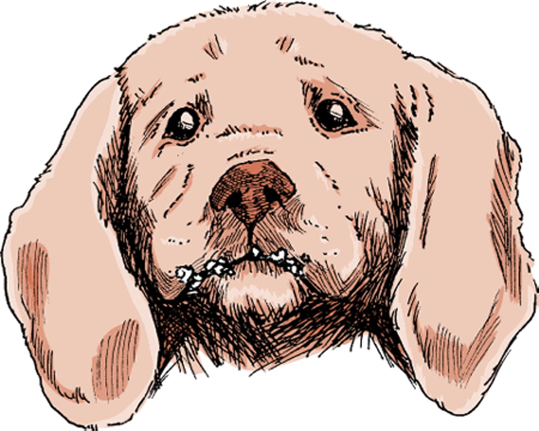
Most warts appear as bumps with a hardened surface resembling a cauliflower. When multiple warts are present they may be sufficiently characteristic to make a working diagnosis. However, there are many things that look like warts and a definitive diagnosis may require identification of the virus or its effects on individual cells (a change known as koilocytic atypia or koilocytosis).
In dogs, there are 3 kinds of warts. The first is canine mucous membrane papillomatosis, which primarily affects young dogs. Multiple warts appear on mucous membranes in the mouth from the lips to (occasionally) the esophagus and on the eyelid and adjacent haired skin. When the mouth is severely affected, chewing and swallowing is difficult. The second kind of wart in dogs is skin warts, which are indistinguishable from the warts that develop on or around mucous membranes. However, they are more frequently solitary and develop on older dogs. Skin warts are common in Cocker Spaniels and Kerry Blue Terriers. The third type found in dogs is called a skin inverted papilloma. In this disease of young adult dogs, warts most commonly develop on the lower abdomen. Infrequently, viral warts in dogs may progress to invasive squamous cell carcinomas.
Warts will eventually go away on their own, although how long this takes varies considerably. A variety of treatments have been suggested, but results vary. Surgical removal is recommended if the warts are sufficiently objectionable. However, because surgery in the early growing stage of warts may lead to recurrence and stimulation of growth, the warts should be removed when near their maximal size or when regressing. Affected dogs may be isolated from susceptible ones, but with the long incubation period (months), many are likely to have been exposed before the problem is recognized. Your veterinarian may recommend medications to help the immune system eliminate your dog's warts.
For More Information
Also see professional content regarding skin tumors.


