


Nervous system, horse

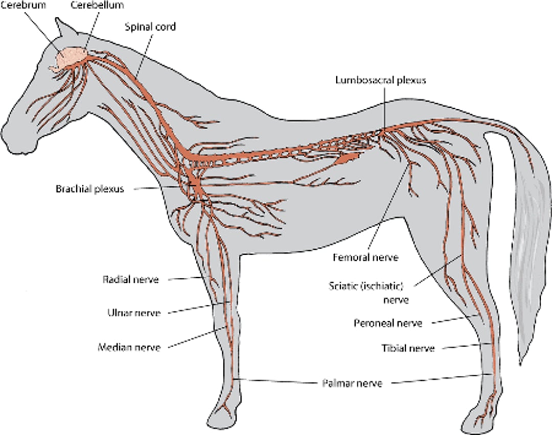
The central nervous system includes the brain and the spinal cord. The brain is divided into 3 main sections—the brain stem, which controls many basic life functions; the cerebrum, which is the center of conscious decision-making; and the cerebellum, which is involved in movement and motor control. The spinal cord of horses is divided into regions that correspond to the vertebral bodies (the bones that make up the spine) in the following order from neck to tail: cervical, thoracic, lumbar, sacral, and caudal (tail) segments. Specialized tissue called the meninges cover the brain and spinal cord, and cerebrospinal fluid surrounds and protects the brain and spinal cord.
The peripheral nervous system is formed by neurons of the cranial and spinal nerves that extend out to the rest of the body.
Neurons
Both the central and peripheral nervous systems contain billions of cells known as neurons. Neurons connect with each other to form neurological circuits. Information travels along these circuits via electrical signals.
All neurons have a center portion called a cell body and 2 types of extensions called dendrites and axons. Dendrites receive signals from other neurons and transmit electrical charges to the cell body. Axons transmit the electrical charges away from the cell body. When the electrical current reaches the end of the axon, the axon releases chemicals called neurotransmitters. Neurotransmitters pass the signal to the dendrites of other neurons, or to muscles or glands.
Neurons in the peripheral nervous system start with pairs of spinal nerves and pairs of cranial nerves. The spinal nerves arise from the spinal cord and extend axons outward into the front and hind legs and to the chest, abdomen, and tail. These nerves subdivide into smaller nerves that cover the entire surface and interior of the body. The cranial nerves include sensory and motor neurons that connect the head and face to the brain.
Types of Neurons
Sensory neurons carry information from the body to the spinal cord or brain stem, and then on to the cerebellum and cerebrum for interpretation. Sensory information includes sensations of pain, position, touch, temperature, taste, hearing, balance, vision, and smell.
Motor neurons carry responses to the sensory information from the spinal cord and brain to the rest of the body. Inside the spinal cord, the axons of motor neurons form bundles known as tracts, which transmit this information to peripheral motor neurons going to muscles in the limbs. Motor neurons are important for voluntary movements and muscle control.
A specialized set of neurons controls and regulates basic, unconscious bodily functions that support life, such as the pumping of the heart and digestion. These neurons make up what is called the autonomic nervous system, which sends axons from the brain stem and spinal cord to various areas of the body such as the heart muscle, the digestive system, and the pupils of the eyes.
For More Information
Also see professional content regarding the nervous system.


