


Diseases that affect the stomach and intestines are common in dogs. They include infectious diseases such as bacterial, viral, and parasitic diseases and noninfectious disorders, such as tumors, bloat, and obstruction.
Canine Parvovirus
Canine parvovirus infection is a potentially fatal viral disease that most often affects puppies or unvaccinated adult dogs. The virus itself is resistant to a number of common disinfectants and may survive for several months or possibly years in contaminated areas. Rottweilers, American Pit Bull Terriers, Doberman Pinschers, English Springer Spaniels, and German Shepherds are at increased risk of disease, but any breed can be affected. With appropriate treatment, 68%–92% of affected dogs will survive the virus.
The virus is transmitted by direct contact with infected dogs or feces. Indirect transmission, such as from objects contaminated by feces, is also an important source of infection. The virus is present in the feces for up to 3 weeks after infection. Recovered dogs may serve as carriers.
After entering the body through the mouth or nose, the virus replicates and spreads to the bloodstream. It attacks rapidly dividing cells throughout the body, especially those in the bone marrow, blood cell-producing tissue, and the lining of the small intestine. Production of the virus in the intestinal lining causes severe damage and bloody diarrhea. Normal intestinal bacteria may enter the damaged tissue and the bloodstream, worsening the disease. Affected dogs can also have decreased numbers of white blood cells, which normally protect the body from infections. This allows for further damage by the virus and secondary bacterial infections. In young puppies infection may also rarely involve the heart, leading to signs of heart failure without digestive signs, such as diarrhea.
Infected dogs may not show signs of illness. Clinical disease may be triggered by stress, such as improper nutrition or boarding, and signs may be worsened by other infections of the digestive system. Prolonged contact with a dog shedding high levels of virus increases the likelihood of becoming infected. An infected dog may be contagious before the onset of signs.
Clinical signs of infection generally develop within 5 to 7 days but can range from 2 to 14 days. Initial signs may be nonspecific (eg, dullness, loss of appetite, fever) with progression to vomiting and bloody diarrhea within 1 to 2 days. Abdominal pain may be a sign the intestines have become blocked, which requires emergency treatment. Severely affected animals may be in shock. On the other hand, inapparent infection is also common. Most dogs recover within a few days with appropriate supportive care; others can die within hours of the onset of signs.
Diagnosis is based on the dog’s history and signs and is confirmed by a positive fecal or blood test. The tests can also detect the newer strain of the virus known as CPV-2c. The fecal test, which detects viral protein, may be negative despite infection if it is done too early in the disease course. Thus, your veterinarian may need to repeat the test if the history and signs support the likely presence of the virus.
Treatment and Control
There is no specific treatment to eliminate the virus. Most dogs recover with appropriate supportive care, which is focused on replacing lost fluids and electrolytes. Oral electrolyte solutions (used to replace sodium and potassium that is lost through the intestines) may be used in mildly dehydrated dogs without a history of vomiting. More severely affected dogs will need intravenous fluids. Most dogs that survive the first 3 to 4 days of disease recover, usually within 1 week. Persistent vomiting can be controlled with prescription medication. Antibiotics may be added in cases where secondary bacterial infection is likely to be present.
Follow your veterinarian’s instructions for your pet’s diet. It was previously thought that food and water should be withheld until vomiting has subsided. However, it is now known that providing nutrition earlier on is associated with earlier improvement, weight gain, and improved gut function. Therefore, veterinarians may place a feeding tube in dogs that will not eat on their own. Once vomiting has stopped for 12–24 hours, frequent, small amounts of a bland diet (such as cottage cheese and rice or a prescription diet) can be slowly introduced. If signs recur after feeding, contact your veterinarian for directions. If food can be tolerated, the bland diet is usually continued for 1 or 2 weeks, after which the dog’s regular diet can be gradually reintroduced.
To limit environmental contamination and spread to other susceptible animals, dogs with confirmed or suspected parvovirus infection must be handled with strict isolation routines (separate housing, protective gowns and gloves for handlers, frequent and thorough cleaning of the area, etc). Contaminated areas should be thoroughly cleaned to remove visible dirt, feces, and other organic matter. Household bleach (diluted to 1 part bleach to 30 parts water) or commercial products labeled for use against parvovirus can inactivate the virus by being applied to the area after cleaning. The same solutions may be used as footbaths to disinfect household footwear. Disinfection of hands, clothing, and the dog’s food and water bowls and toys is recommended.
Vaccination is critical to prevent canine parvovirus infection. Vaccination of pups should begin at 6 to 8, 10 to 12, and 14 to 16 weeks of age, followed by a booster 1 year later and then every 3 years. Follow your veterinarian’s parvovirus vaccination recommendations to protect your pet. In addition, pups should be kept isolated from adult dogs returning from shows or field trials.
As mentioned above, parvovirus can remain viable in the environment for a year or longer. In a kennel, shelter, or veterinary hospital, cages and equipment should be cleaned, disinfected, and dried twice before reuse. The same concepts can be applied to a home situation. Removal of contaminated organic material is important in outdoor areas where complete disinfection is not practical. Disinfectants can be applied outdoors with spray hoses, but they will be less effective than when applied to clean, indoor surfaces. In a home setting, only fully vaccinated puppies or adult dogs should be allowed into a home with a dog recently diagnosed with parvovirus infection.
Inflammation of the Large Intestine (Colitis)
The large intestine (also called colon or large bowel) helps maintain fluid and electrolyte (salt) balance and absorb nutrients; it also temporarily stores feces and provides an environment for normal intestinal bacteria. When the colon is damaged by inflammation, parasites, or other causes, diarrhea is often the result.
Inflammation of the colon (called colitis) may be short- or longterm. In most cases, the cause is unknown; bacterial, parasitic, traumatic, kidney-related, and allergic causes are suspected. Inflammation may be the result of a defect in the function of the immune system in the colon. An exaggerated reaction to dietary or bacterial factors within the intestine, genetic predisposition, or results of previous infectious or parasitic disease have also been implicated.
Animals with inflammation of the colon may strain to defecate and may pass mucus-laden feces, sometimes containing blood. Feces are often of a small volume and a more liquid consistency, with increased frequency. Dogs often have accidents because of increased urgency. Affected dogs can also have pain when defecating. Weight loss and vomiting are rare and much less common than in dogs with diseases of the small intestine. Signs can come and go but tend to worsen with time.
If possible, the cause of the inflammation should be identified and eliminated. Your veterinarian will conduct a physical examination, followed by appropriate tests (which may include taking blood, urine, and fecal samples, abdominal x-rays or ultrasound, endoscopy, or biopsy, as needed). Treatment is based on the cause of inflammation.
Follow your veterinarian’s recommendations for diet. You may be asked to withhold food for 1 or 2 days to “rest” the animal’s digestive system. Once feeding is resumed, dissolvable fiber may be added to the diet. Over time, the fiber dose can be often be reduced or eliminated and a standard dog food substituted without causing a return of the diarrhea. When feeding is first resumed, you may be advised to provide food with a type of protein that your dog has not previously eaten, such as duck, lamb, kangaroo, or venison. This change is to reduce the chance that your pet will have an allergic reaction to the food proteins. There are several other types of diets that veterinarians can recommend for longterm colitis.
Supplementing the diet with fiber improves diarrhea in many animals. However, the addition of fiber alone will not usually resolve signs of large-intestinal diarrhea in dogs. To help the inflammation resolve more rapidly, your veterinarian may add anti-inflammatory medication to the change in diet. An alternative or additional anti-inflammatory medications can be used in dogs that do not respond initially. Some animals require additional short-term use of antidiarrheal medications until inflammation is brought under control, but do not use these medications unless recommended by a veterinarian. Anti-parasitic medications may also be recommended.
Longterm colitis is likely to improve initially, but signs frequently reoccur. Most dogs with inflammatory bowel disease cannot be cured and will need some form of longterm treatment. Many of the inherited causes of longterm colitis have a poor outlook. Please see Congenital and Inherited Disorders of the Digestive System of Dogs for more information.
Constipation
Constipation refers to difficult or infrequent elimination of stool, which is usually dry and hard. It is a common problem in dogs. In most instances, the problem is easily corrected; however, in sicker animals, the condition can be severe. The longer feces remain in the colon, the drier, harder, and more difficult to pass they become. Obstipation is constipation that resists treatment, in which the animal is unable to successfully defecate.
Longterm constipation may be due to an obstruction inside the intestines, constriction from outside the intestines, or because of neuromuscular problems with the colon itself. Obstruction is most common and is due to the dog’s inability to pass poorly digestible, often firm matter (such as hair or bones) that has become mixed with fecal material. A lack of water intake or the reluctance to defecate on a regular basis due to environmental stress or pain that occurs while defecating contributes to the formation of hard, dry feces. In other cases, tumors may block the passage of feces. Constriction may be caused by compression of the colon or rectum by a narrowed pelvic bone (for example, if a broken pelvis heals incorrectly), an enlarged prostate gland or lymph nodes, or cancer. Constipation can also result because of neuromuscular problems, which can be caused by hypothyroidism, dysautonomia, spinal cord disease, pelvic nerve dysfunction, or electrolyte abnormalities. Some drugs may cause constipation as a side effect.
Signs of constipation include straining to defecate and the passage of firm, dry feces. If the passage of feces is hindered by an enlarged prostate or lymph nodes, the feces may be thin or ribbon-like in appearance. Passed feces are often foul-smelling. Some animals are quite ill and also have lethargy, depression, loss of appetite, vomiting, and abdominal discomfort. Your veterinarian can confirm the presence of retained fecal matter by feeling the abdomen and performing a rectal examination. Abdominal x-rays may help establish the cause of fecal retention and indicate whether the feces contain foreign matter, such as bones. Be sure to tell your veterinarian about any tendency your pet has to eat bones, garbage, or other hard matter. Other tests may be needed in cases of longterm constipation or obstipation.
Affected dogs should receive plenty of water. Mild constipation can often be treated by switching to a high-fiber diet, keeping the dog from eating bones or other objects, providing ready access to water, and using appropriate laxatives (usually for a short time only). If laxatives are prescribed, they will be ones suitable for your pet. Laxatives formulated for humans can be very dangerous for animals, especially cats. In more severe cases of constipation, a veterinarian can remove retained feces using enemas or manual extraction while your pet is under general anesthesia. Complete removal of all feces may require 2 or 3 attempts over several days. To prevent recurrence, veterinarians often recommend a high-fiber diet, easy access to water, and frequent opportunities to defecate. Animals with longterm constipation that does not respond to diet changes and medications may require surgery.
Bloat
Bloat (also called gastric dilation and volvulus, or GDV) is a life-threatening emergency. It is caused by the twisting of the stomach along its axis and the accumulation of gas with or without fluid in the stomach.
Bloat tends to primarily affect large, deep-chested dogs. Stress may trigger an acute episode of bloat. Other risk factors include a lean body size, aggressive or fearful behavior, once daily feedings, dry food diet, and eating quickly. The incidence increases with age. Doberman Pinschers, German Shepherds, Standard Poodles, Great Danes, Saint Bernards, Irish Setters, Weimaraners, Standard Poodles, Bassett Hounds, and Gordon Setters are affected most frequently. Dogs that have a parent, sibling, or offspring with the condition also have an increased risk.
Dogs with bloat commonly have eaten a large meal followed by exercise and repeated, unsuccessful attempts to vomit. Signs of bloat may include restlessness, apparent discomfort, rapid breathing, abdominal pain and swelling, repeated dry retching, and excessive drooling. Your veterinarian may note a rapid and weak pulse, pale mucous membranes, and other signs of shock. An irregular heart rate can also develop. Veterinarians usually use x-rays to diagnose stomach rotation, but other imaging techniques can be helpful.
A successful outcome depends on prompt diagnosis and treatment by a veterinarian. The first goals of treatment are to stabilize the animal and decompress the stomach. The dog may require intravenous fluids to counteract shock. The pressure within the stomach will be relieved as soon as possible. This may be done by passing a tube through the mouth into the stomach. Once the tube enters the stomach, gas readily escapes. Excess fluid and food can then be removed via gravity and suction. After the stomach has been decompressed, the veterinarian may rinse it with warm water or saline to remove any remaining debris. If a tube cannot be passed into the stomach, excess gas may be relieved by inserting a large, hollow needle and catheter directly into the stomach through the skin.
Surgery is then performed to assess the condition of the stomach and spleen, to remove any dead tissue, to reposition the stomach to its normal location, and to attach the stomach to the abdominal wall in an attempt to decrease the likelihood that it will twist again. The spleen is removed in some cases. Food is usually withheld for 48 hours after surgery. Drugs may be prescribed to control pain and vomiting, if necessary.
Complications of the surgery include abnormal heart rhythms (arrhythmias), blood infections, severe inflammation of the lining of the abdomen (peritonitis), and a serious clotting disorder called disseminated intravascular coagulation. Approximately 25%–30% of dogs die because of bloat. Immediately seek veterinary care if your dog exhibits signs of bloat; doing so can improve its chance of survival.
If your dog has a tendency to develop bloat, your veterinarian may recommend that it be fed smaller meals more frequently over the course of the day, rather than a few large meals. Excessive exercise should be avoided, especially after eating, to decrease the likelihood of bloat, and consumption of large volumes of water after exercise should be avoided to limit distention of the stomach. In addition, your veterinarian may recommend a surgical procedure to help prevent bloat.
Inflammation of the Stomach (Gastritis)
Gastritis is sudden or longterm vomiting caused by inflammation of the stomach. It can be caused by eating something that irritates or injures the stomach lining, infections, parasites, body-wide illnesses, drugs, or poisons. In cases of acute gastritis, the vomiting is sudden, and the vomited material may contain evidence of whatever the pet ate (grass, for example). Bile, froth, fresh blood, or digested blood that looks like coffee grounds may also be present. Abdominal pain may be signaled by a dog that displays a “praying” position, with the hindquarters raised and chest and forelegs held close to the floor; this position appears to provide some relief. Excessive thirst is often followed by immediate vomiting in dogs with sudden gastritis. Diarrhea may also be noted. Short-term or occasional vomiting is generally not associated with other abnormalities; however, longterm vomiting may be associated with weakness, lethargy, weight loss, dehydration, and electrolyte (salt) imbalance and acid-base disorders. Lymphocytic-plasmacytic gastritis, eosinophilic gastritis, chronic atrophic gastritis, and chronic hypertrophic gastropathy are disorders that cause longterm gastritis.
The diagnosis is typically made by evaluating the dog's history, a physical examination, and response to treatment. Blood, urine, and fecal tests may be necessary, along with x-rays and/or an abdominal ultrasound. Visualizing the stomach with a long, flexible scope (endoscopy) and evaluating tissue samples may be necessary in dogs that have longterm gastritis.
Treatment and control are the same as for vomiting. The outlook depends on the cause of the vomiting and the ability to stop or control it. Short-term gastritis often responds well to fasting and avoiding further consumption of whatever triggered the condition. The outlook for longterm gastritis is variable. Research is ongoing in this area and trials of various diets and medications may provide new treatments in the coming years.
Cancers of the Digestive System
Cancer of the digestive system is uncommon, with stomach tumors representing less than 1% and intestinal tumors less than 10% of all cancers in small animals. The average age of dogs with cancer of the digestive tract is 6–9 years old. Adenocarcinoma and lymphoma are seen most frequently. Belgian Shepherds have an increased risk for stomach cancer. Colorectal tumors are more prevalent in Boxers, German Shepherds, Poodles, Great Danes, and spaniels. No specific cause(s) has been identified for most gastrointestinal tumor types. Tumors in the digestive system in dogs tend to be malignant, which means they are likely to be aggressive and to spread to other parts of the body.
Signs of a possible tumor vary depending on the location and extent of the tumor and associated consequences. Vomiting (sometimes with blood), diarrhea (also with blood), lack of appetite, weight loss, and lethargy are the most common signs. Constipation and straining to defecate are more likely with colon or rectal cancers. Abdominal pain and the accumulation of fluid in the abdomen can suggest that an abdominal infection associated with the rupture of an affected bowel has occurred. Dogs with gastrointestinal cancer may also have signs of anemia, such as pale gums.
A tumor may be detected by a veterinarian when feeling the abdomen and confirmed by contrast x-rays (which use a specialized dye that appears on the x-ray) or by abdominal ultrasound. Bleeding of a tumor may also be found during a rectal examination. Biopsy samples may be taken during abdominal surgery or, possibly, endoscopy. Microscopic examination of the biopsy by a pathologist can confirm the diagnosis.
Surgical removal is usually the preferred treatment. Lymphoma within the digestive tract is typically treated with chemotherapy. Your veterinarian will also attempt to determine the extent of spread of the cancer. The outlook can vary from excellent to poor, depending on the specific type of tumor, whether it has spread to other organs, the number of tumors present, and whether all of the cancer can be removed. The outcome for malignant tumors is usually poor, with an average survival of less than 6 months.
Gastrointestinal Obstruction
In order for an animal to absorb the nutrients in its food, the food must move from the stomach into the intestines. The movement of food out of the stomach can be restricted or stopped due to tumors, foreign objects, polyps, ulcers, and overgrowth of the stomach lining.
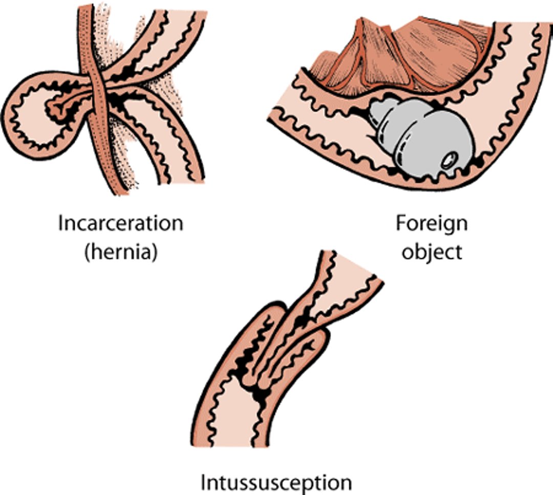
Intestinal obstruction may be partial or complete and may be caused by foreign objects, “telescoping” of the intestine (called an intussusception), bloat, incarceration (such as being constricted in a hernia), certain infections, and tumors.
Gastrointestinal obstruction causes, dog

Signs of small-intestinal obstruction vary depending on its cause, location, duration, and whether it causes partial or complete blockage of the intestinal contents. Signs usually include lethargy, loss of appetite, and vomiting. Diarrhea, weight loss, abdominal pain, abdominal swelling, fever or subnormal body temperature, dehydration, and shock may also occur. The dog may be unable to stop vomiting, which can lead to life-threatening consequences. The intestines first become distended from built-up gas. This is followed by the loss of blood supply to the intestines. Without treatment, death from shock caused by fluid loss may occur within a short period of time.
Obstruction occurring near the beginning of the intestines (closest to the stomach) tends to cause more severe and more frequent vomiting.
Intussusception (telescoping of the intestines) may cause vomiting, abdominal pain, and scant bloody diarrhea. Intussusception is more common in young dogs.
In intestinal incarceration, a loop of bowel becomes stuck through a weak spot in the body wall (hernia), causing it to swell and trap digested food inside. The dog will typically have abdominal pain that rapidly progresses to shock. This occurs because the incarceration of the affected intestine leads to bacterial growth within the stagnant bowel loop and to tissue death, leading to shock.
Young, large-breed dogs are more likely to have an obstruction caused by eating a foreign object. Many dogs with a history of eating inappropriate objects continue that practice even after having experienced discomfort in the past. When you take your dog in for examination, be sure to tell the veterinarian about your pet’s eating habits and any access to string or yarn, fabric, sewing needles, or similar objects. If there are missing objects, such as toys, in your home, this information can be important and should be reported to the veterinarian. Swallowing long, thin foreign objects such as string or thread is much more common in cats than in dogs, but when it occurs these objects can cause more damage than other types of foreign material.
Examination of the abdomen can provide the veterinarian with evidence of pain, peritonitis, organ enlargement, thickened bowel loops, or gas. A rectal examination can provide evidence of eating nonfood objects or blood. Abdominal x-rays may reveal foreign objects, masses, obstruction, abdominal fluid, or bloat. Contrast x-rays or ultrasonography are useful for diagnosing intussusception and some foreign objects. Endoscopic examination employs a tiny camera at the end of a flexible tube. This procedure is useful in identifying foreign objects or tumors in the stomach. If an obstruction is found, the veterinarian may be able to use the endoscope to help remove the object. If the object cannot be removed in this manner, surgery may be needed. Veterinarians may take several sets of x-rays over hours to days to see whether small, smooth foreign objects can pass through the intestinal tract without surgery. However, surgery is usually necessary to alleviate the cause of gastrointestinal obstructions.
Animals with general signs of illness, such as weakness and dehydration, benefit from intravenous fluids. Dogs with an obstruction caused by eating a foreign object tend to do well if diagnosed and treated quickly. Animals with severe complications (such as abdominal infections or low protein counts) are at higher risk for surgical failure. Intussusceptions found near the stomach are associated with a high risk of death.
Gastrointestinal Ulcers
Gastrointestinal ulcers can be caused by several factors, including drugs, tumors, infections, and generalized diseases. The acids and digestive enzymes found within the stomach break down food; the stomach lining must protect the rest of the stomach from these potentially damaging processes. Stomach ulcers result from a breakdown of the normal stomach lining and are aggravated by an increase in hydrochloric acid or pepsin (a digestive enzyme). Conditions that lead to increased acid production or that damage the stomach lining speed up ulcer formation.
Animals with stomach ulcers may have no signs. In other cases, they can have a history that includes vomiting, sometimes with fresh or digested blood, and abdominal discomfort. Dark stools stained with blood and pale gums suggesting anemia may be seen. Dogs may also have a decreased appetite. Some signs may indicate the cause of the ulcer (for example, signs related to kidney failure).
In dogs that have a history of vomiting, abdominal discomfort, loss of appetite, or unexplained weight loss, there are several tests that might be performed by your veterinarian in an attempt to diagnose the cause. These may include a complete blood count, biochemical profile, urinalysis, and evaluation for parasites. Additional blood tests may also be necessary. Abdominal ultrasound scans or x-rays may be used to rule out other conditions. In cases in which the cause is unclear or in those with apparent gastrointestinal disease, endoscopy and biopsy are often recommended.
The goal of ulcer management is to determine the cause of the ulceration and then eliminate or control it. Providing supportive care is also critical. Medication directed at the ulcer itself reduces gastric acidity, prevents further destruction of the stomach lining, and promotes ulcer healing. In general, treatment is continued for 6 to 8 weeks. Dietary management includes the use of bland diets (often prescription food or chicken and rice). Antibiotics may be indicated for some dogs.
Ideally, ulcer healing should be monitored with endoscopy, although costs and the animal’s tolerance for the procedure may limit its use. If ulcers do not respond to appropriate medical management, a biopsy of the stomach and small bowel becomes necessary. Several biopsies may be needed because obvious lesions may not be apparent or may be located sporadically throughout the gut.
The outlook for dogs with gastrointestinal ulcers is good. However, the outlook is poor for those with ulcers associated with renal or liver failure and for animals with cancers, such as stomach carcinoma and gastrinoma. If the ulcer extends through the stomach or intestinal wall, food and digestive fluids can escape into the abdomen. As many as 70% of these dogs can die from this serious complication.
Hemorrhagic Gastroenteritis
Hemorrhagic gastroenteritis is characterized by a sudden onset of vomiting and bloody diarrhea in formerly healthy dogs. The cause is unknown, but it may involve an abnormal response to bacteria. Dogs of either sex or any age may be affected. Young, toy and miniature breeds of dogs appear to be predisposed to this condition. Yorkshire Terriers, Miniature Pinschers, Miniature Poodles, Malteses, and Miniature Schnauzers may be more frequently affected than other breeds.
The disease is often seen in young dogs (an average of 5 years old) and is characterized by a sudden onset of vomiting and bloody diarrhea, loss of appetite, abdominal pain, and depression. The disease is not contagious and may occur without obvious changes in diet, environment, or daily routine. The severe vomiting and diarrhea can lead to shock caused by dehydration. The condition is diagnosed based on blood tests and the presence of signs in predisposed breeds. Veterinarians may also recommend a culture of the feces. Other tests may be required to rule out other conditions that can cause similar signs.
Most dogs respond to supportive veterinary treatment, including fluid treatment and antibiotics. Dogs may develop shock unless fluid support is provided. Follow your veterinarian's recommendation for food and water. Serious complications are uncommon, and most dogs recover from hemorrhagic gastroenteritis.
Inflammatory Bowel Disease
Inflammatory bowel disease is actually a group of digestive system diseases that are recognized by certain persistent signs (see below) and by the presence of inflammation without a known cause. The various forms of the disease are classified by their location and the type of cell that is involved.
The cause of inflammatory bowel disease is unknown. Although food allergies are an unlikely cause in most cases, they may contribute to the development of disease in certain ways (such as causing inflammation through excessive allergic reactions to food, bacteria, or parasites inside the intestine). Inflammation damages the mucosal barrier that protects the intestinal lining, making it even more sensitive to antigens. Persistent inflammation results in thickening and other changes in the lining of the intestine.
Inflammatory bowel disease appears to affect all ages, sexes, and breeds of dogs, though it may be more common in German Shepherds, Yorkshire Terriers, and Cocker Spaniels. Some forms are more common in certain breeds, such as Soft-coated Wheaten Terriers, Basenjis, Norwegian Lundehunds, and Boxers. The average age reported for the onset of disease signs is 6 years in dogs, but it may occur in dogs less than 2 years old. Signs are often present over long periods and sometimes come and go. Vomiting, diarrhea, changes in appetite, and weight loss may occur. Vomiting, dark stools, and abdominal pain are often seen with ulcers and erosion of the stomach and upper portion of the small intestine. If the condition causes excessive protein loss in the feces (protein-losing enteropathy), signs include weight loss, vomiting, diarrhea, swollen abdomen, and fluid retention. Signs of large-intestinal diarrhea, including loss of appetite and watery diarrhea are common.
Inflammatory bowel disease can be difficult to diagnose because many of its signs are found in other diseases as well. Veterinarians often use blood, urine, and fecal tests to rule out other diseases and to identify complications, such as low levels of protein or electrolytes. An abdominal ultrasound may help to identify abnormal sections of the digestive tract. Intestinal changes caused by the disease may be seen using an endoscope in some cases. Tissue biopsies obtained with an endoscope or surgery are necessary for the diagnosis of inflammatory bowel disease.
The goals of treatment are to reduce diarrhea and vomiting, promote weight gain, and decrease intestinal inflammation. If a cause can be identified (such as diet or parasites), it should be eliminated. Modifying the diet, without other treatment, may be effective in some cases. In other cases, changes in diet can enhance medical treatment, allowing for the drug dosage to be reduced or discontinued once signs improve. Glucocorticoids, drugs that are anti-inflammatory and suppress the immune system, are among the drugs most often used in the management of inflammatory bowel disease. Anti-parasitic drugs, certain antibiotics, vitamin supplementation, or other anti-inflammatory drugs may also be recommended.
Your veterinarian may recommend feeding your pet a hypoallergenic or elimination diet. This means providing your pet with a new source of protein and other changes. The recommended diet may be homemade—such as a diet of lamb and rice or venison and rice—or commercial. Commercial diets with these ingredients are usually available from veterinary clinics rather than commercial outlets. The new diet should be the only source of food for a minimum period (often 4 to 6 weeks), and no treats of any kind should be fed unless approved by the veterinarian. Dogs with large-intestinal diarrhea may benefit from diets high in fiber. However, supplementation of dietary fiber alone is rarely effective in severe cases. Your veterinarian will prescribe a diet that is tailored to your pet, its previous diet, and the severity of the disease.
The response to treatment varies between animals, and the outlook is uncertain. The dog's quality of life may be poor. Animals with low protein levels, severe changes on tissue biopsies, or scar tissue in the digestive tract tend to do worse than those without these changes. Relapses of signs are common and are often triggered by diet changes.
Malabsorption
Malabsorption is poor absorption of a nutrient resulting from interference with its digestion, absorption, or both. Interference with food digestion in dogs is typically due to lack of certain enzymes from the pancreas, called exocrine pancreatic insufficiency, whereas most cases of absorption failure are caused by small intestinal disease.
The signs of malabsorption are mainly due to lack of nutrient uptake and loss of nutrients in the feces. Signs typically include longterm diarrhea, weight loss, and altered appetite (loss of appetite or excessive eating). However, diarrhea may be absent even when disease is severe. Weight loss may be substantial despite a good appetite, sometimes characterized by eating of feces or non-food objects. Dogs with malabsorption usually appear healthy in other respects unless there is severe inflammation or cancer. Nonspecific signs may include dehydration, anemia, dark blood in the stools, or fluid retention. A veterinarian may be able to detect thickened bowel loops or enlarged abdominal lymph nodes.
Diagnosing malabsorption can be complex, because longterm diarrhea and weight loss are signs that are common in several diseases, including malabsorption. An exact diagnosis may take more than a single visit. A thorough examination is needed for dogs with signs of malabsorption to determine whether the signs are caused by an underlying generalized or metabolic disease. Certain tests can help determine whether the signs are due to a condition such as inflammatory bowel disease (see above), liver disease, or parasites. The dog’s history is particularly important because it may suggest a specific food allergy, consumption of non-food items, or other sensitivity. Weight loss may indicate malabsorption or protein-losing disease but may also be due to loss of appetite, vomiting, or a non‑digestive disease. There are certain features that help distinguish small-intestinal diarrhea from large-intestinal diarrhea. Suspected large intestine disease in dogs may be further evaluated by a biopsy of the intestinal lining. However, if signs are accompanied by weight loss or large volumes of feces, then the small intestine is probably also affected. Initial tests usually include blood, urine, and fecal tests, x-rays, and an abdominal ultrasound. Specialized blood tests and tissue biopsies may also be necessary.
Treatment of malabsorption involves dietary change, management of complications, and treatment of the cause, if it can be identified. If malabsorption is caused by exocrine pancreatic insufficiency, treatment involves feeding a special low-fiber diet that contains moderate levels of fat or highly digestible fat, very digestible carbohydrate, and high-quality protein. Supplementation with pancreatic extract to provide missing enzymes is also necessary. If the dog’s response to pancreatic replacement treatment is poor, small-intestinal bacterial overgrowth may be suspected. In this case, the dog may be treated with oral antibiotics for about 1 month to reduce the bacterial overgrowth. Effective treatment of small-intestinal disease depends on the nature of the disorder, but when a specific diagnosis cannot be made, treatments may be given on a trial basis.
Dietary modification is an important aspect of the management of small intestinal disease. Your veterinarian may recommend feeding your pet an exclusion diet consisting of a single protein source (one to which your dog has not previously been exposed) as a test when dietary sensitivity is suspected. It is very important that you provide the special diet and prescribed medication(s) for your pet exactly as instructed. Often, owners are tempted to provide a “special treat” not on the diet even though they have been instructed not to do so. Failure to follow the prescribed diet can delay diagnosis and delay the treatment their pet needs. Owners can reward their pets during this time with petting, a new blanket or suitable toy, or some other reward that is not food. Often the best reward for the pet is extended periods of attention.
The outlook is good for dogs with malabsorption if there is a simple cause. For example, 85% of dogs with exocrine pancreatic insufficiency will respond well to treatment with pancreatic enzymes. The outlook is worse for dogs with severe small intestinal disease, cancer, fluid retention caused by low protein levels, severe weight loss, low blood levels of vitamin B12, or a lack of appetite.
For More Information
Also see professional content regarding stomach and intestinal disorders in dogs.


