The skin is the largest organ of the body and, depending on the species and age, may represent 12%–24% of an animal’s body weight. The skin has many functions, including serving as an enclosing barrier and providing environmental protection, regulating temperature, producing pigment and vitamin D, and sensory perception. Anatomically, the skin consists of the following structures: epidermis, basement membrane zone, dermis, appendageal system, and subcutaneous muscles and fat.
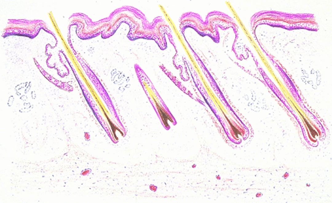
Skin with hair
The skin, with hair. Illustration by Dr. Gheorghe Constantinescu. |

Epidermis in Animals
The epidermis is composed of multiple layers of cells consisting of keratinocytes, melanocytes, Langerhans cells, and Merkel cells.
Keratinocytes function to produce a protective barrier. They are produced from columnar basal cells attached to a basement membrane. The rate of cell mitosis and subsequent keratinization are controlled by a variety of factors, including nutrition, hormones, tissue factors, immune cells in the skin, and genetics. Keratinocytes play a key role in the skin immune system and in regulating the growth and renewal of cells. The dermis may also exert significant control over the growth of the epidermis. It has been hypothesized that photoperiod and reproduction cycles may affect the epidermis in animals. Glucocorticoids decrease mitotic activity; disease and inflammation also alter normal epidermal growth and keratinization.
As keratinocytes migrate upward, they undergo a complex process of programmed cell death or keratinization. The goal of this process is to produce a compact layer of dead cells called the stratum corneum, which functions as an impermeable barrier to the loss of fluids, electrolytes, minerals, nutrients, and water, while preventing the penetration of infectious or noxious agents into the skin. The structural arrangement of keratin and the lipid content of the skin are critical to this function. The vitamin D precursor, 7-dehydrocholesterol, is formed in the epidermis. The epidermis is thickest in large animals. The stratum corneum is continuously shed or desquamated.
Melanocytes are located in the basal cell layer, outer root sheath, and ducts of sebaceous and sweat glands. They are responsible for the production of skin and hair pigment (melanin). Production of pigment is under hormonal and genetic control. Melanocytes provide constitutive pigmentation, the genetically programmed pigment and facultative pigmentation, that occurs as a result of stimulation from hormones, UV light, inflammation, etc.
Langerhans cells are mononuclear dendritic cells that are intimately involved in regulating the immune system of the skin. They are damaged by excessive UV light exposure and glucocorticoids. Antigenic and allergenic material is processed by these cells and transported to local and nodal T cells to induce hypersensitivity reactions. Epidermal proteins may also conjugate with exogenous haptens, rendering them antigenic.
Merkel cells are specialized sensory cells associated with skin sensory organs, eg, whiskers and tylotrich pads. These are slow-adapting mechanoreceptors. These cells may also influence cutaneous blood flow and sweat production, coordinate keratinocyte proliferation, and stimulate stem cell growth of hair follicles.
Basement Membrane Zone in Animals
The basement membrane zone serves as a site for attachment of basal epidermal cells and as a protective barrier between the epidermis and dermis. A variety of skin diseases, including several autoimmune conditions, can cause damage to this zone. Vesicles are an example of a damaged basement membrane zone. The basement membrane zone has important functions:
anchors the epidermis to the dermis
maintains a functional and proliferative epidermis
maintains tissue architecture
participates in wound healing
acts as a physical barrier
regulates nutritional support between the epidermis and underlying connective tissue
Dermis in Animals
The dermis is a mesenchymal structure that supports, nourishes, and to some degree, regulates the epidermis and appendages. The dermis consists of ground substance, dermal collagen fibers, and cells (fibroblasts, melanocytes, mast cells, and occasionally eosinophils, neutrophils, lymphocytes, histiocytes, and plasma cells). Blood vessels responsible for thermoregulation, nerve plexuses associated with cutaneous sensation, and both myelinated and unmyelinated nerves are present in the dermis. Motor nerves are primarily adrenergic and innervate blood vessels and arrector pili muscles. Except in horses, apocrine glands do not appear to be innervated. Sensory nerves are distributed in the dermis, hair follicles, and specialized tactile structures. The skin responds to the sensations of touch, pain, itch, heat, and cold.
Appendageal System in Animals
These structures grow out of (and are continuous with) the epidermis and consist of hair follicles, sebaceous and sweat glands, and specialized structures (eg, claw, hoof). The hair follicles of horses and cattle are simple, ie, the follicles have one hair emerging from each pore. The hair follicles of dogs, cats, sheep, and goats are compound, ie, the follicles have a central hair surrounded by 3–15 smaller hairs all exiting from a common pore. Animals with compound hair follicles are born with simple hair follicles that develop into compound hair follicles.
The growth of hair is controlled by a number of factors, including nutrition, hormones, and photoperiod. The growing stage of the hair is referred to as anagen, and the resting stage (mature hair) is referred to as telogen. The transitional stage between anagen and telogen is catagen. Animals normally shed their hair coat in response to changes in temperature and photoperiod; most animals undergo a shed in the early spring and early fall.
The size, shape, and length of hair is controlled by genetic factors but may be influenced by disease, exogenous drugs, nutritional deficiencies, and environment. Hormones have a significant effect on hair growth. Thyroxine initiates hair growth, and glucocorticoids inhibit hair growth.
The primary functions of the hair coat are to provide a mechanical barrier, to protect the host from actinic damage, and to provide thermoregulation. In most species, trapping dead air space between secondary hairs conserves heat. This requires that the hairs be dry and waterproof; the cold-weather coat of many animals is often longer and finer to facilitate heat conservation. The hair coat can also help cool the skin. The warm-weather coat of animals, particularly large animals, consists of shorter thicker hairs and fewer secondary hairs. This anatomic change allows air to move easily through the coat, which facilitates cooling. The hair coat also helps conceal or camouflage the animal.
Sebaceous glands are simple or branched alveolar, holocrine glands that secrete sebum into the hair follicles and onto the epidermal surface. They are present in large numbers near the mucocutaneous junction, interdigital spaces, dorsal neck area, rump, chin, and tail area; in some species, they are part of the scent-marking system. For example, in cats, sebaceous glands are present on the face, dorsum, and tail in high concentration; cats mark territories by rubbing their face on objects and depositing a layer of sebum laced with feline facial pheromones. Sebum is a complex lipid material containing cholesterol, cholesterol esters, triglycerides, diester waxes, and fatty acids. Sebum is important to keep the skin soft and pliable and to maintain proper hydration; it gives the hair coat sheen and has antimicrobial properties.
Sweat glands (epitrichial [formerly apocrine] and atrichial [formerly eccrine]) are part of the thermoregulatory system. Atrichial glands are only present in the foot pads. Epitrichial glands are not present on footpads or on the planum nasale. The evaporation of sweat from the skin is the primary body cooling mechanism for horses and primates and, to a lesser degree, pigs, sheep, and goats. There is some clinical evidence to suggest that limited sweating occurs in dogs and cats, and that it may have a minor role in cooling the body. Dogs and cats thermoregulate primarily by panting, drooling, and spreading saliva on their coats (cats). Cats also sweat through their paws, especially when excited; this is most commonly seen as wet paw prints on surfaces, eg, examination tables.
Subcutaneous Muscles and Fat in Animals
The “twitch muscle” (panniculus carnosus) is the major subcutaneous muscle. The subcutaneous fat (panniculus adiposus) serves many functions, including insulation; reservoir for fluids, electrolytes, and energy; and shock absorber.