The schedule for determination of pregnancy varies among breeding farms. One schedule is as follows: 1) days 14–18—check for pregnancy and twins; if open, mare can be rebred on days 19–20; 2) days 25–30—evaluate normal embryo development (heartbeat present at 24–25 days), recheck for twins; 3) days 40–60—evaluate normal fetal development; 4) fall check—confirm mare is still pregnant.
Palpation Per Rectum for Pregnancy Determination in Horses
The pregnant cervix should be tightly closed and elongated with a prominent portio vaginalis 14–21 days after ovulation. Uterine tone increases and the uterine wall thickens so that by 14–18 days, the endometrial folds can no longer be readily palpated per rectum.
The conceptus develops in a recognizable pattern of size and shape, allowing estimation of age based on palpable characteristics. In maiden and barren mares, at 25–28 days of gestation, a careful, experienced examiner may be able to feel the embryonic vesicle ventrally at the base of one uterine horn, as a bulge 3.5 cm in diameter. At 30 days, the uterine horns are small with pronounced tone, and the conceptus can be felt as a ventral bulge 4 cm in diameter and positioned at the base of the gravid uterine horn. The uterine wall is thin over the expanding conceptus. At 42–45 days, the conceptus occupies about half of the gravid uterine horn and is 5–7 cm in diameter. By 48–50 days, the enlargement of the conceptus begins to involve the uterine body and is 6–8 cm in diameter and 8–10 cm long.
At 60 days, nearly the entire gravid horn and half of the uterine body are occupied by conceptus, but the nongravid horn remains small with considerable tone. The 60-day conceptus is 8–10 cm in diameter and 12–15 cm long. After 85 days, the turgidity of the conceptus decreases so that the fetus becomes palpable. At 90 days, the conceptus fills the entire uterus, and the cranial portion of the uterus may extend over the brim of the pelvis into the abdominal cavity.
After 100–120 days of gestation, the gravid uterus is positioned cranial to the pelvic brim in the abdominal cavity. The ovaries are positioned cranially and ventrally and closer together because of the ventral traction exerted by the enlarging uterus on the broad ligament. After 150 days of gestation, the ovaries are not routinely felt per rectum. During midgestation, the gravid uterus may be difficult to reach, because it is positioned ventrally in the abdomen. But as the conceptus/gravid uterus enlarges, its dorsal surface comes back into reach in late gestation. One should always confirm that two uterine horns with palpable endometrial folds and ovaries cannot be identified in the pelvic canal before making a midterm pregnancy diagnosis.
Ultrasonography for Pregnancy Determination in Horses
The spherical shape of the equine embryo and the characteristic pattern of development of the fetal membranes permit accurate estimation of stage of gestation by ultrasonography until 45 days after ovulation. The embryo may first be imaged in the uterus with a 5–10 MHz linear transducer at 9–10 days as a round anechoic yolk sac 4 mm in diameter. The spherical conceptus moves throughout the lumen of the uterine horns and uterine body from day 6–16. The early conceptus is seen to have a bright white (echogenic) line on the dorsal and sometimes the ventral aspect of its image that is called a specular reflector, which is an artifact seen when the ultrasound beam strikes the wide, smooth surface of the conceptus at a 90° angle. This specular reflector, embryonic motility, and linear growth rate may help differentiate an early (<16 day), motile embryo from some uterine cysts.

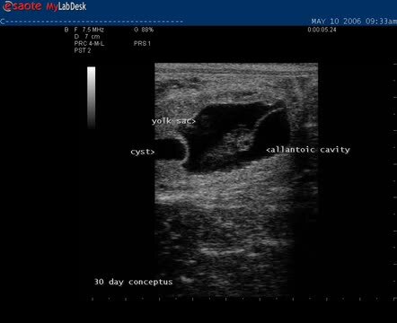
At day 17–18, the conceptus has a characteristic “guitar-pick” shape. At day 21, the embryo proper can be seen in the ventral aspect of the yolk sac, and by 24 days the allantoic cavity is visible as an anechoic compartment ventral to the embryo proper. At day 25, a heartbeat should be present in the embryo proper. As the allantoic cavity enlarges, the yolk sac comprises a decreasing proportion of the conceptus. The position of the allantois and relative sizes of the developing allantoic cavity and the regressing yolk sac can indicate the stage of gestation between days 24 and 45. At 30 days, the allantoic cavity occupies approximately one-half of the conceptus, so that the size of the yolk sac is equal to that of the allantoic cavity. At 45 days, the only visible fluid cavity is the allantoic cavity, and the fetus appears to be suspended from the dorsal wall of the uterus by its umbilical cord and is positioned in dorsal recumbency.
Endocrine Tests for Pregnancy Determination in Horses
Cells from the chorionic girdle of the conceptus invade the endometrium to form endometrial cups that produce equine chorionic gonadotropin (eCG). Increased serum concentrations of eCG 40–120 days after ovulation indicate the presence of endometrial cups. Concentrations of eCG may remain increased until 120 days, even if fetal death occurs (false-positive). A false-negative result will be obtained if eCG assay is used as a pregnancy test in a pregnant mare before 40 or after 120 days of gestation.
Estrone sulfate is produced by the fetus and is a good indicator of fetal viability. Plasma and urine concentrations of estrone sulfate are increased after 60 days and 150 days, respectively, of pregnancy. Either endocrine assay can be used properly as a pregnancy test only if the breeding and ovulation dates are known.
For More Information
Also see pet health content regarding routine health care for horses and the breeding and reproduction of horses.