


Lymphoid Tumors of the Skin
Canine extramedullary plasmacytomas (atypical histiocytomas, cutaneous neuroendocrine tumors, reticulum cell sarcomas, cutaneous nodular amyloidosis) are relatively common cutaneous tumors. Although their derivation was long debated, neoplastic cells characteristically express cytoplasmic immunoglobulin and may produce primary amyloid, leaving little doubt as to their lymphoplasmacytic origin. These tumors of dogs and, rarely, cats are most frequently identified on the head (including ears, lips, and oral cavity) and extremities of mature adult to senior animals. Cocker Spaniels, Airedales, Scottish Terriers, and Standard Poodles are most at risk.
The tumors are generally small (< 5 cm in diameter) and sometimes pedunculated. Most of these tumors are locally confined, and complete but conservative surgical excision is the treatment of choice. Infrequently, extracutaneous plasmacytomas may be locally invasive or multiple (or both), especially when they occur in the oral cavity. Recurrence has also been correlated with the presence of amyloid. Treatment for these tumors targets local control using localized techniques described above. For recurrent, invasive tumors, more aggressive attempts at excision or electroporation may be required. When tumors are multiple or when surgical excision is not feasible, radiation therapy appears to be the best secondary treatment. For tumors resistant to radiation and for those animals unable to receive radiation therapy, systemic treatment with administration of chemotherapeutic agents, including melphalan, chlorambucil, cyclophosphamide, and glucocorticoids, have been recommended and have yielded long-term survival.

Courtesy of Dr. Alice Villalobos.
Cutaneous lymphosarcoma may occur as a disease in which the skin is the initial and primary site of involvement, or it may be secondary to systemic, internal disease. ( See also Lymphoma in Dogs; see Bovine Leukosis; and see Feline Leukemia Virus.) Cutaneous lymphosarcoma is uncommon but has been identified in all domestic species. In general, two distinct forms are recognized—an epitheliotropic form (in which there is infiltration by malignant lymphocytes into the epidermis and adnexa) and a nodular, nonepitheliotropic form. Both forms usually express surface and cytoplasmic antigens characteristic of T cells; this, along with the frequent identification of at least small foci of epitheliotropism in many cases of nonepitheliotropic forms in dogs and cats, suggest they may be variants of the same tumor.

Courtesy of Dr. Alice Villalobos.

Courtesy of Dr. Alice Villalobos.

Courtesy of Dr. Alice Villalobos.
Epitheliotropic cutaneous lymphosarcoma (ECL, mycosis fungoides) is the most frequently recognized form of cutaneous lymphosarcoma in dogs and arguably cats. It is a disease of middle-aged and older dogs, and Poodles and Cocker Spaniels may be predisposed.
Classically, the lesions progress from patch to plaque to tumor; however, one or any combination of these three primary lesions may be present. For example, a form of ECL known as pagetoid reticulosis has minimal to no dermal involvement, and cutaneous lesions always appear as erythematous patches. Another common feature of the disease in dogs is the presence of areas of alopecia due to follicular atrophy caused by infiltration of neoplastic cells into the outer sheath and lumen of hair follicles. Although most cases are associated with diffuse cutaneous involvement, forms limited primarily to mucous membranes or the footpads have been identified. Because of the variable clinical appearance of this tumor, diagnosis based on clinical features can be very difficult, and early stages can be confused with allergic, autoimmune, endocrine, infectious, or seborrheic diseases. Most cases are limited to the cutis until late in the course of the disease. With concurrent leukemia, ECL is known as Sézary syndrome.
In dogs, ECL is a slow to moderately progressive disease for which a number of therapies have been attempted. To date, all appear more effective in improving the clinical features of the disease than in prolonging an affected dog’s life. Mechlorethamine (nitrogen mustard) has been used in the past as a topical therapy, but because large areas of a dog’s body may be affected (including the mucous membranes) and because of its sensitizing potential in people, it is infrequently used. The disease is often transiently responsive to steroids. Chemotherapeutic agents, such as combinations of adriamycin, chlorambucil, cyclophosphamide, doxorubicin, and vincristine, are variably effective. Lomustine (an alkylating nitrosourea), retinoids with and without glucocorticoids, and high-dose linoleic acid supplementation (high percent safflower oil at 3 mL/kg, PO, every 24 hours for 3 consecutive days each week) may occasionally achieve partial or complete remission.
Studies show that a lymphocyte T-cell immunomodulator (LTCI), which signals the differentiation of late stage CD4 lymphocytes and causes apoptosis of malignant T cells, is a reasonable adjunctive therapeutic agent to administer, as an intratumor injection or given subcutaneously and may improve the clinical course in some cases.
In cats, ECL is rare and tends to develop in older animals. Lesions often follow a defined progression, appearing initially as a crusty plaque that is variably pruritic. Biopsies of early lesions are often diagnosed as lymphocytic mural folliculitis. In many cases in which this diagnosis is applied, the lesions evolve into unequivocal cutaneous lymphosarcoma. In contrast to ECL in dogs, epitheliotropism is often extremely subtle in cats. Little is known about treatment or whether treatment used in dogs would be effective in cats; however, since the availability of LTCI for use in cats with feline leukemia virus, it can be offered as a viable option.
Nonepitheliotropic cutaneous lymphosarcoma (NECL) is the most recognized form of cutaneous lymphosarcoma in all domestic animals but dogs and cats. In dogs, NECL is most common in middle-aged or older animals. Lesions are nodules or plaques that most commonly develop on the trunk. They generally are multiple, although solitary lesions may be noted, especially in cats. In many cases, NECL is grossly indistinguishable from the tumor stage of ECL. A definitive diagnosis is important because NECL in dogs is generally more aggressive than ECL, and systemic involvement occurs commonly and early in the course of the disease.
Various modes of treatment, including excision, chemotherapy, and less frequently radiotherapy, have been used as monotherapy and in combination. Excision is the best choice when the disease is limited to a solitary tumor, and complete cures have occasionally been obtained. Excision, laser ablation, cryosurgery, or electroporation in more diffuse forms infrequently elicit longterm remissions. Chemotherapy or chemoimmunologic protocols used for other forms of canine lymphosarcoma should be considered as palliative. Typical remission time is ~8 months.
In cats, NECL is a rare disease of middle-aged or older animals. The role of feline leukemia virus remains undefined. The lesions are plaques or nodules that may be solitary or multiple, alopecic or haired, and ulcerated or lined by an intact epidermis. Feline NECL is aggressive; even when complete excision of a solitary nodule is attempted, recurrence is common. No definitive therapy is known; however, administering a combination of lomustine, steroids, linolenic acid (oil of evening primrose), and intralesional or subcutaneous injections of LTCI may be of some value.
In horses, NECL (nodular lymphosarcoma, subcutaneous lymphosarcoma, lymphohistiocytic lymphosarcoma) may be recognized at any age but is most common in young and middle-aged animals. Firm, nonulcerated nodules are most common in the subcutaneous fat of the ventral body surface.
Microscopically, two types of nodular lymphosarcoma are recognized in horses. The most common type consists of a mixture of histiocytes and small, well-differentiated lymphocytes, occasionally with plasmacytoid features; the second type consists of a monomorphic population of large atypical lymphocytes, with only occasional histiocytic cells.
Differentiation between these two forms is important because most cases of cutaneous lymphosarcoma in horses with a monomorphic pattern of cells have internal involvement, and the disease progresses rapidly. In contrast, the lymphohistiocytic form seldom is associated with internal involvement, and affected horses may live for years. As the lymphohistiocytic form progresses, the nodules tend to become more frequent on the ventral cervical regions. In many cases, euthanasia may be warranted when pharyngeal involvement induces dyspnea. Because of the expense of cytotoxic drugs, treatment is generally limited to glucocorticoids administered orally or intralesionally; remission, if induced, is usually short term.
In cattle, cutaneous lymphosarcoma is a disease of young animals (generally < 4 years old). It is one of the sporadic bovine leukosis syndromes that is not transmissible. The term sporadic bovine leukosis is usually reserved for calf, cutaneous, and thymic types of lymphoma, which are defined by the young age of occurrence and the distribution of tumors. The cause or causes are not known. Only lymphomas caused by bovine leukemia virus infection should be termed leukosis or enzootic bovine leukosis. There may also be lymphosarcomatous conditions that do not fall into either the sporadic or enzootic bovine leukosis categories (ie, adult multicentric lymphoma with sporadic occurrence of unknown etiology). Cutaneous lymphosarcoma of young cattle is presumed not to be associated with bovine leukemia virus infection and presently has an unknown etiology. The lesions are typically nodular, involve the dermis or subcutaneous fat, and are often ulcerated. There is no known therapy.
Cutaneous Mast Cell Tumors

Courtesy of Dr. Alice Villalobos.

Courtesy of Dr. Alice Villalobos.

Courtesy of Dr. Alice Villalobos.
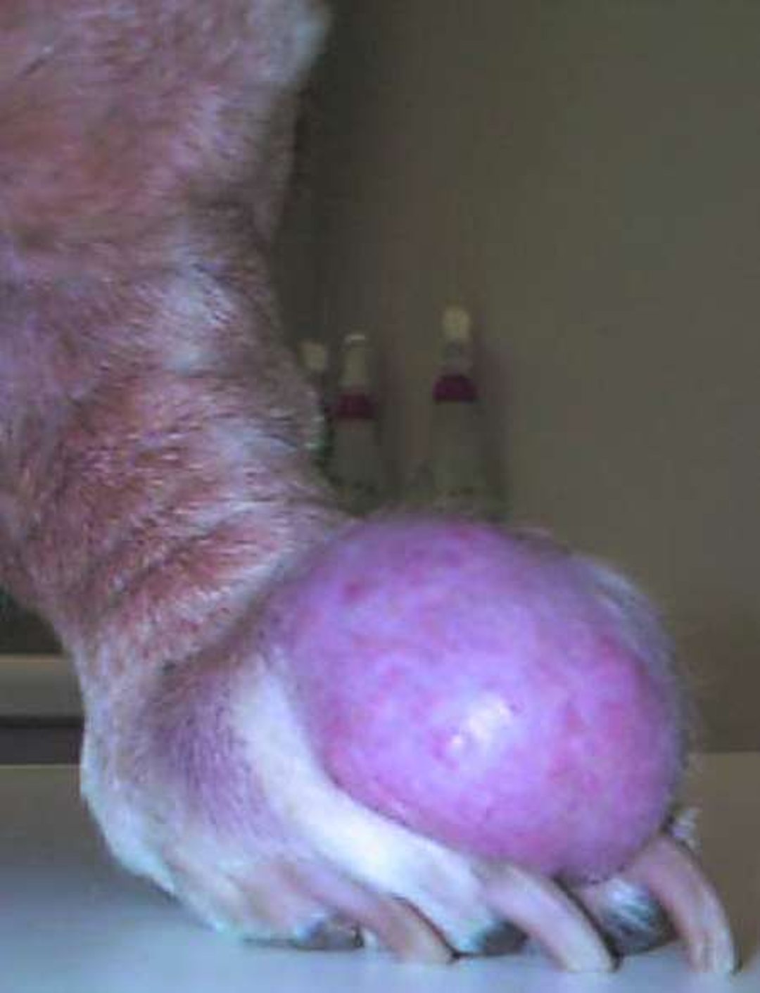
Mast cell tumors and mastocytoma are the most frequently recognized malignant or potentially malignant cutaneous neoplasms of dogs (16–19% of all canine skin tumors). In addition, visceral and leukemic forms can occur. A viral etiology has been speculated but remains controversial. Tumors may be seen in dogs of any age (typically 8–10 years). They may develop anywhere on the body surface as well as in internal organs, but the limbs (especially the posterior upper thigh), ventral abdomen, and thorax are the most common sites; ~10% are multicentric. However, studies suggest that multiple tumors do not carry a worse prognosis when they are all adequately excised if grade 1 or low grade. Initial size at the time of surgery is highly predictive of outcome; tumors >3 cm in diameter correlate with decreased survival time. Tumors that are incompletely excised, grow rapidly, or arise from mucocutaneous junctions, muzzle, ventrum, prepuce, or subungual areas or in animals with adverse clinical signs at diagnosis are associated with more aggressive biological behavior.
Many breeds appear to be predisposed, especially Boxers and Pugs (in which tumors are often multiple), Rhodesian Ridgebacks, and Boston Terriers. The tumors vary markedly in size, and clinical appearance alone cannot establish a diagnosis. Most commonly, they appear as raised, nodular masses that may be soft to solid on palpation. Although they often seem encapsulated, mast cell tumors in dogs are seldom discrete. Rather, they consist of a highly cellular center surrounded peripherally by a halo of smaller numbers of mast cells that palpate as normal skin.
Dogs can also develop clinical signs associated with the release of vasoactive products from the malignant mast cells. The most common clinical sign is gastroduodenal ulceration that may be present in up to 25% of cases.
Cytologic evaluation of fine-needle aspirates or impression smears, with Giemsa and or toluidine blue stain, can be used to identify the characteristic cytoplasmic granules to establish the diagnosis of mast cell tumors in dogs and cats. All skin tumors should be examined by fine-needle aspiration cytology before excision to exclude mast cell tumor. If the surgeon is aware that the tumor is of mast cell origin, a surgical plan for wide and deep excision will yield the best results. All mast cell tumors should be submitted for biopsy evaluation to determine margins and grade, because cytology is not a substitute for histopathology to correlate with prognosis. However, certain cytologic features can be used to identify high-grade tumors.
Two systems of histopathologic grading have been defined, the Bostock system of 1973 and the Patnaik et al system of 1984. To avoid confusion, it is essential to know which system is being used. The commonly used Patnaik grading system has the following designations: grade 1 = low grade, grade II = intermediate grade, grade III = high grade. Up to 80% of canine mast cell tumors are classified as grade II and are further divided into subgroups of low grade II (mitotic index ≤5) and high grade II (mitotic index >5) along with characteristics that may predict clinical behavior in this large category.
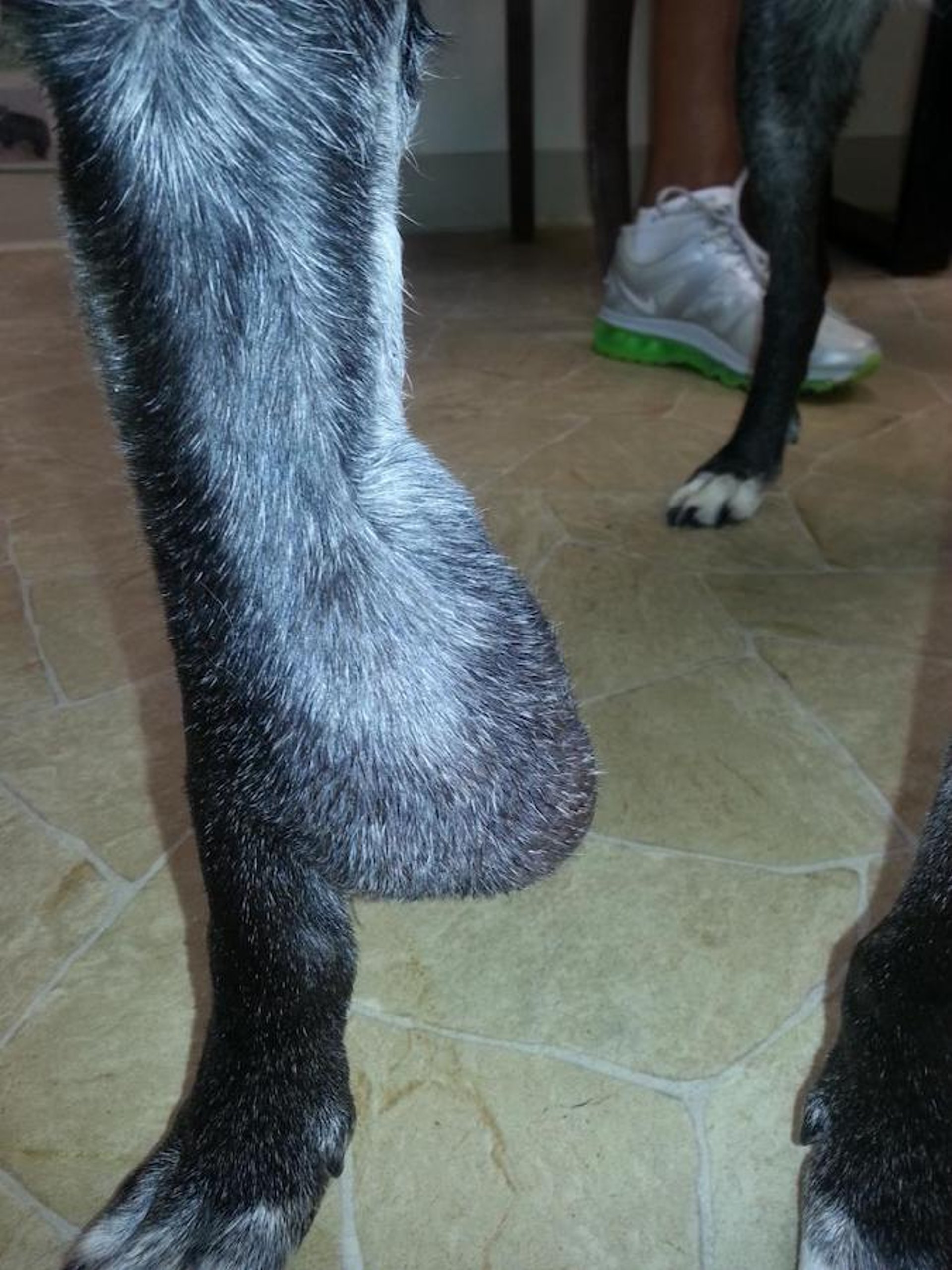
Although there is believed to be a benign variant of canine mast cell tumor, there is no clinical or microscopic way to identify it. In addition, small mast cell tumors may remain quiescent for long periods before becoming aggressive. A subcutaneous variant of canine mast cell tumor, which does not have a primary infiltrative dermal involvement, occurred in ~10% of biopsied tissues in one study. This variant appeared most frequently in the hind limb, with 66% having incomplete excision, most commonly at the deep margin. These tumors have intermediate histologic grade, a lower rate of recurrence (only 9%), and extended mean survival times, with only a 6% rate of metastases.
Up to 65% of incompletely excised mast cell tumors do not recur. This suggests that their biological behavior is not always aggressive and that aggressive treatment may not always be warranted. Specialized stains can be used to distinguish tumor grades more adequately.
Proliferation marker expression can help determine the likelihood that an incompletely removed mast cell tumor will recur or metastasize. The combination of Ki-67 >23/grid; proliferating cell nuclear antigen (PCNA/AgNOR) count >54; mutations in exon 8 and exon 11 of c-Kit gene; and increased PCNA levels are prognostic for local recurrence and a poor prognosis. Correlation of test results with survival times can be useful; however, it is not always a reliable predictor of the outcome. There is agreement on cytologic features for high-grade mast cell tumors: ≥7 mitotic figures/10 high-power fields (hpf), 3 multinucleated cells/10 hpf, 3 bizarre nuclei/10 hpf, and karyomegaly in 10% of the cells. These high-grade features confer a prognosis of a 4-month survival time vs >2 years for low-grade mast cell tumors. Because of the difficulty of subcategorizing canine mast cell tumors, all should be treated as at least potential malignancies. The Mast Cell Tumor Panel to determine prognosis and treatment guidance can be requested for grade II tumors at the Diagnostic Center for Population and Animal Health at Michigan State University.
Treatment depends on the clinical stage of the disease and the predicted aggressive biological behavior. For stage I tumors (a solitary tumor confined to the dermis without nodal involvement), the preferred treatment is complete excision with a wide margin; at least 3 cm of healthy tissue surrounding all palpable borders and one facial plane underlying the tumor should be removed in an attempt to excise both the nodule and its surrounding halo of neoplastic cells. Intraoperative cytology (examination of impression smears at the excised tissue margins) can guide the surgeon, who should continue to remove tissue until the margins are adequate and free of mast cells.
If histologic evaluation suggests that the tumor extends beyond the surgical margins, re-excision or tumor bed excision should be attempted. Radiation therapy is an option. It may be curative if the remaining tumor is small or microscopic. Combination or multimodal treatments with intralesional chemotherapy and or local hyperthermia therapy or electrochemotherapy along with systemic chemotherapy including tyrosine-kinase inhibitors, may be more effective than radiation alone to control biologically and locally aggressive mast cell tumors.

Courtesy of Dr. Alice Villalobos.
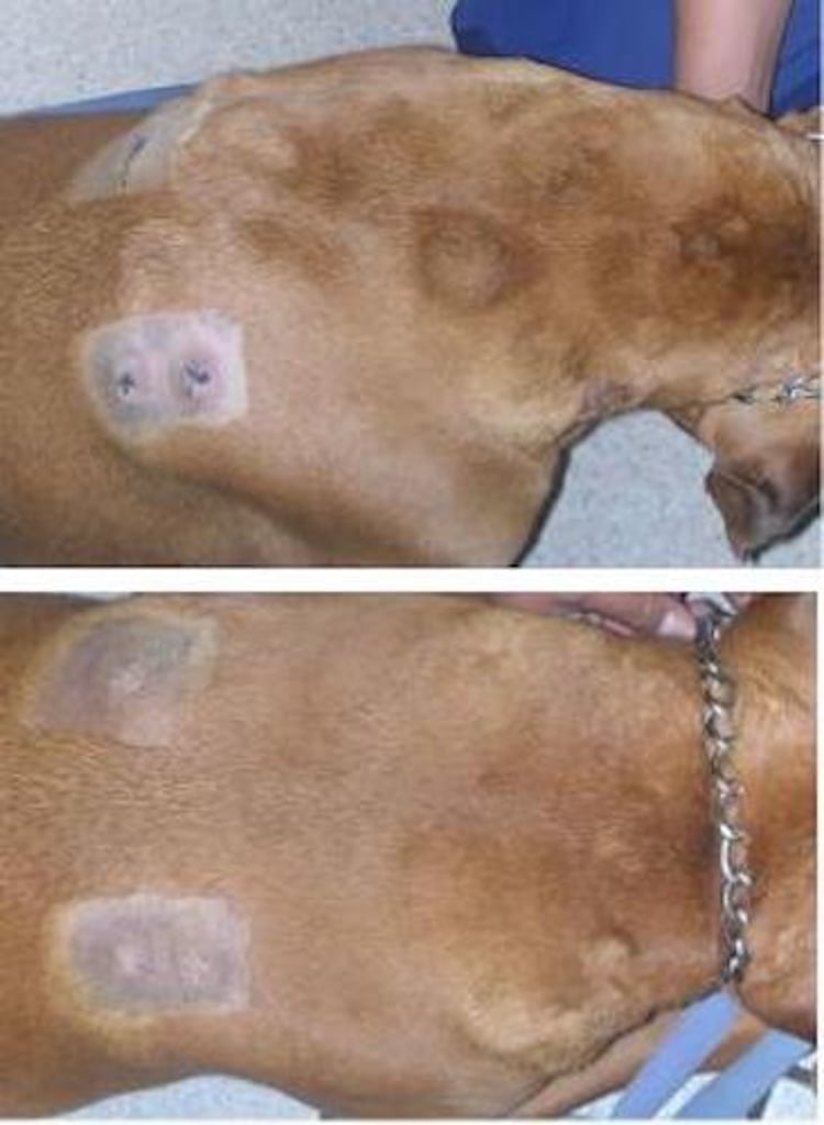
There is no agreed-upon treatment for stage II–IV mast cell tumors; however, administration of multikinase inhibitors in combination with cytotoxic chemotherapy requires dose reduction to avoid adverse events. Standard care options for stage II tumors (a solitary tumor with regional lymph node involvement) include excision of the mass and the affected regional node (if feasible), prednisone, and radiotherapy, administered either singly or in combination. Triamcinolone or dexamethasone sodium phosphate, injected evenly into the tissues of the tumor bed at the time of surgery or postoperatively as a follow-up series, may also help reduce recurrence.
Intraoperative radiation therapy or follow-up external beam therapy is still considered the highest standard of care; however, it is often declined because of cost. Injections into the tumor bed after incomplete excision using hypotonic, deionized, or distilled water has been debated. Treatment of stage III (multiple dermal tumors with or without lymph node involvement) or stage IV (any tumor with distant metastasis or recurrence with metastasis) tumors is generally considered palliative. One recommended treatment is administration of prednisone (2 mg/kg, PO, every 24 hours for 5 days, followed by a maintenance dose of 0.5 mg/kg, PO, every 24 hours) or intralesional injections of triamcinolone (1 mg/cm diameter of tumor, every 2 weeks). Treatment with H1- and H2-receptor antagonists for the peripheral and gastric effects of histamine, respectively, is indicated for animals with systemic disease or clinical signs referable to histamine release. Chemotherapy with vinca alkaloids (vinblastine, vincristine), chlorambucil, lomustine, L-asparaginase, and cyclophosphamide has also been used with variable effectiveness, ranging from 20–90%. Prednisone and vinblastine used as adjuvant chemotherapy to incomplete surgical resection conferred improvement over historical survival data regarding surgery alone, yielding a 57% 1- and 2-year disease-free state and a 45% survival rate at 1 and 2 years for dogs with grade III tumors. In 19 dogs administered a high dose of lomustine every 21 days, 42% of mast cell tumors showed measurable responses, ranging from stable to partial, with one complete response.
Neutropenia appears 7 days after treatment, with neutrophil counts of 1,500 cells/mcL. Chemotherapy doses must be lowered if used in combination with multikinase inhibitors. Lomustine at metronomic doses administered daily along with multikinase inhibitors may be helpful and may avoid the adverse events associated with high-dose lomustine. A new antineoplastic agent containing tigilanol tiglate, which induces necrosis after intralesional injection may benefit patients with nonresectable, nonmetastatic subcutaneous mast cell tumors located on the limbs. Tumors must be < 8 cm3 in volume. Intratumoral injection of tigilanol tiglate will cause wound formation, pain, and lameness, in addition to vomiting and tachycardia.

Courtesy of Dr. Rachel Jones.
Small-molecule multikinase inhibitors such as masitinib mesylate and toceranib phosphate are available to treat mast cell tumors and have been shown to be very helpful in management of difficult cases. They inhibit the c-Kit tyrosine kinase receptor, an activated or mutated proto-oncogene associated with the development of mast cell tumors. Tissue sampling to determine the biological aggressiveness of mast cell tumors and to look for the presence of the mutated tyrosine kinase c-Kit receptor has been recommended before starting treatment, although evidence shows that many mast cell tumors respond regardless of their c-Kit status.
A study of 202 client-owned dogs with or without prior treatment, having measurable cutaneous grade II or III mast cell tumors without nodal or visceral metastasis, found that masitinib (12.5 mg/kg, PO, every 24 hours) was a relatively safe and beneficial treatment option. Another clinical trial found that toceranib (3.25 mg/kg, PO, every 48 hours), a receptor tyrosine kinase inhibitor, results in inhibition of Kit phosphorylation in canine mast cell tumors and was of clinical benefit with continued use. Note that the toceranib dosage stated on the package insert may lead to unacceptable toxicity. Veterinary oncologists are the most informed sources for clinical application using these novel tyrosine kinase inhibitor drugs against mast cell tumors, which may also benefit other malignancies.
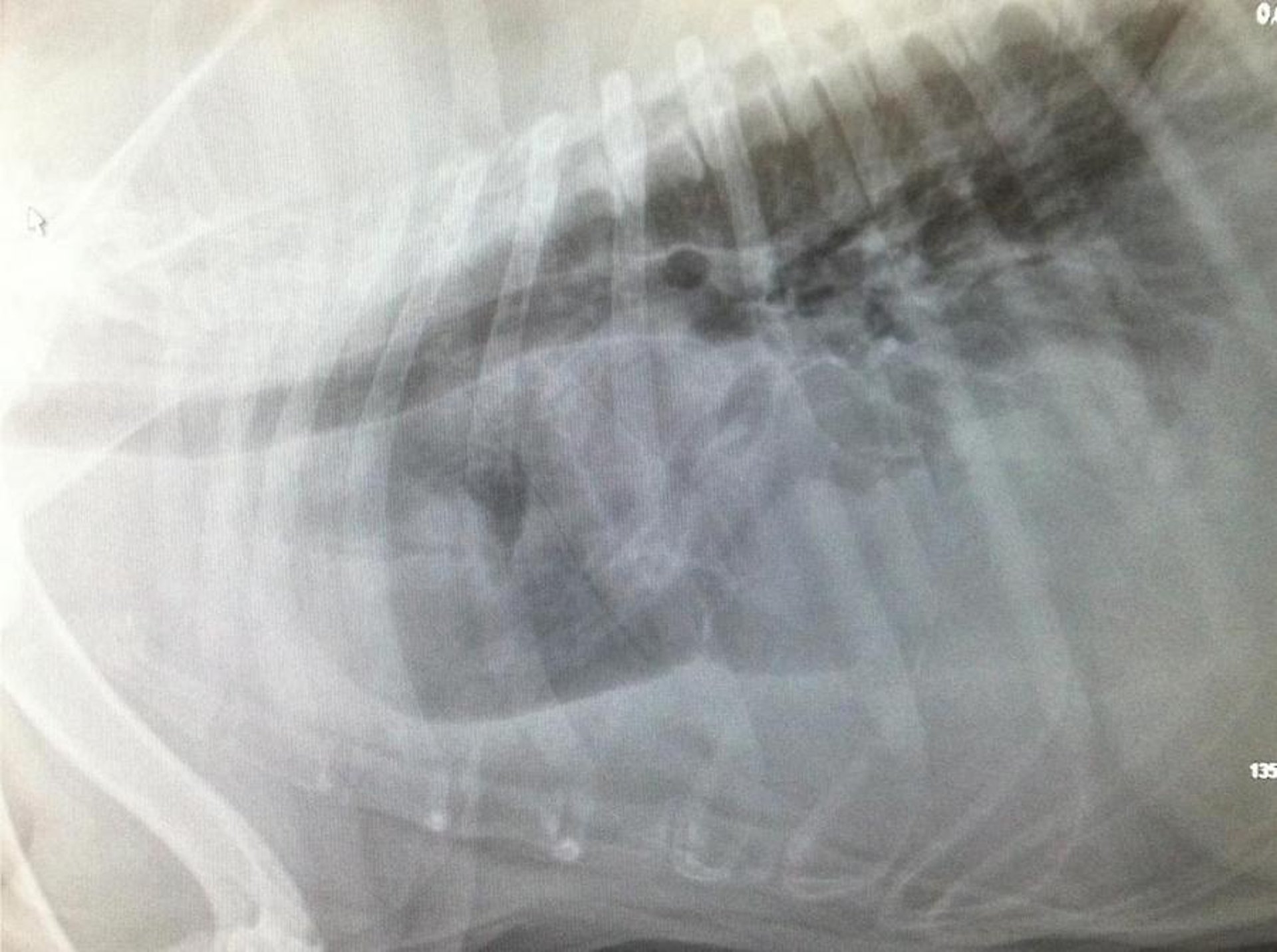
In cats, cutaneous mast cell tumors are the second-most common skin tumor; however, the disease is seen only occasionally in practice. In addition to cutaneous tumors, primary splenic, systemic, leukemic, and GI forms have been recognized. Two distinct variants of the cutaneous form occur—a mast cell type analogous to, but not identical with, cutaneous mast cell tumors in dogs, and a histiocytic type unique to cats. Feline cutaneous mast cell tumors may be either solitary or multiple. Primary splenic, systemic, recurrent, and multiple tumors (five or more) are associated with a guarded prognosis. Some cats with primary splenic mast cell tumors may survive 1–2 years post splenectomy. Combination surgery and chemotherapy may help cats with intestinal mast cell tumors survive >1 year.

Courtesy of Dr. Alice Villalobos.
The mast cell type is most common. It is found primarily in cats >4 years old and may develop anywhere on the body but most commonly on the head and neck. The tumors are single, alopecic nodules, generally 2–3 cm in diameter, that occasionally extend into the subcutaneous fat. Lymphoid nodules are common; eosinophils are rare. Unlike mast cell tumors in dogs, those in cats are generally benign; atypia and clinical behavior are poorly correlated. Surgical excision is the treatment of choice; 30% of tumors recur after surgery, and some metastasize. Cryotherapy may be a good option to treat multiple recurrent small lesions to avoid anesthesia. Recurrent tumors may respond to chemotherapy, radiation therapy, and novel small-molecule targeted therapy.
The histiocytic type of cutaneous mast cell tumor in cats is recognized primarily in Siamese cats < 4 years old. Lesions may develop anywhere on the body and appear as multiple (miliary), small (generally 0.5–1 cm in diameter), firm, subcutaneous papulonodules. Usually, the older the cat, the fewer the lesions. This variant may be difficult to distinguish morphologically from a granulomatous inflammatory response. Because some of these tumors are reported to resolve spontaneously, treatment may not be necessary.
In horses, mast cell tumors are uncommon and generally benign, although metastasis has been reported and should be considered. There has been debate as to whether mast cell tumors are actually a neoplastic process or an unusual inflammatory response; however, they are currently considered a neoplastic process caused by a gain of function mutation of the Kit proto-oncogene. Lesions may develop anywhere on the body but are most common on the head and legs. Typically, there is a single, solitary mass in the dermis or subcutaneous fat that rarely expands to involve the underlying musculature.
Erythema and wheal formation (Darrier sign) is not a clinical feature of equine mastocytoma. Most affected horses are male, with an median age at diagnosis of 7 years (range, 1–18 years).[1] The tumor begins as a nodule composed of a generally monomorphic proliferation of mast cells. As the lesion evolves, the mast cells are limited to aggregates in a fibrous stroma that surrounds large foci of liquefactive necrosis containing numerous eosinophils. In the late stages, the necrotic foci undergo dystrophic mineralization, and mast cells may be very difficult to identify. Once mineralization occurs, the lesion is gritty on sectioning. The classification system used to grade mast cell tumors in dogs is unreliable in horses because of the variable histologic appearance of tumors in this species. Alopecia and ulceration are variable features.
A variant of cutaneous mast cell tumor is seen in newborn foals, in which the lesions may become generalized but regress over time, suggesting an equine equivalent of urticaria pigmentosa in people.
Conventional therapy for equine metastatic mast cell cancer is suboptimal. Excision is the treatment of choice; however, lesions have been reported to metastasize occasionally. Affordable protein tyrosine kinase inhibitors or small molecule drugs, such as masitinib mesylate and toceranib phosphate that selectively target mutated forms of the c-Kit tyrosine kinase receptors, may become available.
In pigs and cattle, mast cell tumors are rare. In pigs, mast cell tumors most appear as discrete, solitary, cutaneous nodules. Most of these tumors are benign but disseminated, and leukemic variants do occur. In cattle, most mast cell tumors are malignant and characterized by multiple cutaneous nodules often accompanied by systemic involvement; purely cutaneous forms have been recognized occasionally.
Tumors with Histiocytic Differentiation
Tumors with histiocytic differentiation comprise a group of poorly defined skin diseases all characterized by a proliferation of histiocytes (tissue macrophages) in the absence of any known stimulus.

Courtesy of Dr. Alice Villalobos.
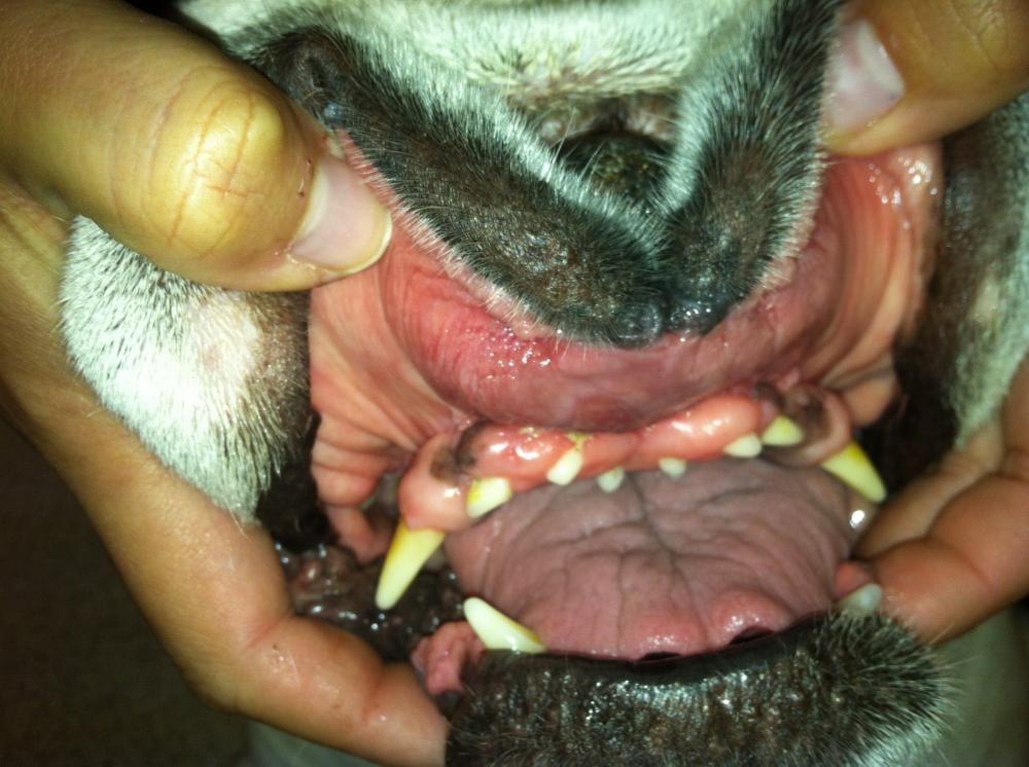
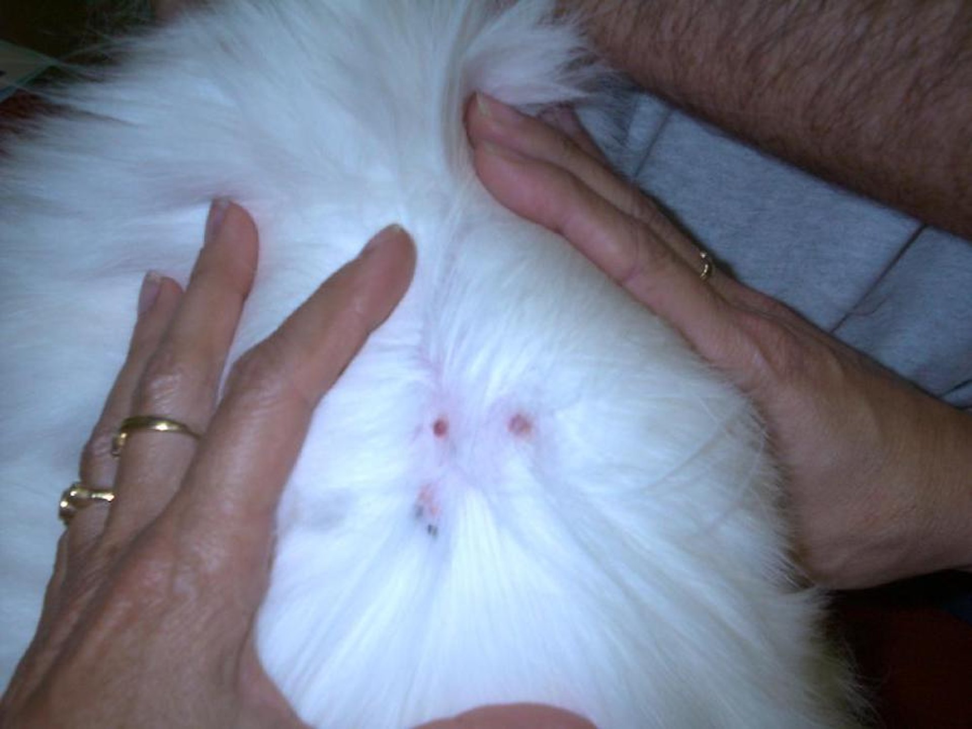
Cutaneous histiocytomas are common in dogs and rare in goats and cattle; it is debatable whether they are found in cats. Strong immunohistochemical evidence suggests that in dogs they are derived from Langerhans (intraepidermal antigen processing) cells. These tumors are typically seen in dogs < 3.5 years old but can be seen at any age. English Bulldogs, Scottish Terriers, Greyhounds, Boxers, and Boston Terriers are most at risk. The head (including the pinnae) and limbs are the most common sites of involvement. These classic "button tumors" appear as solitary, smooth, pink, raised nodules that are generally covered by alopecic skin, or they may be ulcerated. They are freely movable. Although a common neoplasm, histiocytomas are not always easy to diagnose histologically and can be confused with granulomatous inflammation, mast cell tumors, plasmacytomas, and cutaneous lymphosarcomas. Canine histiocytomas should be considered benign, and most resolve spontaneously within 2–3 months without treatment. Surgical excision is optional once the diagnosis is established (which can often be made via cytologic evaluation).
In goats and cattle, histiocytomas are extremely rare and behave the same as those in dogs. Histiocytomas have also been reported in young cats; however, they most likely represent the histiocytic form of mast cell tumor in cats.
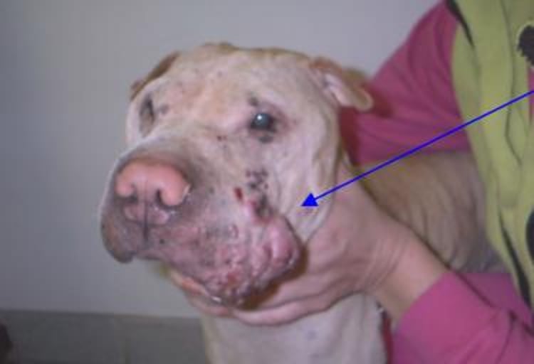
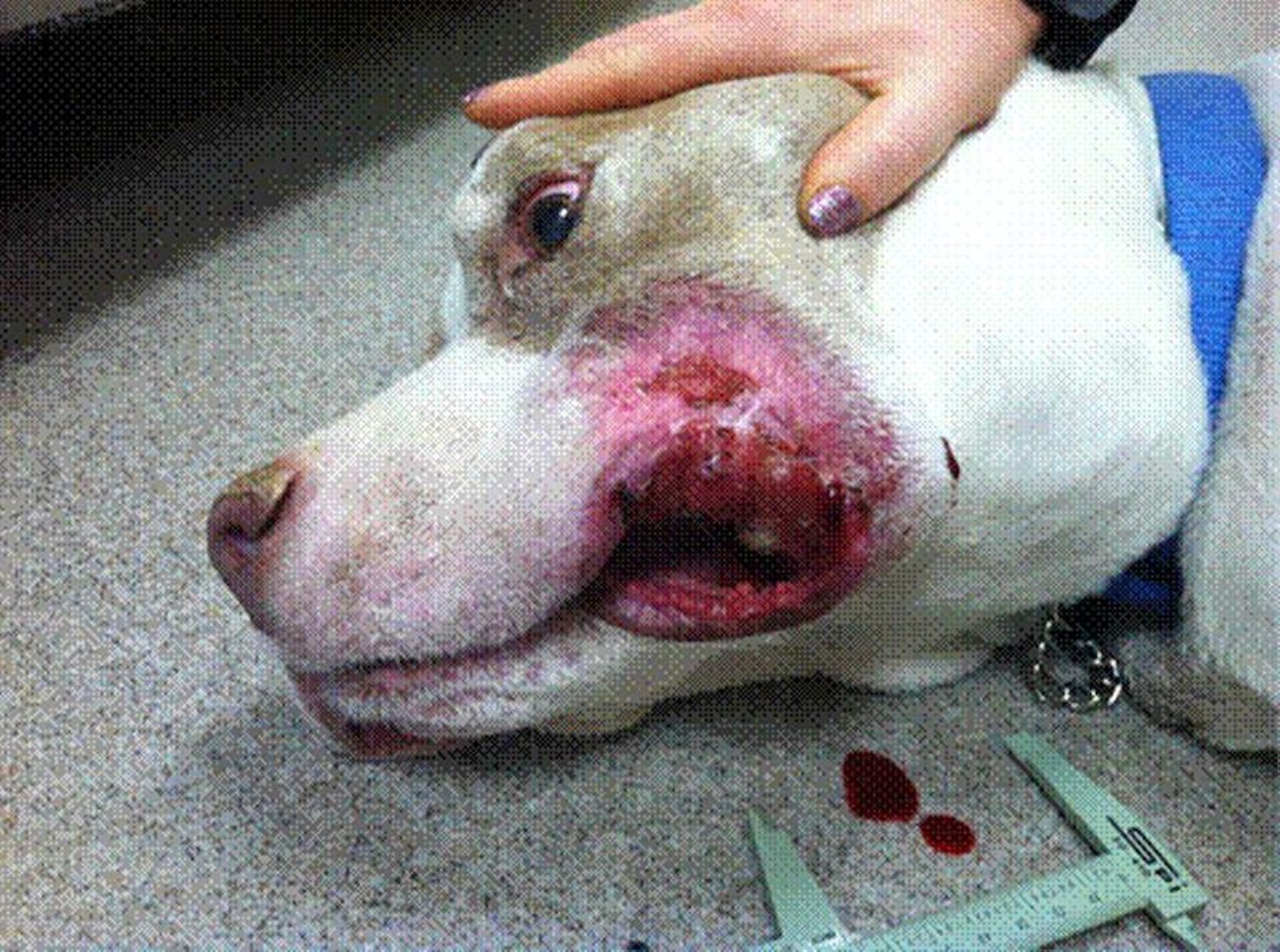
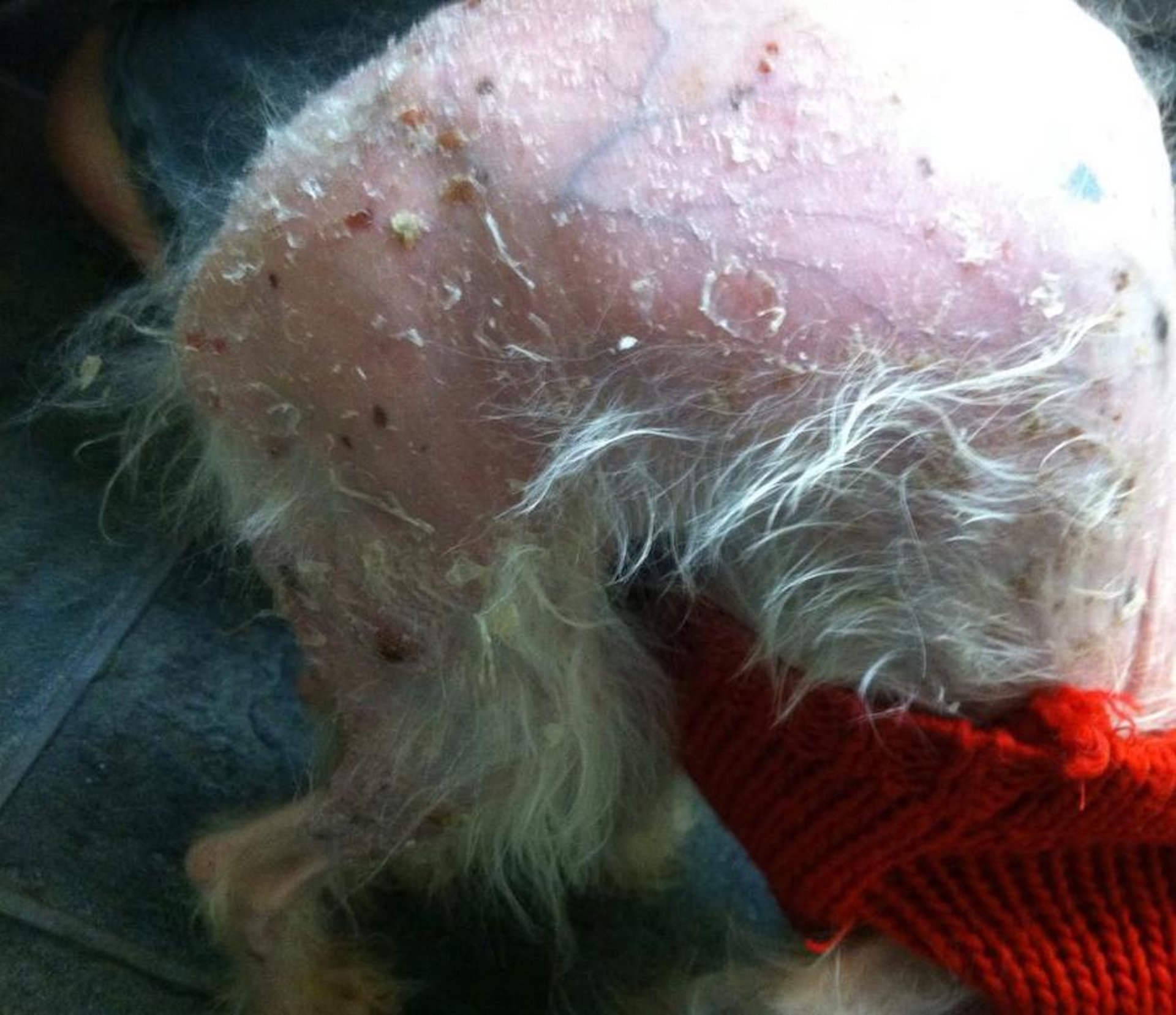
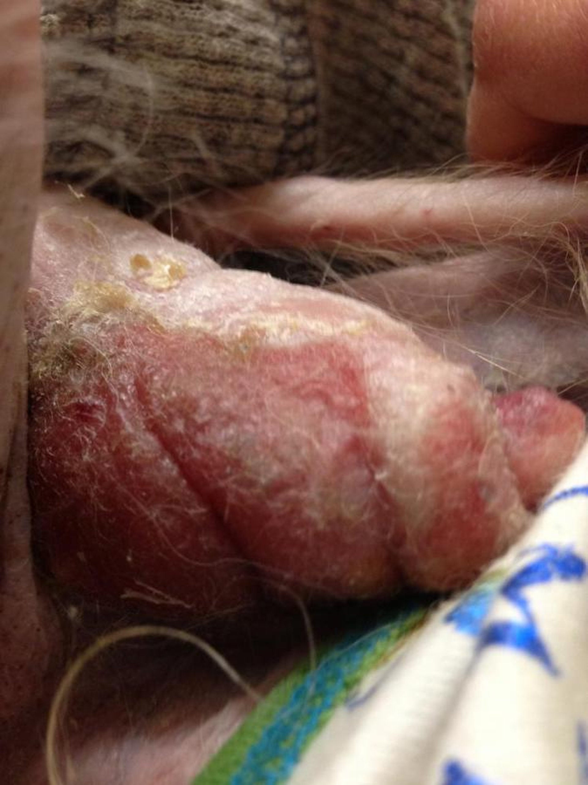
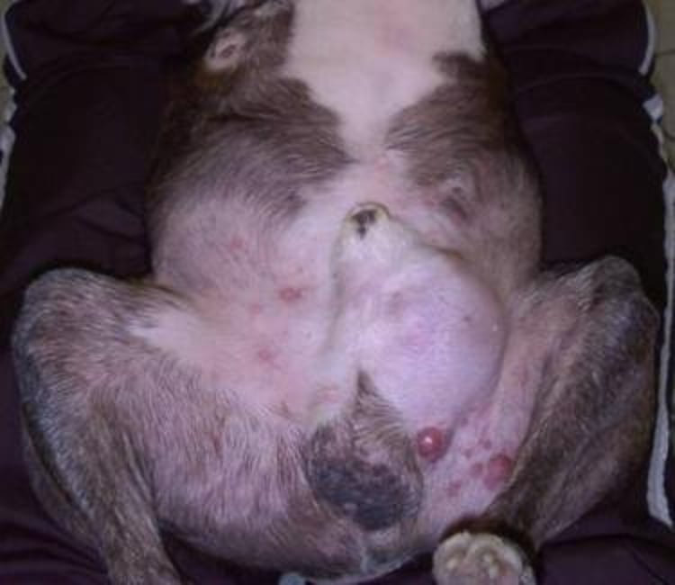
Cutaneous histiocytosis is associated with development of numerous plaques and nodules involving the dermis or subcutaneous fat. It is rare in dogs and can develop at any age but is most common in young adults. Chinese Shar-Pei, Collies, Border Collies, Shetland Sheepdogs, Briards, Bernese Mountain Dogs, Golden Retrievers, and German Shepherd Dogs may be predisposed. Chinese Shar-Pei have excessive skin folding and severe cutaneous mucinosis due to an accumulation of dermal hyaluronan, a protein associated with angiogenesis and tumor cell motility. The nodules and plaques tend to wax and wane, and the extremities and trunk are involved most commonly. Lesions also occur on the face and nasal planum, and swelling of the nares may cause difficulty breathing. Lesions are nonpruritic, and larger lesions may ulcerate. Cutaneous histiocytosis seldom involves internal organs.
Various forms of treatment have been attempted, including administration of systemic glucocorticoids and a combination of glucocorticoids and chemotherapy and more recently in combination with masitinib and other tyrosine kinase inhibitors. Leflunomide without steroids has been administered, as well as azathioprine and cyclosporine. Response is variable; the lesions in some dogs respond rapidly and permanently, whereas in others, lesions are either transiently improved or unchanged.
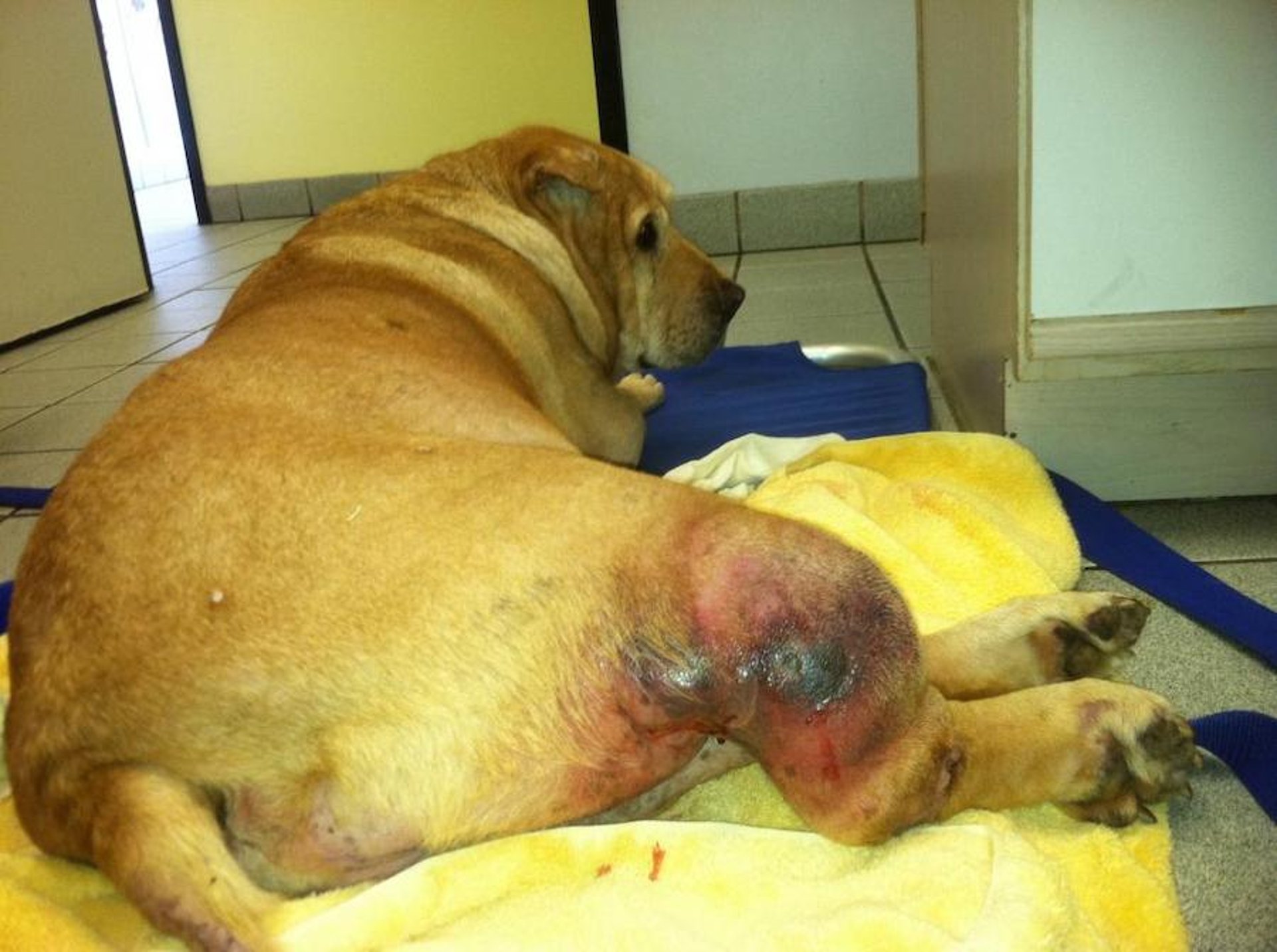
The histiocytoses of Bernese Mountain Dogs are systemic, familial disorders of unknown etiology with two manifestations: a more indolent and generally cutaneous form known as systemic histiocytosis and a more aggressive form in which skin lesions are rare, known as malignant histiocytosis. Malignant histiocytosis has been infrequently identified in other canine breeds. In systemic histiocytosis, males (mean age at onset 4 years) are affected more often than females. There are multiple cutaneous nodules, papules, and plaques involving the skin (especially of the scrotum), nasal mucosa, and eyelids. The lesions are poorly circumscribed and variably alopecic and may be ulcerated; they develop in waves and slowly regress, only to recur several months later. Clinical signs tend to become more severe with each new wave of eruptions. Although the skin is the primary target organ, lesions may also develop in other organs, including lymph nodes, spleen, and bone marrow. The disease may be episodic in its clinical presentation, but it is progressive and eventually fatal. A commercially available histiocytic malignancy assay can evaluate tumor biopsy or cytology samples for copy number aberrations to confirm diagnosis.
Courtesy of Dr. Alice Villalobos.
Malignant histiocytosis occurs in male Bernese Mountain Dogs (mean age at onset 7 years) and, less frequently, in other canine breeds. The lungs, lymph nodes, and liver are the most common organs affected, and the disease tends to spare the skin. Grossly, the lesions appear as large, solitary, firm masses that may cause pleural effusion or efface large portions of affected internal organs. The disorder is rapidly progressive and does not wax and wane as does systemic histiocytosis. Few dogs survive >6 months.
Various chemotherapeutic regimens mentioned above have been administered to treat both forms; hopefully, trametinib, administered in combination with CCNU (lomustine), vincristine, or other combinations of antineoplastic drugs, will be of benefit. Bovine thymosin fraction 5 may help induce remissions, especially in the systemic form. However, despite efforts to extend survival times, both forms of the disease are ultimately fatal.
Transmissible Venereal Tumors
Also see Canine Transmissible Venereal Tumor. Transmissible venereal tumors can also develop initially on haired skin due to inoculation via cutaneous injuries.


