


Exposures to illicit or abused drugs in pet animals can be accidental, intentional, or malicious. Occasionally, drug-sniffing dogs also ingest these substances. Because of the illegal nature of illicit or abused drugs, owners may provide inaccurate, incomplete, or misleading exposure histories. Illicit drugs are often adulterated with other pharmacologically active substances, making the diagnosis even more difficult.
In suspected cases of exposure to illicit or abused drugs, an attempt should be made to gather information about the animal’s environment; amount of exposure; and time of onset of clinical signs and their type, severity, and duration. These questions will help include or exclude possible exposure to an illicit or abused drug. Illicit and abused drugs are often known by street names that vary from area to area. A call to a local police station, or animal or human poison control center, may help identify the illicit substance if its street name is known. Most human hospitals, emergency clinics, and some veterinary diagnostic laboratories have illicit drug screens available and can check for the presence of illicit drugs or their metabolites in different body fluids. The presence of a parent drug or its metabolites in blood or urine may help confirm exposure in suspect cases. The laboratories should be contacted for information on the types of samples needed and time required to complete the screens or tests.
Commonly available over-the-counter drug test kits may help exclude a suspected case of illicit drug toxicosis. They are designed to detect drug metabolites in the urine and can detect most commonly available illicit or recreational drugs such as amphetamines, cocaine, marijuana, opiates, and barbiturates. The sensitivities and specificities of these test kits may vary. The kits are inexpensive, efficient, and easy to use, but the instructions provided with each kit should be followed carefully for best results.
Amphetamines and Related Drugs
Amphetamines and their derivatives are CNS and cardiovascular system stimulants commonly used in people for suppression of appetite, narcolepsy, attention deficit disorder, parkinsonism, and some behavior disorders. Some commonly encountered amphetamines or related drugs are benzphetamine, dextroamphetamine, lisdexamfetamine, pemoline, methylphenidate, phentermine, diethylpropion, phendimetrazine, methamphetamine, and phenmetrazine. Amphetamines sold on the street have common names such as speed, bennies, or uppers. Commonly used adulterants are caffeine, ephedrine, or phenylpropanolamine.
Pharmacokinetics and Toxicity:
Amphetamines are rapidly absorbed in the GI tract, reaching peak plasma concentrations in 1–2 hr. Sustained-release formulations have a delayed absorption and relatively longer half-life. The plasma half-life of amphetamines depends on the urinary pH. With an alkaline pH, the half-life is 15–30 hr; with an acidic pH, the half-life is 8–10 hr. The acute oral LD50 of amphetamine in rats and mice is 10–30 mg/kg. In people, deaths have been reported after ingestion of methamphetamine at 1.3 mg/kg.
Pathogenesis:
Amphetamine stimulates the release of norepinephrine, affecting both α- and β-adrenergic receptor sites. Amphetamine also stimulates catecholamine release centrally in the cerebral cortex, medullary respiratory center, and reticular activating system. It increases the amount of catecholamine at nerve endings by increasing release and inhibiting reuptake and metabolism. The neurotransmitters affected in the CNS are norepinephrine, dopamine, and serotonin.
Clinical Findings and Diagnosis:
Clinical signs of amphetamine and cocaine toxicosis are similar and difficult to differentiate clinically. The only difference may be the longer duration of clinical signs of amphetamine toxicosis because the half-life of amphetamine is longer than that of cocaine. The most commonly reported signs are hyperactivity, aggression, hyperthermia, tremors, ataxia, tachycardia, hypertension, mydriasis, circling, head bobbing, and death.
Diagnosis is as for cocaine (see below) and relies mostly on owner knowledge of exposure. Most amphetamines and related drugs or their metabolites are detectable in the stomach contents and urine. They are difficult to detect in plasma unless large amounts have been ingested.
Treatment:
Phenothiazines are preferred to control CNS signs in amphetamine toxicosis (see below for cocaine toxicosis). Other anticonvulsants, such as diazepam, barbiturates, or isoflurane, may be used if needed. Acidifying the urine with ammonium chloride (25–50 mg/kg/day, PO, qid) or ascorbic acid (20–30 mg/kg, PO, SC, IM, or IV) may enhance amphetamine elimination in the urine. However, this should be done only if acid-base status can be monitored. Cyproheptadine (1.1 mg/kg, PO or per rectum) may also be given once or twice (6–8 hr apart) for serotonin syndrome (disorientation, muscle stiffness, agitation). Heart rate and rhythm ( see Treatment: for using β blockers to treat tachycardia), body temperature, and electrolytes should be monitored and treated as needed.
Cocaine
Cocaine (benzoylmethylecgonine) alkaloid is obtained from the leaves of the coca plant, Erythroxylon coca and E monogymnum. Some common street names for cocaine are coke, gold dust, stardust, snow, C, white girl, white lady, baseball, and speedball (cocaine and heroin). Cocaine alkaloid from coca leaves is processed into cocaine hydrochloride salt, then reprocessed to form cocaine alkaloid or free base (a process called free-basing or base balling), which is colorless, odorless, transparent, and more heat stable. Free-base cocaine is also called crack, rock, or flake. Cocaine is cut (diluted) several times before it reaches the user. Xanthine alkaloids, local anesthetics, and decongestants are some of the most common adulterants.
Cocaine is a schedule II drug approved for human use. Its medical uses are restricted to topical administration as a local anesthetic on mucous membranes of the oral, laryngeal, and nasal cavities. However, it is mostly used as a recreational drug.
Pharmacokinetics and Toxicity:
Cocaine is absorbed from most routes. Orally, it is better absorbed in an alkaline medium (ie, intestine). In people, ~20% of an oral dose is absorbed. The reported plasma half-life is 0.9–2.8 hr. Cocaine is extensively metabolized by liver and plasma cholinesterases to several inactive metabolites that are primarily excreted in the urine. The acute LD50 of cocaine hydrochloride administered IV in dogs is 13 mg/kg; the LD100 is 12 mg/kg in dogs and 15 mg/kg in cats. The oral LD50 in dogs is believed to be 2–4 times more than the IV dose.
Pathogenesis:
Cocaine acts on the sympathetic division of the autonomic nervous system. It blocks the reuptake of dopamine and norepinephrine in the CNS, leading to feelings of euphoria, restlessness, and increased motor activity. Cocaine can also decrease concentrations of serotonin or its metabolites. Topical use of cocaine causes vasoconstriction of small vessels. Hyperthermia in cocaine toxicosis may develop either due to increased heat production from muscular activity or due to decreased heat loss from vasoconstriction.
Clinical Findings and Diagnosis:
CNS excitation, hyperactivity, shaking, ataxia, panting, agitation, mydriasis, nervousness, seizures, tachycardia, hypertension, acidosis, or hyperthermia characterize cocaine toxicosis. CNS depression and coma may follow CNS excitation. Death may be due to hyperthermia, cardiac arrest, or respiratory arrest. Some nonspecific chemistry changes may include hyperglycemia and increased CK and liver-specific enzymes.
Diagnosis is based on a history of exposure and the presence of characteristic clinical signs. Identification of cocaine in plasma, stomach contents, or urine can confirm exposure. Differential diagnoses include amphetamines, pseudoephedrine, ephedrine, caffeine, chocolate, metaldehyde, strychnine, tremorgenic mycotoxins, lead, nicotine, permethrin (cats) and other pesticides, and encephalitis.
Treatment:
The objectives of treatment are GI decontamination, stabilization of CNS and cardiovascular effects, thermoregulation, and supportive care. Animals with clinical signs should be stabilized first before attempting decontamination. Emesis can be induced in a recent exposure if the animal is asymptomatic and has the ability to guard its airway via a gag reflex. This should be followed by administration of activated charcoal with a cathartic. If the animal’s condition contraindicates induction of emesis (eg, presence of CNS signs or extreme tachycardia), a gastric lavage with a cuffed endotracheal tube to reduce the risk of aspiration should be performed. A dose of activated charcoal with a cathartic should be left in the stomach after the lavage.
Controlling the CNS signs may require use of more than one anticonvulsant. Clinical signs of CNS excitation can be controlled with diazepam; however, the effects of diazepam are short-lived, and repeated administration may be needed. Phenothiazine tranquilizers such as acepromazine (0.05–1 mg/kg, IV, IM, or SC, repeated as needed) or chlorpromazine (0.5–1 mg/kg, IV or IM) also usually work well to control the CNS effects. However, phenothiazines should be used cautiously, because they may lower the seizure threshold. If phenothiazines are ineffective, phenobarbital at 3–4 mg/kg, IV, or pentobarbital, IV to effect, could be used. If CNS signs are uncontrolled by the preceding measures, a gas anesthetic such as isoflurane may be useful.
Blood pressure, heart rate and rhythm, ECG, and body temperature should be monitored frequently and treated as needed. Propranolol at 0.02–0.06 mg/kg, IV, tid-qid, or other β-blocking agents such as esmolol (0.2–0.5 mg/kg, slow IV over 1 min, or 25–200 mcg/kg/min as a constant-rate infusion) can be used to control tachycardia. After CNS and cardiovascular effects have been stabilized, IV fluids should be administered, and electrolyte changes and acid-base status monitored and corrected as needed. Treatment and monitoring should continue until all clinical signs have resolved.
Ecstasy (MDMA or 3,4-Methylenedioxymethamphetamine)
Ecstasy is a semisynthetic psychoactive designer drug (developed by street chemists with minor structural changes in parent compounds) with hallucinogenic and amphetamine-like properties. Street names include Adam, XTC, E, Roll, X, or Love Drug. A typical dose may be 75–150 mg. MDMA is believed to cause excessive release of serotonin. It also binds to serotonin transporter (a protein), which is responsible for removing serotonin from the synapse. The overall effect is increased serotonin and serotonergic effects. MDMA also affects dopamine and norepinephrine. Studies in rodents indicate that use of MDMA can lead to permanent serotonergic neuronal injury.
In pets, MDMA toxicosis is not common. It is usually acute and occurs from accidental ingestion (powder, pills, or capsules). Clinical signs develop within 30 min to 2 hr after ingestion and may consist of sympathomimetic effects (CNS excitation, agitation, hyperactivity, pacing, hyperthermia, tachycardia, hypertension, seizures [just like amphetamines]), sedation, or signs thought to be related to hallucination (vocalization, disorientation, muscle rigidity). The half-life in people is 7.6–8.7 hr. Treatment is similar to that for amphetamine toxicosis ( see Treatment:). For serotonergic effects (agitation, muscle rigidity, nervousness), antiserotonergic drugs such as cyproheptadine can be tried (1.1 mg/kg, PO, repeated once in 6–8 hr).
Marijuana

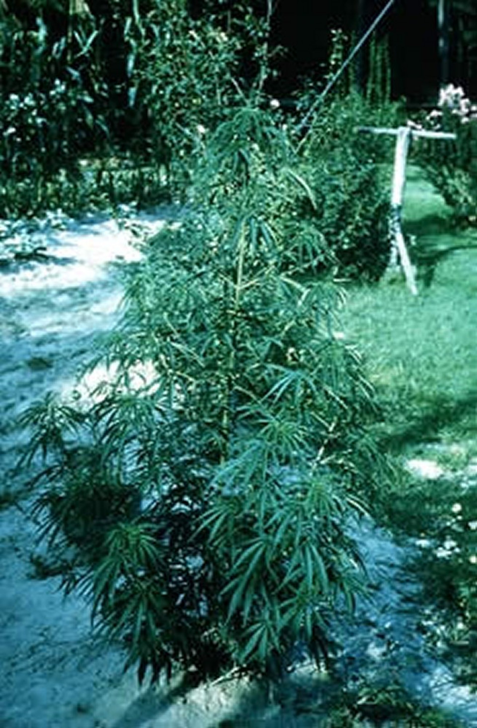
Courtesy of Dr. Cecil Brownie.
Marijuana refers to a mixture of cut, dried, and ground flowers, leaves, and stems of the leafy green hemp plant Cannabis sativa. Several cannabinoids are present in the plant resin, but delta-9-tetrahydrocannabinol (THC) is considered the most active and main psychoactive agent. The concentration of THC in marijuana varies between 1%–8%. Hashish is the resin extracted from the top of the flowering plant and is higher in THC concentration than marijuana. Street names for marijuana include pot, Mary Jane, hashish, weed, grass, THC, ganja, bhang, and charas. Pure THC is available by prescription under the generic name dronabinol. A synthetic cannabinoid, nabilone, is also available. Marijuana or hashish sold on the streets may be contaminated with phencyclidine, LSD, or other drugs.
Marijuana is a schedule I controlled substance mostly used by people as a recreational drug. It is also used as an antiemetic for chemotherapy patients and to decrease intraocular pressure in glaucoma patients. Some clinicians advocate the use of dronabinol as an appetite stimulant, but the dysphoric effects of this drug outweigh any benefit of appetite stimulation.
Synthetic marijuana is a designer drug in which different herbs or incense or other leafy materials are sprayed with laboratory-synthesized chemicals. The use of synthetic marijuana produces psychoactive effects similar to those of THC. It is often claimed to be natural, safe, and legal. Spice, K-2 , skunk, and moon rocks are some of the common street names. Clinical signs of toxicosis from ingestion of synthetic marijuana in dogs can be more severe and last longer than those of THC.
Pharmacokinetics and Toxicity:
The most common route of exposure is oral. After ingestion, THC goes through a substantial first-pass effect. It is metabolized by liver microsomal hydroxylation and nonmicrosomal oxidation. In dogs, clinical signs begin within 30–90 min and can last up to 72 hr. THC is highly lipophilic and readily distributes to the brain and other fatty tissues after absorption. The oral LD50 of pure THC is 666 mg/kg in rats and 482 mg/kg in mice. However, clinical effects of marijuana are seen at much lower doses than this.
Pathogenesis:
THC is believed to act on a unique receptor in the brain that is selective for cannabinoids and is responsible for the CNS effects. Cannabinoids can enhance the formation of norepinephrine, dopamine, and serotonin. They can also stimulate release of dopamine and enhance γ-aminobutyric acid turnover.
Clinical Findings and Diagnosis:
The most common signs of marijuana toxicosis are depression, ataxia, bradycardia, hypothermia, vocalization, hypersalivation, vomiting, diarrhea, urinary incontinence, seizures, and coma.
Diagnosis is based on a history of exposure and typical clinical signs. THC is difficult to detect in body fluids because of the low levels found in the plasma. Urine testing at human hospitals or using an over-the-counter marijuana drug test kit in the early course of exposure may help confirm the diagnosis. Marijuana toxicosis can be confused with ethylene glycol (antifreeze, see Ethylene Glycol Toxicosis) or ivermectin toxicosis; hypoglycemia; benzodiazepine, barbiturate, or opioid overdose; intervertebral disc problems; or head trauma.
Treatment:
Treatment consists of supportive care. If the exposure is recent and there are no contraindications, emesis should be induced and activated charcoal administered. Comatose animals should be monitored for aspiration pneumonia, given IV fluids, treated for hypothermia, and rotated frequently to prevent dependent edema or decubital ulceration. Diazepam can be given for sedation or to control seizures. Treatment and monitoring should continue until all clinical signs have resolved (up to 72 hr in dogs). For cases of synthetic marijuana toxicosis, in addition to the preceding treatment options, use of IV lipid emulsion solution may be considered.
Opiates
The term opiate initially referred to all naturally occurring alkaloids obtained from the sap of the opium poppy (Papaver somniferum). Opium sap contains morphine, codeine, and several other alkaloids. Currently, opioid refers to all drugs, natural or synthetic, that have morphine-like actions or actions mediated through opioid receptors. Structurally, opioids can be divided into five classes. Some of the common agents within each class are 1) phenanthrenes—morphine, heroin, hydromorphone, oxymorphone, hydrocodone, codeine, and oxycodone; 2) morphinan—butorphanol; 3) diphenylheptanes—methadone and propoxyphene; 4) phenylpiperidines—meperidine, diphenoxylate, fentanyl, loperamide, and profadol; and 5) benzomorphans—pentazocine and buprenorphine. Some of the widely used opioids in veterinary medicine include tramadol, buprenorphine, fentanyl, loperamide (antidiarrheal), and hydromorphone. The use of meperidine is no longer common.
Opioids are used primarily for analgesia. In addition, they are used as cough suppressants and to treat diarrhea. Occasionally, opioids are used for sedation before surgery and as a supplement to anesthesia.
Pharmacokinetics and Toxicity:
Opioids are generally well absorbed after oral, rectal, or parenteral administration. Some more lipophilic opioids are also absorbed through nasal, buccal, respiratory (heroin, fentanyl, buprenorphine), or transdermal (fentanyl) routes. For some opioids, there is variable reduction in bioavailability because of a first-pass effect when given orally. Opioids generally undergo hepatic metabolism with some form of conjugation, hydrolysis, oxidation, dealkylation, or glucuronidation. Because cats are deficient in glucuronidase, the half-life of some opioids in cats may be prolonged. After absorption, opioids are rapidly cleared from blood and stored in kidney, liver, brain, lung, spleen, skeletal muscle, and placental tissue. Most of the opioid metabolites are excreted through the kidneys.
Toxicity of opioids in animals is highly variable. In dogs, morphine administered at 100–200 mg/kg, SC or IV, is considered lethal. The estimated lethal dose of codeine in human adults is 7–14 mg/kg; in infants, 2.5 mg of hydrocodone has been lethal.
Pathogenesis:
The effects of opioids are due to their interaction with opiate receptors (μ, κ, δ, σ, and ε) found in the limbic system, spinal cord, thalamus, hypothalamus, striatum, and midbrain. Opioids may be agonists, mixed agonist-antagonists, or antagonists at these receptors. Agonists bind and activate a receptor, whereas antagonists bind without causing activation.
Clinical Findings:
The primary effects of opioids are on the CNS, respiratory, cardiovascular, and GI systems. Commonly reported clinical signs of toxicosis include CNS depression, drowsiness, ataxia, vomiting, seizures, miosis, coma, respiratory depression, hypotension, constipation/defecation, and death. Some animals—especially cats, horses, cattle, and swine—can show CNS excitation instead of CNS depression.
Diagnosis:
Diagnosis of opioid toxicosis is based on history of exposure and the types of clinical signs (CNS and respiratory depression) present. Plasma opioid levels are usually not clinically useful. Urine may be used to determine exposure to opioids using some of the over-the-counter illicit drug kits (manufacturer’s instructions should be followed). Opioid toxicosis should be differentiated from ethylene glycol, ivermectin, benzodiazepine, barbiturate, and marijuana toxicosis, as well as hypoglycemia-inducing conditions.
Treatment:
Clinical signs can be reversed with the opiate antagonist naloxone. The dosages in different species are dog and cat, 0.04–0.16 mg/kg, IV, IM, or SC; rabbit and rodent, 0.01–0.1 mg/kg, SC or IP; horse, 0.01–0.02 mg/kg, IV. Administration of naloxone should be repeated as needed (hourly), because its duration of action may be shorter than that of the opioid toxicity being treated. Animals should be closely monitored for respiratory depression and ventilatory support provided if needed. Other signs should be treated symptomatically. Dysphoric reactions (vocalization, agitation, restlessness, and excitation) can be treated with diazepam or other benzodiazepines. For serotonin-like syndrome (disorientation, muscle rigidity, agitation) induced by some opioids, cyproheptadine (1.1 mg/kg, PO or per rectum) once or twice (6–8 hr apart) can be tried.


