


Courtesy of Dr. Raffaele Roncalli.
The tropical warble fly, or torsalo, one of the most important parasites of cattle in Latin America, is distributed between southern Mexico and northern Argentina. Larval stages are found in many hosts, including cattle, sheep, goats, pigs, buffalo, dogs, cats, rabbits, and humans. Cattle and dogs are infected most commonly. D hominis is thought to initiate the lesion that gives rise to lechiguana, a disease of cattle.
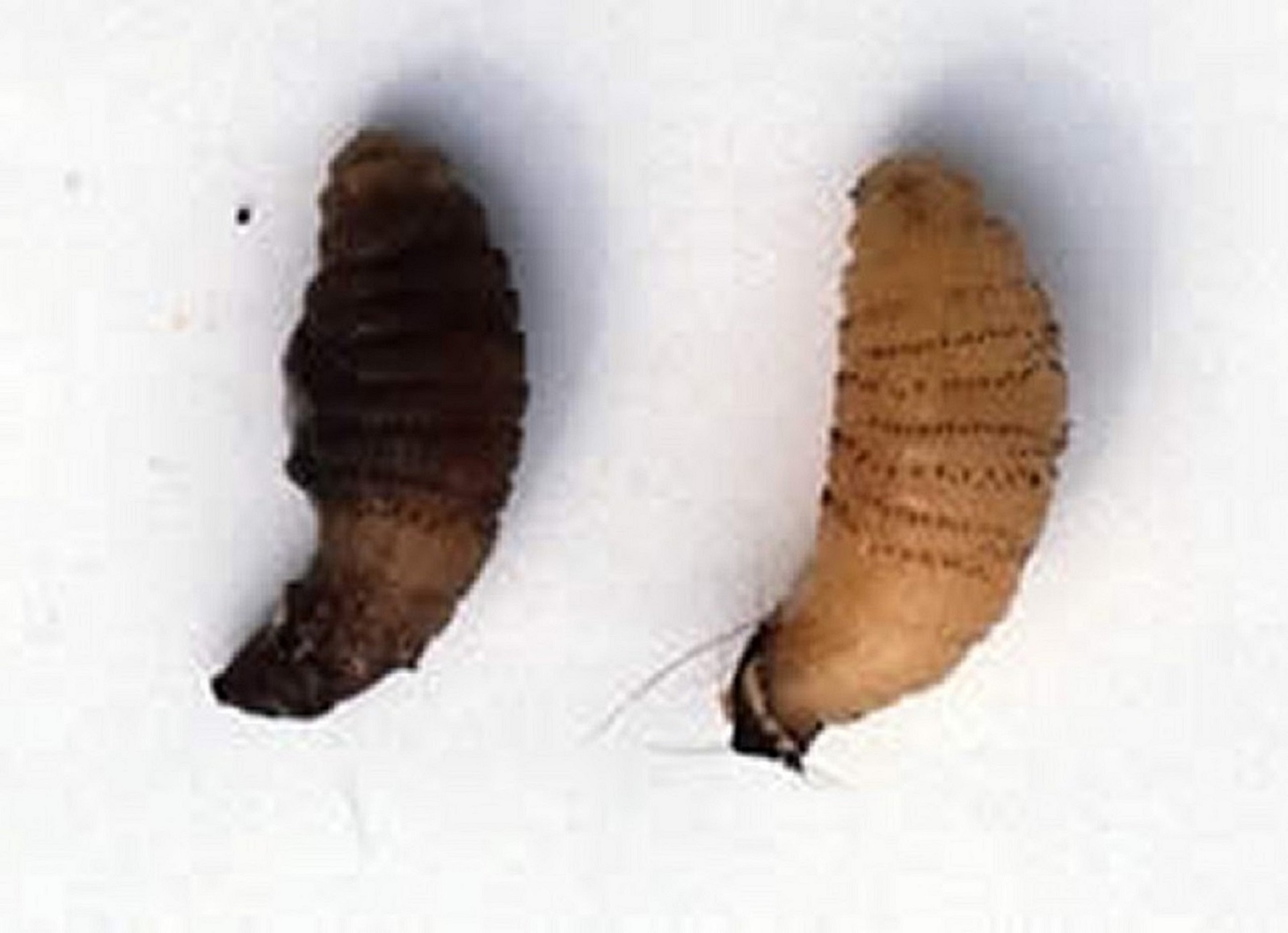
Life Cycle of Dermatobia hominis

Courtesy of Dr. Raffaele Roncalli.
The adult D hominis fly is 12–15 mm long and has a short lifespan (1–9 days). The adult fly fastens its eggs to different types of insects (49 have been described as vectors of D hominis in Latin America; most are mosquitoes or muscoid flies), which then transport the eggs to warm-blooded hosts, where they hatch as the insects feed. The larvae penetrate the skin of the animal within a few minutes of hatching and remain in the subcutaneous tissue for 4–18 weeks. During this period, the larvae grow within warbles that have breathing holes. When mature, the larvae leave the host and drop to the ground, burrow, and pupate. After the pupal period, which lasts 4–11 weeks, the flies emerge as adults. The complete life cycle takes 11–17 weeks.
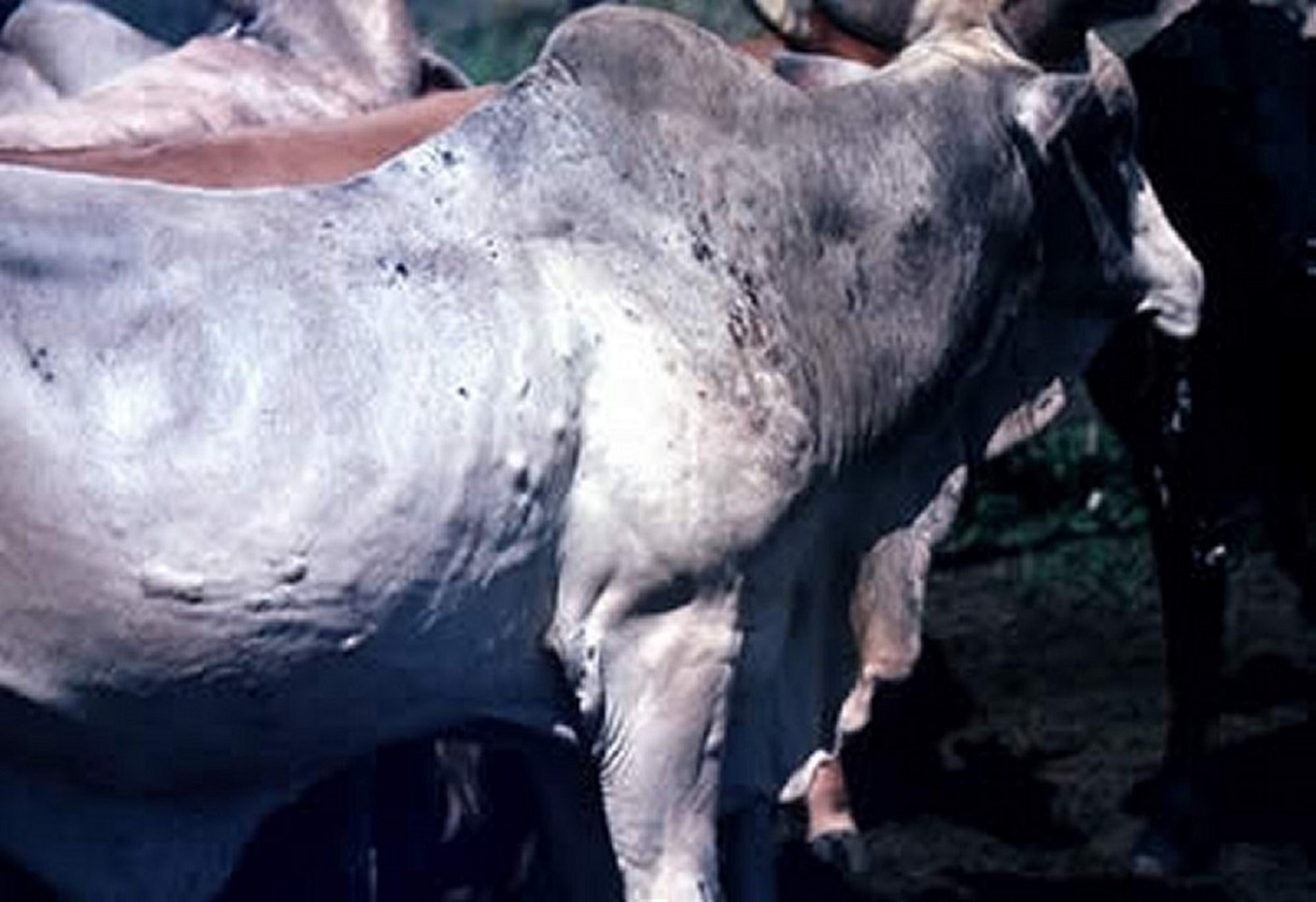
Clinical Findings and Pathogenesis of Dermatobia hominis Infestation

Courtesy of Dr. Raffaele Roncalli.
Larval penetration of the skin is accompanied by pain and local inflammation, and pus gradually forms. Infested hides are condemned at slaughter, and production of milk and meat is decreased. In 1982, it was estimated that cattle infested with D hominis resulted in a yearly decrease in weight of 40.6 g/larva. In 2014, combining losses due to decreased weight gain and hide damage, infestation by D hominis in cattle raised in Brazil was estimated to cost producers US$383 million annually. Efforts to characterize and produce antigenic proteins that confer immunity against D hominis have led to identification of a candidate vaccine that has 90% efficacy in immunized cattle.
Treatment and Control of Dermatobia hominis Infestation
Different topical and systemic insecticides in various formulations are available to treat D hominis infestations in cattle. Generally, D hominis is susceptible to systemic organophosphates and macrocyclic lactone endectocides, which may be approved and available locally.
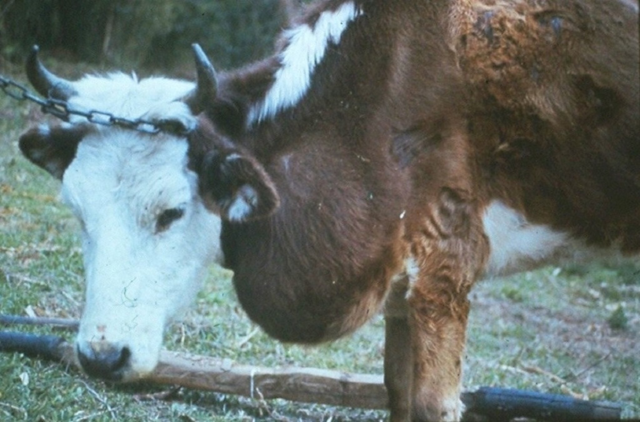
Lechiguana

Courtesy of F. Riet-Correa.
Lechiguana (bovine focal fibrogranulomatous proliferative panniculitis) is a sporadic, chronic disease of cattle that, thus far, has been reported only from southern and southeastern Brazil, in areas where infection by D hominis is common. It is characterized by large, firm, subcutaneous swellings that develop rapidly, mainly in the scapulae and adjacent areas (chest, neck, shoulders, and ribs). Most affected cattle have only one swelling; occasionally, however, two swellings occur. The regional lymph nodes are enlarged and, without treatment, may become enormous.
The bacterium Mannheimia granulomatis has been recovered from lechiguana lesions and is considered causative. The lesion that gives rise to lechiguana is initiated by D hominis larvae. M granulomatis is consistently recovered from lechiguana lesions, and it is thought to be largely responsible for the characteristic tissue changes. The involvement of a myofibroblast-like cell population that expresses mRNA for type I collagen is suggested to be associated with the increase of collagen deposition. In addition, macrophages activated by M granulomatis induce fibroblast proliferation. The habitat or source of M granulomatis is not known. It has not been recovered from cattle without lechiguana.
Histologically, lechiguana lesions consist of focal proliferations of fibrous tissue infiltrated by plasma cells, eosinophils, lymphocytes, and sometimes neutrophils. The primary lesion is an eosinophilic lymphangitis, which results in eosinophilic abscesses, with occasional rosettes containing bacteria in the center. The subcutaneous, tumorous mass produced may attain a size of up to 40 × 50 cm in 2 months. Without treatment, death occurs after 3–11 months, probably because of inanition resulting from the enormous swellings.
When well established, the disease is clinically obvious. Diagnosis is confirmed by recovery of M granulomatis and observation of the characteristic histopathologic lesions.
Administration of chloramphenicol (3 g, IM, every 24 hours for 5 days) or danofloxacin mesylate (1.25 mg/kg, IM, every 24 hours for 3 days) results in rapid decrease of swellings, with almost complete regression in 30 days. Susceptibility testing should be conducted before other antimicrobials are administered.
Key Points
Detection of Hypoderma-specific antibodies at the beginning of migration by the larvae enables early systemic treatment to avoid tissue damage.
Late treatments against Hypoderma in severely affected cattle can provoke adverse effects resulting from the massive liberation of the enzymatic content of first instars.
Macrocyclic lactones are highly effective and their use in coordinated treatment campaigns has almost eradicated bovine hypodermiasis in several countries.
For More Information
Boulard C. Durably controlling bovine Hypodermosis. Vet Res. 2002;33:455-464. doi:10.1051/vetres:2002032
Hassan M, Khan MN, Abubakar M, Waheed HM, Iqbal Z, Hussain M. Bovine hypodermosis—a global aspect. Trop Anim Health Prod. 2010;42(8):1615-1625.


