

Probably there are more clinical cases of coccidiosis diagnosed in cattle than in any other mammalian species. The parasite is one of the most important causes of diarrhea in calves and young growing stock, often decreasing production. Death is a potential outcome. In consequence, coccidiosis is more studied in cattle than in all other production animals except poultry.
Etiology and Pathogenesis of Coccidiosis of Cattle
More than 20 Eimeria spp have been identified in the feces of cattle worldwide. The species include Eimeria alabamensis, E auburnensis, E bovis, E brasiliensis, E bukidnonensis, E canadensis, E cylindrica, E ellipsoidalis, E pellita, E subspherica, E wyomingensis, and E zuernii. However, only four (E alabamensis, E auburnensis, E bovis, and E zuernii) are usually associated with clinical disease. Other Eimeria spp have been shown experimentally to be mildly or moderately pathogenic but are not considered important pathogens. Mixed infections are common and may increase pathogenicity. All the bovine Eimeria spp are host-specific.
Typically, the life cycle of bovine coccidia involves two asexual reproducing generations and then a sexual reproducing generation that releases resistant oocysts into the feces. Under suitable conditions of oxygen, moisture, and temperature, infective sporulated oocysts are produced in days. With E alabamensis, E auburnensis, E bovis, and E zuernii, the first asexual generation is in the distal half of the small intestine, usually with the second asexual generation and sporogony generation mainly confined to the cecum and colon.
Each of the Eimeria spp usually localizes itself in specific parts of the intestinal tract, and, within that location, each species may invade different cells. Pathogenic Eimeria spp such as E bovis and E zuernii can damage the distal small intestine, cecum, and colon. The less pathogenic E ellipsoidalis infects the small intestine.
An infection of 1,000 oocysts can lead to the destruction of 24 billion host intestinal cells. Some experimental studies have shown that the weight reduction in young calves was maintained throughout at least their first grazing season. Coccidiosis is a self-limiting disease, and spontaneous recovery without specific treatment is common once the multiplication stage of the coccidia has passed.
Epidemiology of Coccidiosis of Cattle
Clinical infection occurs at about 1–2 months of age but can occur up to 1 year of age. Because coccidiosis does not typically occur in the first 3 weeks of life, it is not considered part of the neonatal diarrhea complex in calves. Rarely, cases have been reported in adults, usually singly, probably because of some other underlying problem. However, calving cows contribute to the infection level by increased oocyst shedding. Cases in Britain increased after the mid-1990s, possibly because of removal of the previous routine inclusion of coccidiostats in calf feeds.
Bovine coccidiosis occurs indoors and, slightly less often, outside. Infection is from contaminated environments, particularly when conditions are moist and warm. Cattle confined to feedlots are susceptible to coccidiosis throughout the year. In many countries, it occurs year round, but there is some seasonality, with less disease in the winter.
In the northwestern and midwestern US, cattle infection is higher in the summer, fall, and spring, and lower in midwinter and early summer. In Canada, winter coccidiosis occurs after a prolonged cold spell or after a sudden change to severe weather. In Queensland, Australia, weaned cattle tend to be more affected in dry years, possibly because of stress. In the UK, most cattle infections occur in the summer, with the next most in the spring, fall, and winter. In the UK, the commonest month for disease is June; the least common is January (US) and February (UK). In Scandinavia, E alabamensis occurs after calf turnout. In North America, summer coccidiosis and winter coccidiosis occur in range cattle, probably resulting from severe weather stress and crowding around a limited water or feed source, which concentrates the hosts and parasites within a restricted area. A CNS form of coccidiosis occurs in Canada and northern US, during or just after severe cold weather in midwinter, but it has not been reported elsewhere.
In many outbreaks of infection, intercurrent diseases or nutritional problems are present. Calves with concurrent enteric infections (eg, giardiasis) may be more severely affected than calves with coccidial infections alone. Environmental and management factors, such as weather, housing, feeding practices, and how animals are grouped, are important in determining the expression of clinical coccidiosis in cattle. Often, there is an underlying stress involvement. During or after infection, cattle may become more susceptible to other diseases, especially respiratory disease.
Coccidial immunity involves both cellular and humoral elements. The cellular component appears more important in preventing further clinical infections. After exposure to Eimeria spp, immunity is good, but during the animal’s life, a few parasites develop, thereby maintaining resistance. There can be a periparturient rise in oocyst numbers, allowing contamination of calving areas. Immunity is only mounted to the specific Eimeria spp to which the animal is exposed. Initial infection appears immunosuppressive and reduces neutrophil function, thereby making the host more susceptible to other infections or increasing their severity. Corticosteroid injections allow the parasite to complete its life cycle and oocysts to be produced.
Clinical Findings of Coccidiosis of Cattle
The prepatent period forE bovis and E zuernii is about 15–21 days, with a patent period of about 11 days (5–17 days). The prepatent period forE alabamensis is 8 days, with a patent period of 6 days.
Most infected calves show no clinical signs. The animals appear healthy but have some oocysts in the feces. Subclinical infection results in decreased growth that may not be regained. This is the most common coccidial effect, and there are no other clinical signs. In subacute infections, the main clinical sign is soft or loose feces, usually about 3 weeks (16–23 days) after infection for E bovis and E zuernii, but 3–4 days after infection for E alabamensis. The animals appear slightly dull with a poor coat. Growth rate is decreased.
More severe cases are associated with inappetance, lethargy, diarrhea (possibly containing blood), slight signs of abdominal discomfort, and tenesmus (with large intestine involvement). Growth rate is reduced, with poor feed conversion. Very severe cases may be associated with dehydration, often bloody diarrhea, anemia (rare), muscle weakness, recumbency, and death. In so-called winter coccidiosis, CNS signs occur. Chronic infections tend to be associated with poor growth, staring coat, and soiled hair in the perineal region. Such wasting and debility occurs most especially with E bovis. The decreased weight gain can remain for at least the first grazing season.

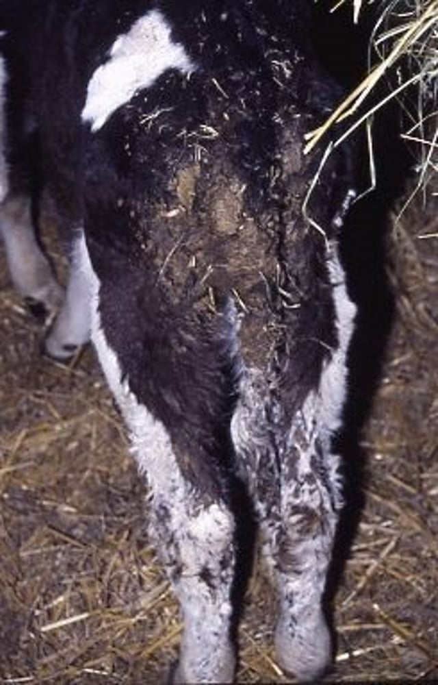
Photograph of a cow with chronic coccidiosis. Note the fecal matting on legs, hindquarters, and tail; poor body condition; and staring coat.
Courtesy of Dr. Anthony Andrews.

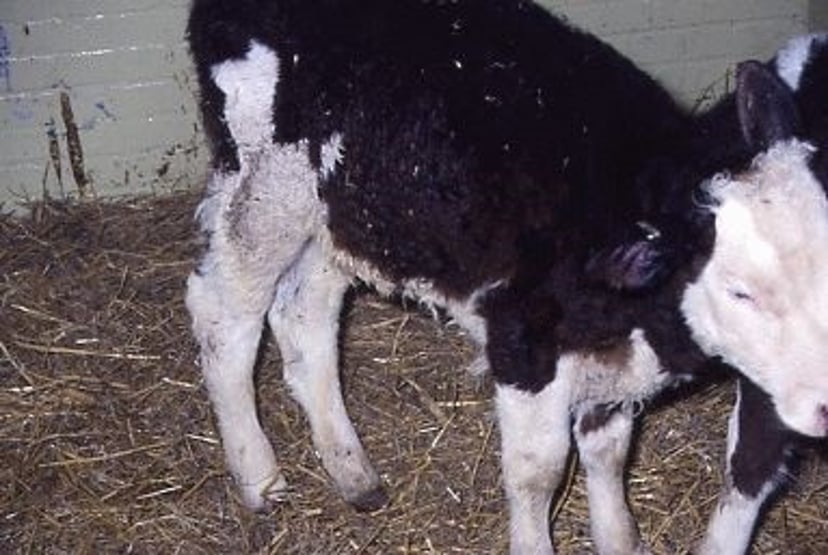
Photograph of a calf persistently infected (PI) with bovine viral diarrhea virus (BVDV), showing dullness and loss of hair with chronic coccidiosis.
Courtesy of Dr. Anthony Andrews.

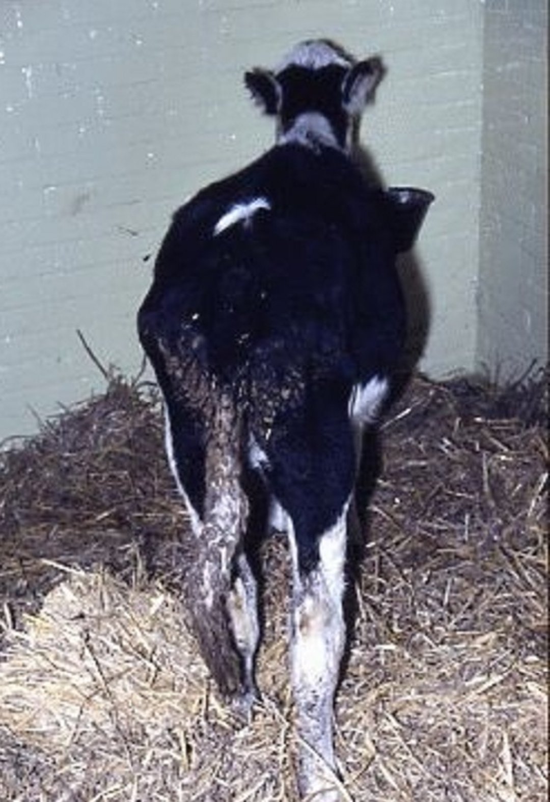
Photograph of a calf with typical subacute coccidiosis. Note the tacky feces and poor-quality coat.
Courtesy of Dr. Anthony Andrews.
In North America, CNS signs occur almost exclusively in midwinter; CNS signs (eg, muscular tremors, hyperesthesia, clonic-tonic convulsions with ventroflexion of the head and neck, nystagmus) and a high mortality rate (80%–90%) occur in some calves with acute clinical coccidiosis. Affected calves may die < 24 hours after the onset of dysentery and CNS signs, or they may live for several days, commonly in a laterally recumbent position with mild opisthotonos. As they have not been reported in experimental clinical coccidiosis in calves, CNS signs may be unrelated to the dysentery or, indeed, even to coccidiosis.
Postmortem Lesions
The small intestine may show visible white areas of meronts and pinpoint hemorrhages. There is diffuse inflammation of the last part of small intestine, cecum, and large intestine with E bovis and E zuernii. The mucosa is thickened, congested, and edematous with petechial or larger hemorrhages. Small raised white areas of schizonts may occur at villous tips in the distal ileum. Mucosal scrapes contain many gamonts and oocysts. Histologic examination shows loss and shedding of the epithelial surface, villous atrophy in the small intestine, oocysts, and gamonts. Similar lesions may occur in the cecum and large intestine.
Diagnosis of Coccidiosis of Cattle
History
Clinical signs
Presence of oocysts in feces
Diagnosis is based on the history (age of cattle, suitably contaminated environment, stressed animals, previous problems), clinical signs, fecal sampling, speciation, and postmortem findings. Quantitative oocyst counts on individual rectal samples from at least five calves in a pen, some with and without clinical signs, can be helpful to confirm coccidiosis as a cause of clinical disease. There is considerable difficulty in interpreting oocyst counts because of the many nonpathogenic species and timing of sampling. Often, however, 2,500–10,000 oocysts/g in several members of a pen, some with clinical signs, may be an indication. Species identification is required for future control strategies even if treatment has taken place before receipt of the results.
On postmortem examination, visual changes are often not apparent, and smears of intestinal contents or lining show various coccidial development stages.
Taking a fecal sample should allow most other common differential diagnoses to be ruled out. Salmonellosis may be detected by bacteriologic culture. Occasionally, other pathogenic bacteria may be detected. Clostridium perfringens types A, B, C, and E are detected by presence in smears. Possible toxin identification may be undertaken. Coronavirus and rotavirus may need to be ruled out, dependent of calf age, by use of ELISA. Bovine viral diarrhea virus may be found in feces, but blood tests are more useful. Cryptosporidia can be detected by microscopic examination with staining or by ELISA. Helminth eggs can be seen. Toxins may be indicated by the history and then require further investigation. Nutritional deficiencies and malnutrition may be detected by feeding history but may require blood samples for specific problems. Other management problems can usually be detected by careful history-taking and observation.
Treatment of Coccidiosis of Cattle
Because coccidiosis is self-limiting, cattle without clinical signs need not be treated. When treatment is necessary, those showing clinical signs should be separated to ensure they receive their medication and any necessary individual attention. This also decreases overall oocyst contamination for the group. Ideally, clinically affected calves should be moved to a separate pen and provided extra bedding, good-quality feed, and water. It should be ensured that the feed and water troughs are not contaminated. The in-contact group should also be moved to clean accommodation if possible. If not, extra bedding should be given and action taken to prevent fecal contamination of feed and water troughs. Any vacated pens should be thoroughly cleaned and disinfected, and a fallow period is recommended before further occupation.
Once calves are ill, treatment has a variable effect, dependent on the extent of intestinal damage. Because feed and water intake may be decreased, all treatments for ill calves should be administered individually.
Diclazuril (1 mg/kg, PO, once) or toltrazuril (15 mg/kg, PO, once) administered to all affected calves and others in the group decreases oocyst production. Some affected calves may still show clinical signs, but oocyst counts decrease.
Sulfadimethoxine (50 mg/kg, as a drench or in water, for 1 day and then 25 mg/kg, every 24 hours for 4 days) is used in cattle in North America. Sulfamethazine (sulfadimidine) is also used (247.5 mg/kg, in the drinking water or as a drench, for 1 day, then 124 mg/kg, every 24 hours for 3 days). Sulfaquinoxaline (13 mg/kg, in the drinking water or by drench if not drinking sufficiently, for 3–5 days) is approved by the US FDA for treatment of coccidiosis. It is reported to be particularly useful for weaned calves that develop bloody diarrhea after arrival at a feedlot. Parenteral sulfonamide treatment may be indicated to control development of secondary bacterial enteritis or pneumonia, which may occur in calves with coccidiosis during very cold weather.
Decoquinate (1 mg/kg, PO in feed, every 24 hours for 28 days) treats coccidiosis in calves; using a 60 g/kg premix at 1.67 kg/ton of feed provides the recommended treatment dose of 100 mg/kg of feed, allowing 500 g of feed for a 50-kg calf for 28 days. However, if calf feed intake is less, higher premix concentrations are necessary. Decoquinate may prevent decreased neutrophil function due to the parasite.
Amprolium (10 mg/kg, PO in the drinking water, feed, or by drench, every 24 hours for 5 days) is used as a treatment course. Drench is preferable when calves are ill. It is approved by the US FDA and also in some non-EU countries. Use of amprolium in calves is controversial because it can cause thiamine deficiency and associated neurologic disease. After treatment with amprolium, it is advisable to provide thiamine.
Estradiol and progesterone help to enhance cellular immunity and decrease the debilitating effects of E bovis.
Besides use of coccidiostats, fluid therapy or blood transfusions may be needed in cases of dehydration, dysentery, and anemia. Corticosteroids are contraindicated because they increase shedding of oocysts and have induced clinical disease in subclinically infected calves.
Control and Prevention of Coccidiosis of Cattle
Coccidiosis is difficult to reliably control because oocyst numbers rapidly increase in suitable conditions. However, it is mainly a management problem, usually from overcrowding and a contaminated environment. Thus, calves should be kept as unstressed as possible. As part of preventive management and control, ensure all calves enter and are kept in a clean, disinfected environment with good ventilation. The calves should have a satisfactory stocking density to avoid overcrowding, with all feed troughs and water sources raised to decrease fecal contamination. An all-in/all-out system should be used for groups entering and moving pens. Those in groups should remain together. Lying, feeding, and watering areas inside and outdoors must be well drained. Generous or extra bedding should be provided to decrease oocyst contamination. Stressful events (eg, mixing groups, routine procedures, feed changes) should be kept to a minimum. Potentially stressful changes should not be made when there are, or are anticipated, marked weather fluctuations.
Some ammonia-based disinfectants kill the oocysts but can only be used in areas vacated by animals. On many production farms, coccidiosis is only controllable by medication. Medication should be used for as short a period as possible, only when oocyst exposure is anticipated, and continuing no longer than needed to allow effective immunity to develop. Thus, control of coccidiosis in North American feeder calves brought into a crowded feedlot depends on management of population density, appropriate feed bunks, and use of chemotherapeutics.
Management methods should be developed to decrease the reliance on preventive medication. Some medicines are licensed in various countries for the control of calf coccidiosis, but many other products used appear to have some efficacy although they are unlicensed in cattle. The ideal coccidiostat suppresses the full development of the life cycle of the coccidia, allows immunity to develop, and does not interfere with production performance.
Diclazuril (1 mg/kg, PO, once) or toltrazuril (15 mg/kg, PO, once) administered 14 days after animals are moved into group housing can effectively prevent diarrhea due to coccidiosis. Preventive in-feed medication is used in many countries, but some medicines are not approved.
Sulfadimidine (sulfamethazine) in the feed (25–35 mg/kg for ≥15 days) effectively controls coccidiosis in calves. Sulfaquinoxaline (13 mg/kg, PO in the drinking water, every 24 hours for 3–5 days) is approved by the US FDA for coccidiosis control. Sulfadimethoxine (50 mg/kg, PO in the drinking water, for 1 day and then 25 mg/kg for 4 days) is used in cattle in North America. A combination of sulfonamide and chlortetracycline has provided early protection to calves but may not be conducive to decreasing antimicrobial use.
Amprolium (5 mg/kg, PO in the drinking water or in feed, every 24 hours for 19–21 days) can be used as a preventive program. Provision of thiamine after treatment should be considered.
Most countries permit decoquinate in the feed to suppress oocyst production. It is most effective in preventing coccidial infections when fed continually in dry feed at 0.5 mg/kg (using a 60 g/kg premix at 833 g/ton of feed provides the recommended treatment dose of 50 mg/kg of feed); decoquinate-containing feeds should be provided continually for at least 28 days.
Monensin (0.3–0.9 mg/kg, PO in feed, every 24 hours for 31 days) is an effective coccidiostat and growth promotant in calves and growing cattle. It is used in the feed at 16.5 or 30 g/ton for 31 days. Postweaning coccidiosis in beef calves is controlled using monensin administered via intraruminal continuous-release devices.
Lasalocid is related to monensin and is also an effective coccidiostat for ruminants. Doses of 0.75–3 mg/kg are effective in preventing experimental coccidiosis. Mixing lasalocid in the milk replacer of calves beginning at 2–4 days of age is an effective way to control coccidiosis. It can also be administered at 40 mg/kg in calf starter ration, beginning at 2 days and continuing for 12 weeks. Lasalocid is also effective as a coccidiostat when fed free-choice in salt at a level of 0.75% of the total salt mixture. A concentration of 1 mg/kg is recommended as being effective and rapid when outbreaks of coccidiosis are anticipated.
Decoquinate, lasalocid, and monensin at the manufacturer’s recommended levels are equally effective. Oregano oil may assist in coccidiosis control.
No vaccines are available, partly because of the number of pathogenic species and a lack of complete understanding of immunity to the parasite.


