


Dental caries involving the peripheral cementum is common in large animals. Rarely, the disease progresses and involves enamel and dentine. These carious lesions are usually related to diets that lead to a decrease in the oral pH—eg, silage or forage with a high proportion of soluble carbohydrate, such as oaten hay. A water supply with a low pH can cause the decay of peripheral cementum. Cementum decays at a pH of 6.7; for enamel decay, the pH must be 5.5 or less. Changing the diet or the water source can correct the problem in most cases (1).

Courtesy of Dr. Jack Easley.
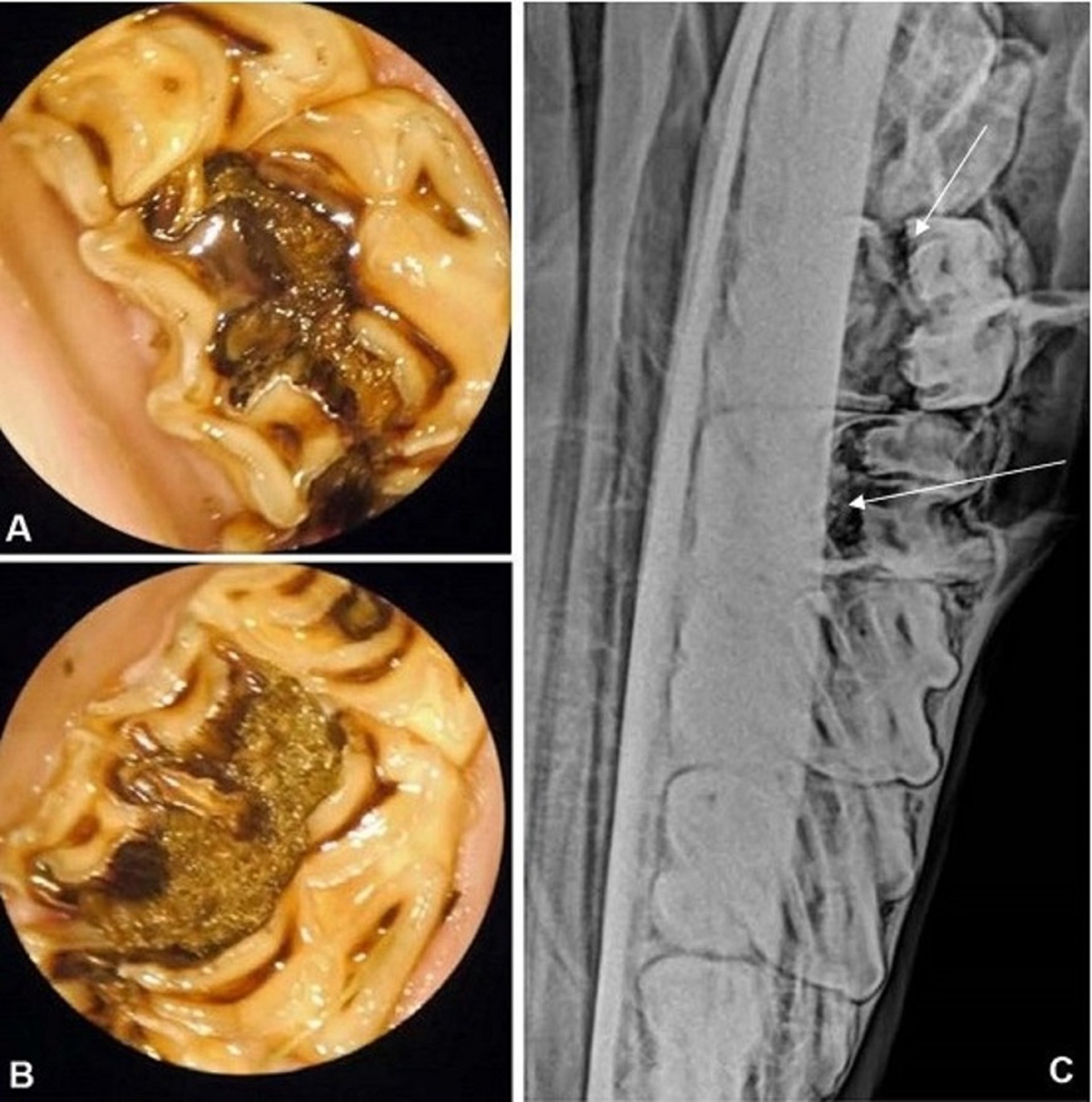
Another form of dental caries that occurs in horses involves the two deep infundibula of the upper cheek teeth. Infundibular cement hypoplasia predisposes the infundibula to become filled with feed material that can ferment and produce an acidic environment. As these affected teeth continue to erupt, the weakened decayed crown can fracture midsagittally through the mesial and distal infundibula, leading to oral discomfort and periapical disease. Crown restorative techniques have been used to delay the progress of infundibular caries; after the tooth has fractured, however, oral extraction is the treatment of choice.
Reference
Jackson, K., Kelty, E., Staszyk, C. and Tennant, M. (2019) Peripheral caries and disease of the periodontium in Western Australian horses: An epidemiological, anatomical and histopathological assessment. Equine Vet J. 51, 617- 624.
For More Information
Dixon PM, Gerard MP. Oral cavity and salivary glands. In: Equine Surgery, 5th edn. Eds: A. J. Auer, J. A. Stick, J.M. Kummerle and T. Prange, Elsevier, St. Louis, Missouri. 2018; 440-473.
Rice MK, Henry TJ. Standing intra-oral extractions of cheek teeth aided by partial crown removal in 16 horses (2010-2016). Equine Vet J. 2017;1-6.
Suske A, Poschke A, Schrock P, Kirschner S, Brockmann M, Staszyk C. (2016) Infundibula of equine maxillary cheek teeth. Part 1: Development, blood supply and infundibular cementogenesis. Vet J. 209; 57-65.
Horbal A. Smith S. Dixon PM. (2019) A Computed Tomographic (CT) and Pathological Study of Equine Cheek Teeth Infundibular Extracted from Asymptomatic Horses. Part 1: Prevalence, Type and Location of Infundibular Lesions on CT Imaging. Front Vet Sci. Vol. 6: article 124; 1-10.


