


Tooth resorption is a common disease in feline patients. Dental radiographs are imperative for determining the type and stage of tooth resorption, which ultimately affects which treatment can be performed. Treatment is accomplished by surgical extraction or crown amputation of the affected teeth and is aimed at eliminating the source of pain and discomfort for the cat.
Tooth resorption, formerly known as cervical line lesion and feline odontoclastic resorptive lesion, refers to an inflammatory process resulting in progressive loss of destruction of mineralized dental tissues (ie, the enamel, dentin, and cementum) and eventually the entire tooth structure, in one or more teeth. Tooth resorption is painful and a major cause of tooth loss. Tooth resorption may occur physiologically but is typically pathological and primarily affects cats.
Etiology and Pathogenesis of Tooth Resorption in Small Animals
Resorption of tooth structure occurs through the action of odontoclasts—cells virtually identical to osteoclasts.
Odontoclasts are cells that target the tooth and cause resorption of the tooth structure. Osteoclasts are responsible for the remodeling of bone. Both cells cause damage to tooth and bone, leading to eventual loss or remodeling of the tooth and its attachment to the surrounding alveolar bone.
Tooth resorption can occur on the external or internal tooth surface (external or internal resorption). Odontoclastic activity can be stimulated by inflammation, pressure from adjacent structures, and orthodontic tooth movement; as a result of normal processes, such as exfoliation of deciduous teeth; or in the absence of these processes (idiopathically).
In cats, tooth resorption begins with a loss of the normal periodontal ligament architecture and focal damage to the cementum that covers the root surface. Microscopic areas of root resorption often repair uneventfully in cats.
Tooth resorption from any cause occurs through the action of odontoclasts that remove tooth structure, creating a resorptive lacuna. In many but not all lesions, concomitant osteoblast and cementoblastic activity replaces the lost tooth with bone or cementum.
If repair does not occur, the resorption progresses into dentin and extends coronally into the crown of the tooth, where it undermines the enamel to cause clinically apparent defects on the tooth surface (at the cervical region or "neck" of the tooth).
In general, no known specific etiology is associated with tooth resorption. Inflammation from periodontitis is known to cause external resorption and is most likely responsible for tooth resorption in areas of periodontal disease. However, the etiology of idiopathic tooth resorption affecting multiple (possibly all) teeth in cats has not yet been identified.
Nutritionally, excessive intake of dietary vitamin D has been hypothesized as one possible cause among many others; ultimately, there is no true identified cause of tooth resorption in cats.
Epidemiology of Tooth Resorption in Small Animals
Tooth resorption occurs sporadically in many species (including humans) and most frequently occurs in domestic cats.
It has been reported that > 60% of cats will have evidence of tooth resorption within their lifetime (1). An increased incidence of tooth resorption is observed in older cats as well as purebred cats.
Clinical Findings and Lesions of Tooth Resorption in Small Animals
The clinical appearance of tooth resorption greatly varies between cats. The most common clinical finding is the loss of tooth structure. Frequently, this occurs at the cementoenamel junction.
Clinically, an affected tooth can have evidence of inflammation associated with the gingiva of an affected tooth. This can then progress to obvious loss of the tooth structure (see resorptive lesions radiograph and photograph).

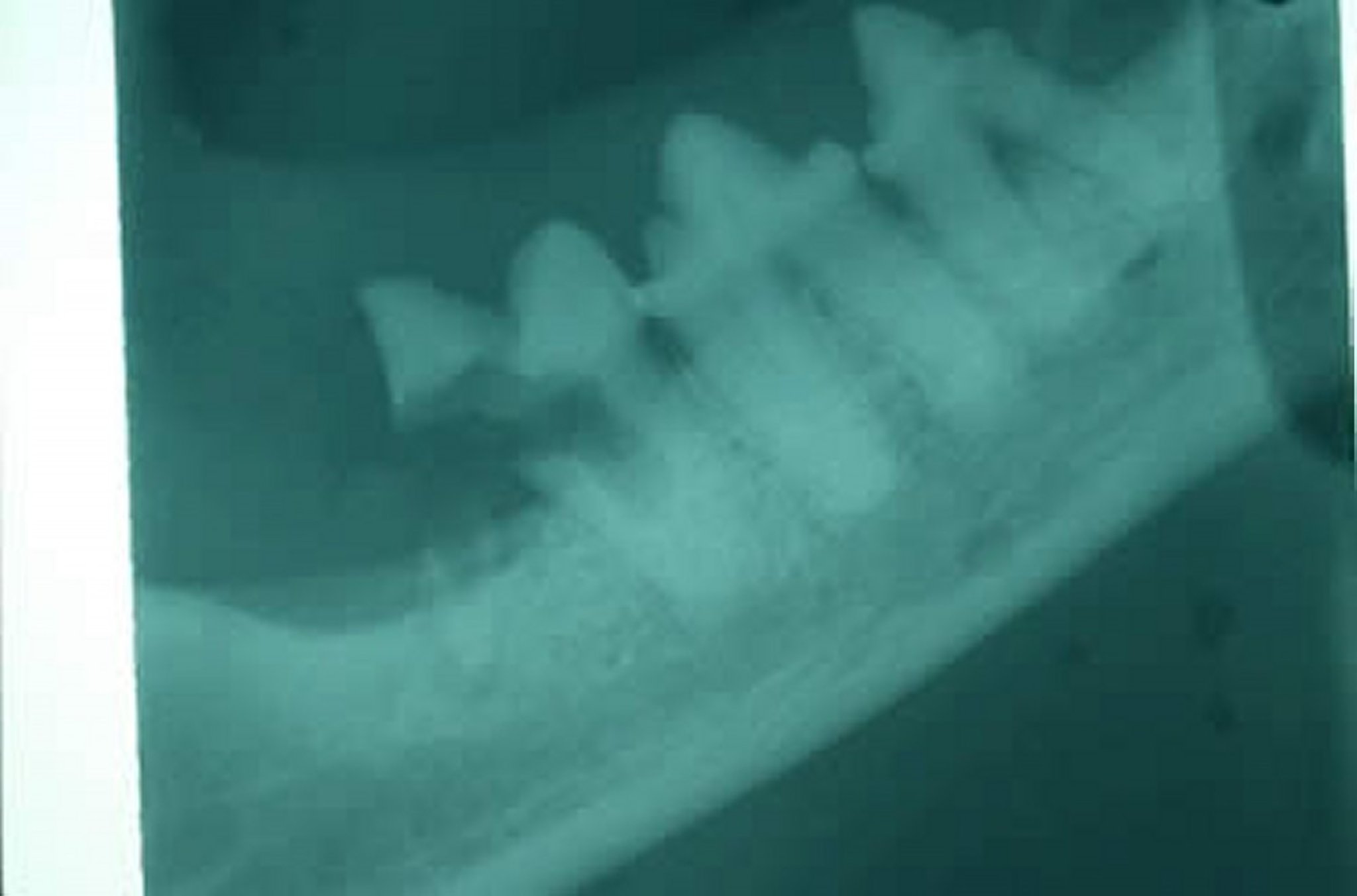
Courtesy of Dr. Ben Colmery III.
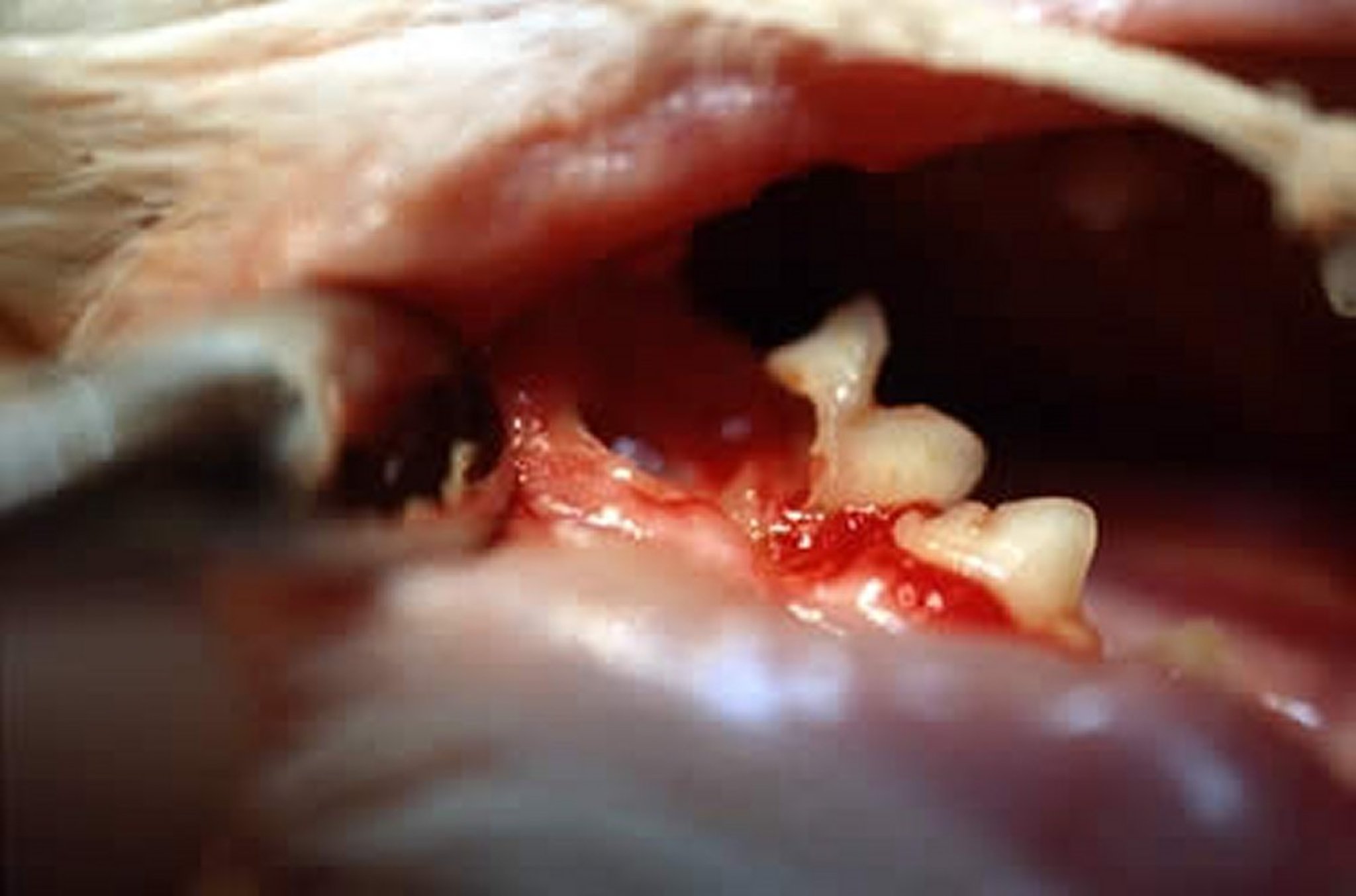
Courtesy of Dr. Ben Colmery III.
In cats, the mandibular third premolar (the first cheek tooth) is often the first tooth affected. While any tooth can be affected by tooth resorption, the left and right mandibular third premolar teeth (307 and 407) appear to be the most frequently affected within the feline oral cavity. In dogs, premolar and molar teeth are commonly involved.
Small lesions on the enamel of the tooth crown usually begin somewhere on the root surface but can progress toward the crown and appear at the gingival margin as inflamed granulation tissue filling a defect. The margin of the defect frequently has a sharp ledge of enamel. At this stage, the visible part of the lesion is small, with most of the defect affecting the roots.
Tooth resorption can be characterized by severity (stage) and radiographic appearance of the periodontal ligament space (type).
The 5 stages of tooth resorption describe the portions of the tooth that are affected and to what extent the tooth is affected:
Stage 1 lesions affect the cementum or cementum and enamel but have not yet progressed into the dentin.
Stage 2 lesions affect the dentin but have not yet progressed into the pulp cavity.
Stage 3 lesions affect the pulp cavity, but most of the tooth retains its integrity.
Stage 4 lesions have substantial crown or root damage, with most of the tooth having lost its integrity.
Stage 5 lesions have remnants of dental hard tissue visible only as irregular radiopacities, and gingival covering is complete.
Based on the presence or absence of the periodontal ligament space, lesions can be categorized radiographically into 3 types:
Type 1: A focal or multifocal radiolucency is present in the tooth with otherwise normal radiopacity and normal periodontal ligament space (inflammatory resorption).
Type 2: There is narrowing or disappearance of the periodontal ligament space (dentoalveolar ankylosis) in at least some areas and decreased radiopacity of part of the tooth (replacement resorption, moth-eaten "ghost" roots).
Type 3: Features of both type 1 and type 2 are present in the same tooth.
Tooth resorption lesions exposed to the oral cavity may cause discomfort. Lesions limited to root surfaces are unlikely to cause discomfort or other clinical signs unless they are associated with resorption of bone adjacent to the tooth resorption (eg, resorption caused by painful inflammation from periodontal or endodontic disease).
Diagnosis of Tooth Resorption in Small Animals
Clinical evaluation and periodontal probing
Radiographic evaluation
Stage 1 lesions in tooth resorption are typically identified on clinical examination only. Because the loss of tooth structure is confined to the enamel or cementum in this stage, there is no radiographic evidence of disease.
Using the explorer end of a periodontal probe-explorer to examine the crown of the tooth is imperative to help identify early tooth resorption lesions.
Marginal gingivitis of individual teeth in the absence of periodontitis may indicate an early subgingival lesion.
Meticulous examination of the cementoenamel junction is vital to the identification of these early-stage lesions. In addition, magnification (eg, surgical loupes) can make early lesions more easily identifiable.
Lesions present under the free gingival margin can be identified by a dental explorer.
Larger lesions are identified by their typical gross appearance on the tooth surface or recognized radiographically (see external resorption photograph and internal resorption radiograph). Dental radiography is used to identify the type of tooth resorption present as well as the stage and severity of the hard tissue loss.

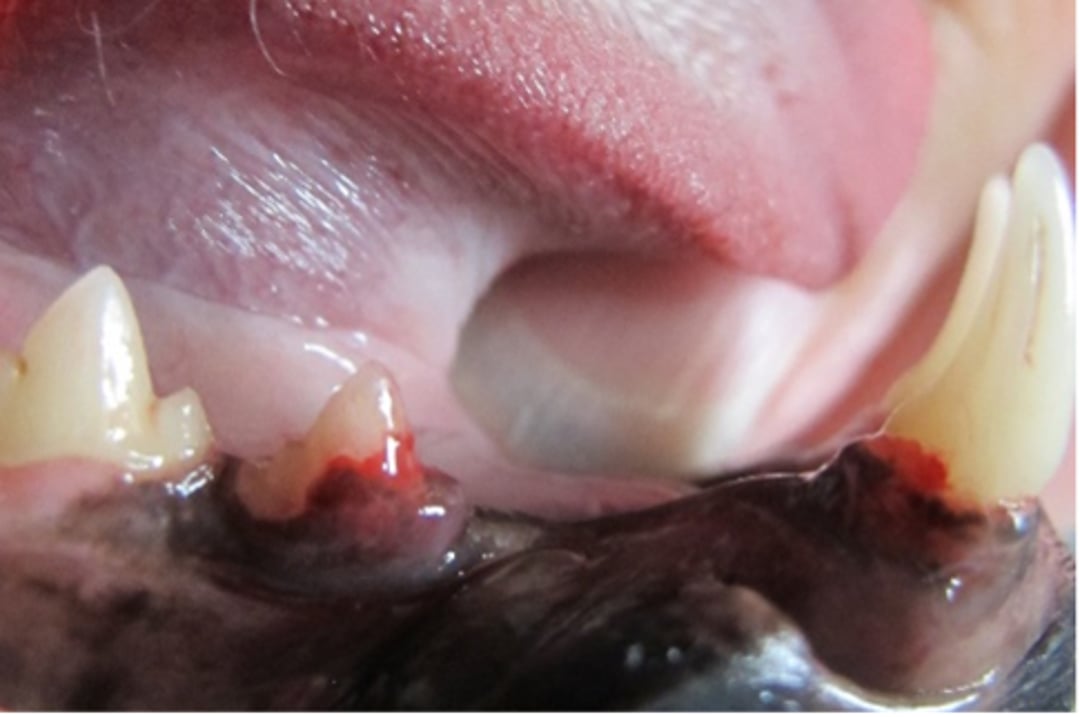
Courtesy of Dr. Brenda Mulherin.

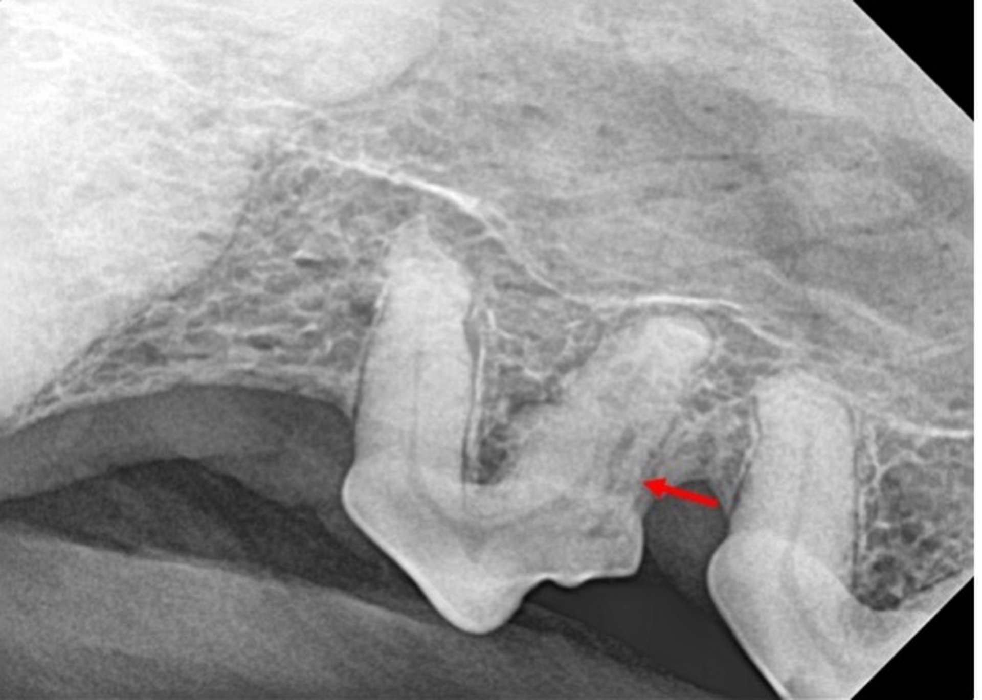
Courtesy of Dr. Brenda Mulherin.
Internal tooth resorption may sometimes clinically appear as pinkish discoloration of the crown. More frequently, internal tooth resorption is only identifiable radiographically as round- to oval-shaped areas of decreased radiopacity.
Treatment and Prevention of Tooth Resorption in Small Animals
Dental extraction
The accepted treatment for tooth resorption is surgical extraction of the teeth that are exposed to the external environment. This is why paying close attention to the cementoenamel junction is important to identify and treat early lesions. Tooth resorption is a progressive disease; therefore, early diagnosis and treatment of lesions exposed to the external environment are vital to improving the patient's quality of life.
Most teeth affected with resorptive lesions should be extracted. Surgical crown amputation with intentional root retention of already-resorbing dental tissues can be performed on patients with radiographically confirmed type 2 lesions in the absence of periodontitis, endodontic disease, and stomatitis.
For inflammatory resorptive lesions caused by marginal periodontitis, oral hygiene measures can slow the progression of hard tissue destruction. Root canal therapy or extraction of primary endodontically compromised teeth (eg, tooth fracture) prevents resorption of the apex caused by apical periodontitis.
Idiopathic lesions cannot be prevented, because their etiology is unknown.
Once a patient has been identified with radiographic or clinical evidence of tooth resorption, annual anesthetized oral examinations with full mouth radiographs should be performed to monitor for progression of disease and early intervention with removal of affected teeth.
Key Points
Tooth resorption is a progressive disease with no identified etiology.
Treatment is aimed at removal of the affected tooth or teeth.
The 5 stages of tooth resorption involve the integrity of the tooth structure; the 3 types of tooth resorption involve the radiographic assessment of the periodontal ligament space.
References
Lommer MJ, Verstraete FJ. Prevalence of odontoclastic resorption lesions and periapical radiographic lucencies in cats: 265 cases (1995-1998). J Am Vet Med Assoc. 2000;15;217(12):1866-1869. doi:10.2460/javma.2000.217.1866
For More Information
Teeth Abnormalities and Related Procedures: AVDC classification of tooth resorption. American Veterinary Dental College.
Also see pet health content regarding dental disorders of cats.


