Abdominal ultrasonography results are shown below:
Observations and Impressions:
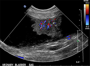
- At least 3 florid-like urinary bladder masses are seen in the dorsal and ventral wall. The largest measures 4.3cm x 1.5cm x 2.4cm, with pinpoint hyperechoic specks and marked vascularity on color flow Doppler. This is consistent with primary urinary bladder neoplasia, likely a transitional cell carcinoma (TCC). The proximal urethra and prostate look normal.
- Hepatic hypoechoic nodules - nodular hyperplasia or extramedullary hematopoiesis or metastatic tumor nodules.
- Hyperechoic splenic and hepatic nodules - suspect myelolipomas.
- The medial iliac lymph nodes are normal.
- The rest of the abdomen is normal.