

Maldigestion or malabsorption disorders usually manifest as chronic, persistent GI signs, including vomiting, weight loss, small- and/or large-intestine diarrhea, or a combination of these factors. There are many potential etiologies, both heritable and acquired, and most are associated with inflammatory bowel disease (IBD). Congenital conditions may have specific breed predilections.
Soft Coated Wheaten Terriers have a high incidence of protein-losing enteropathy (PLE), which may occur alone or in association with protein-losing nephropathy. Dogs with PLE have both food hypersensitivities and IBD, but the pathogenesis is not clear. The demonstration of increased fecal alpha1-protease inhibitor concentrations can help confirm abnormal protein loss through the intestinal tract; this screening test is more useful in animals < 3 years old, however. A definitive diagnosis is based on intestinal and renal histopathology. Despite hypoallergenic diet trials and immunosuppressive treatment directed at IBD and/or glomerulonephritis, the prognosis is poor.
Gluten-sensitive enteropathy has been shown to be inherited in an autosomal recessive manner in Irish Setters. However, the pathogenesis in dogs appears to be different from that of celiac disease in humans. Clinical signs include chronic intermittent diarrhea and weight loss. One case has been described in a horse. The wheat sensitivity is both confirmed and treated through the feeding of gluten-free diets.
Basenjis have been diagnosed as carriers of a severe form of lymphocytic-plasmacytic enteritis called Basenji enteropathy; the mode of inheritance, however, is not known. The stomach may also be affected. Clinical signs include diarrhea and weight loss, with hyperglobulinemia. Diagnosis is based on histopathologic examination of GI biopsies, usually obtained through endoscopy. Treatment trials with immunosuppressive drugs and hypoallergenic diets are usually unsuccessful, unless aggressively initiated early in the disease.
Lymphangiectasia is a malformation of the intestinal lymphatic system that results in PLE, with a 50% incidence in Norwegian Lundehunds in the US. Other affected breeds include Yorkshire Terriers, Maltese, Rottweilers, and Chinese Shar-Pei. Lymph vessels become dilated, secreting lymph into the intestines and thereby leading to hypoproteinemia, lymphopenia, and lipogranulomatous inflammation of surrounding tissues. Lymphangiectasia is diagnosed through exclusion of other protein-losing diseases and confirmed by histopathology of the small-intestine wall. Most affected animals respond to a combination of dietary manipulation and the administration of anti-inflammatory doses of glucocorticoids. Many patients with lymphangiectasia are underweight as a result of PLE. Diets should contain minimal fat, be energy dense, and be easily digestible. Although remission of clinical signs may be achieved, the longterm outcome is usually poor.
Exocrine pancreatic insufficiency (EPI) has a higher incidence in German Shepherd Dogs and rough Collies than in other dog breeds; however, it has been diagnosed in many breeds. Disease is due to pancreatic acinar atrophy, which is due to immune-mediated destruction and infiltration of lymphocytes. In German Shepherd Dogs and Pembroke Welsh Corgis, genome-wide association studies have identified several major-histocompatibility-complex haplotypes associated with EPI, suggesting a complex mode of inheritance. The lack of pancreatic enzymes results in an osmotic diarrhea, in which steatorrhea is a prominent feature. Affected animals either fail to gain weight or, if EPI is acquired later in life, show dramatic weight loss. Diagnosis is based on the measurement of serum trypsin-like immunoreactivity; validated tests are available for both dogs and cats. Treatment involves exogenous replacement of pancreatic enzymes and the feeding of highly digestible diets. Cobalamin deficiency is also common in dogs with EPI, and cobalamin supplementation should be included in treatment regimens.
Granulomatous colitis, previously called histiocytic ulcerative colitis, has been diagnosed in Boxers and French Bulldogs, with sporadic cases reported in a few other breeds. The disease is characterized by granulomatous inflammation of the colonic mucosa in association with infection by adherent and invasive Escherichia coli. An autosomal recessive defect in the immune system that predisposes the animal to E coli infection is suspected.
Clinical signs arise in animals < 4 years old and include diarrhea, hematochezia, tenesmus, and weight loss. Diagnosis is based on histologic examination of colonic mucosal biopsies. Successful remission has been achieved in patients treated with enrofloxacin; however, treatment has been unsuccessful when E coli was resistant to fluoroquinolones. Because multidrug-resistant strains of E coli have been identified in cases of granulomatous colitis, susceptibility testing is recommended before the initiation of antimicrobial treatment.

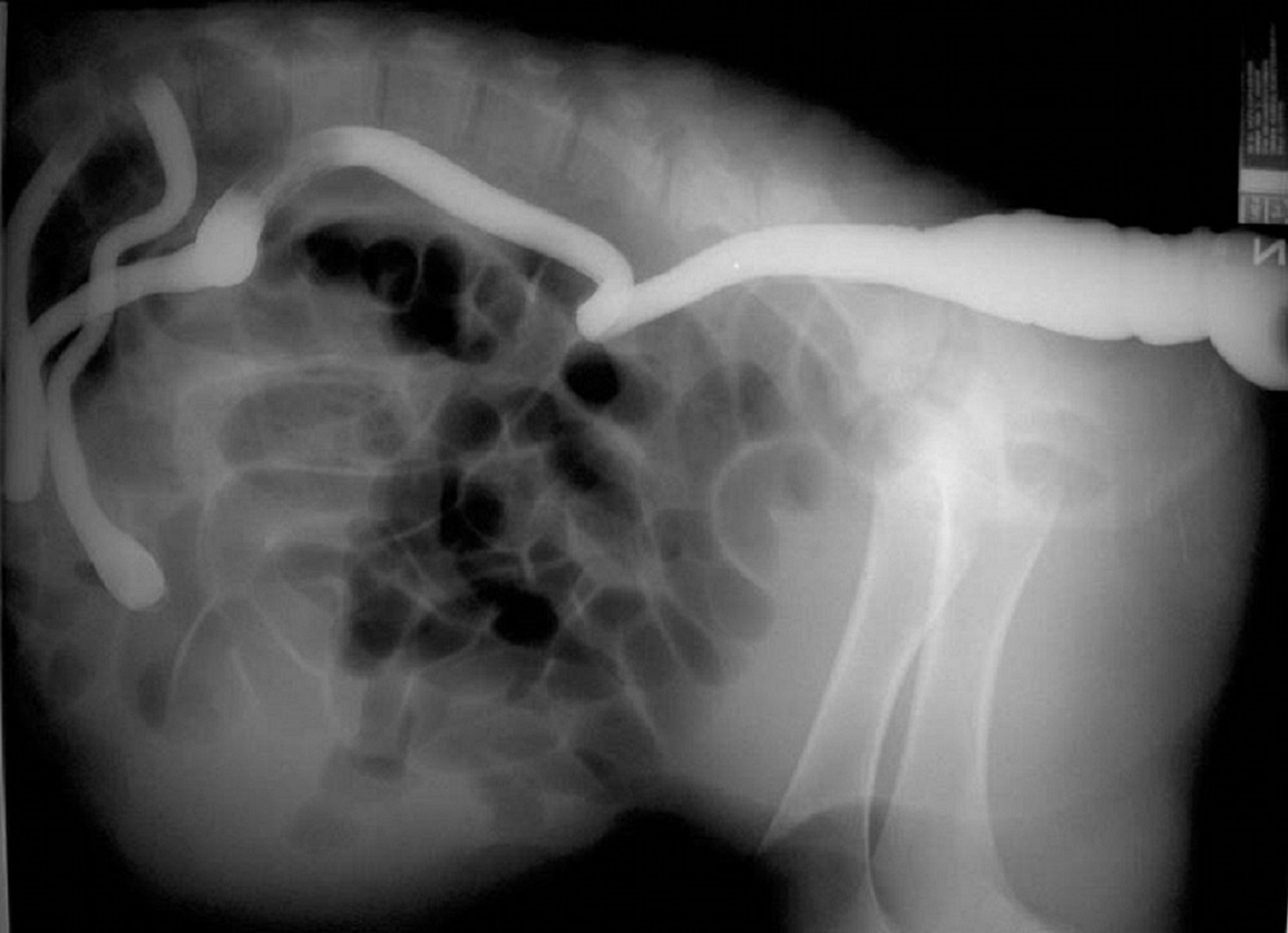
This lateral radiograph (with contrast) shows atresia coli in a calf. Contrast material has been administered rectally and continues orally only to the colon, where it stops in a blind end.
Courtesy of Dr. Sameeh M. Abutarbush.
Ileocolonic aganglionosis, or overo lethal white syndrome, occurs in white foals with blue eyes produced by overo-overo matings. The condition is fatal. It is due to mutation in the endothelin B receptor and is inherited in an autosomal recessive manner. Although foals appear normal at birth, they rapidly develop clinical signs of colic and meconium impaction due to hypoinnervation of the intestinal tract, which results in lack of motility. Diagnosis can be confirmed at necropsy by the lack of ganglia in the colon. It has been estimated that nearly 90% of overo horses are heterozygotes and therefore carriers of the recessive allele responsible for overo lethal white syndrome. Adult overo horses can be screened for genetic status before breeding to decrease incidence.
Congenital atresias of either the small- or large-intestine tracts have been described in most domesticated animal species. Atresia coli has been reported in several foals and calves of differing breeds. Affected calves will nurse, but they may experience abdominal distention and colic after feeding because the intestinal tract is not completely patent. Digital rectal examination often reveals mucoid contents with no feces present in the rectum. Atresia ilei in Swedish Highland cattle and atresia jejunae in Jersey cattle are likely due to autosomal recessive inheritance. These conditions are invariably fatal.

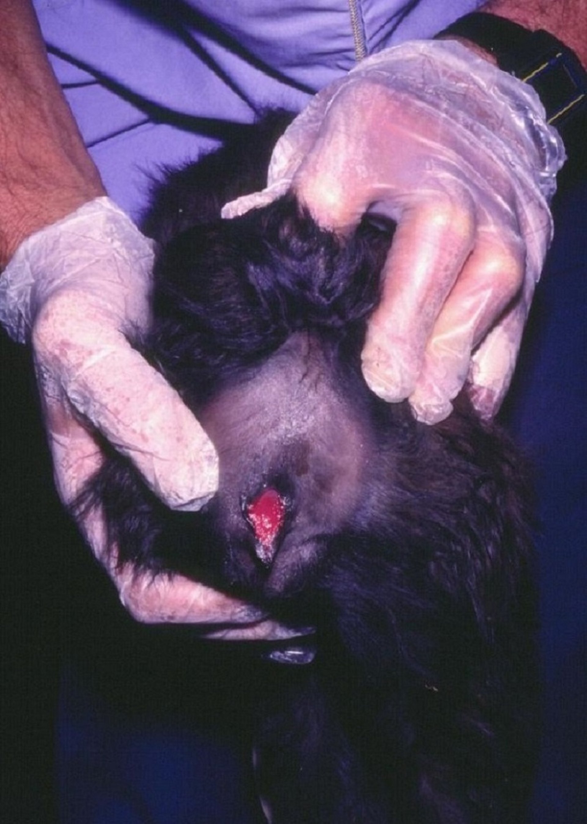
This photograph shows atresia ani in an alpaca cria. The vaginal mucosa (dark pink tissue) is visible in the vulvar opening, but there is no rectal opening present dorsal to the vulva.
Courtesy of Dr. Lisa Pearson.
Atresia ani results when the dorsal membrane separating the rectum and anus fails to rupture. Clinical signs are apparent at birth and include tenesmus, abdominal pain and distention, retention of feces, and absence of an anal opening. The condition is rare in dogs, but it has been reported in several breeds, including Toy Poodles and Boston Terriers, with a higher incidence in females. Concurrent rectovaginal fistulas may occur. Euthanasia is recommended in large animals. In small animals, postoperative fecal incontinence may be a complication of surgical intervention.
Segmental aplasia of the rectum, or rectal atresia, occurs when the rectum terminates in a blind pouch before reaching the anus. Surgical correction is difficult because the location of the terminal section varies, and iatrogenic damage to nerves in the area may occur.
Enteric duplications are very rare; only a few cases are described in several species. They may include the colon or rectum. Duplications may communicate with the lumen of the patent tract or be separate (duplication cysts). Affected animals may be diagnosed by identification of an abdominal mass, or they may show GI signs. Diagnosis is based on evaluation using ultrasonography or contrast CT. The condition can be corrected by surgical removal of the duplication; some cases, however, have multiple concurrent abdominal developmental anomalies that preclude complete surgical correction.
Rectourethral fistulas have been reported primarily in English Bulldogs. They are diagnosed clinically on the basis of simultaneous urination from both the urogenital and anal orifices, along with a history of chronic urinary tract infections. Diagnosis is based on cystourethroscopy with concurrent colorectal infusion of contrast material, or on contrast CT. Surgical correction is curative.
Rectovaginal fistulas are fistulous tracts that connect the vagina and rectum and usually occur in conjunction with an imperforate anus. Passage of feces through the vulva is suggestive. Diagnosis may be confirmed by barium enema, which outlines the extension of the defect into the vagina, or by evaluation with video endoscopy. Identification of the fistula, surgical correction, and reestablishment of the normal anatomical structures are imperative. Prognosis is usually guarded. Complications are common and include fecal and urinary incontinence.
Urinary and fecal incontinence often occur in Manx cats as sequelae of heritable spina bifida.


