Campylobacter spp can cause GI disease in humans and several animal species. Infections in humans are frequently acquired through ingestion of undercooked chicken, unpasteurized dairy products, or undercooked beef. Direct contact with the feces of infected animals can also be a source of zoonotic transmission. Clinical signs include diarrhea, fever, and abdominal discomfort; treatment is supportive and in some cases includes antimicrobial therapy. Diagnosis of campylobacteriosis is via culture, which can be challenging, or by PCR assay in conjunction with clinical signs.
Campylobacter spp are spiral, microaerophilic, gram-negative bacteria that can cause gastroenteritis in humans and animals. Several Campylobacter spp are zoonotic. Many domestic animals, including dogs, cats, sheep, pigs, ferrets, mink, monkeys, and several species of laboratory animals, can develop acute gastroenteritis after ingesting Campylobacter spp. (Also see Bovine Genital Campylobacteriosis, Zoonotic Diseases, and Avian Campylobacter Infection.)
Infection with Campylobacter jejuni is one of the most common causes of gastroenteritis in humans worldwide, and C jejuni is the most extensively studied Campylobacter species.
Etiology of Enteric Campylobacteriosis
Campylobacter spp are thin (0.2–0.8 mcm × 0.3–5 mcm), gram-negative, motile, curved rods that exhibit a characteristic corkscrew darting motility by virtue of their single flagellum. The cells are generally S-shaped or curved; however, occasionally they are long (8 mcm) spiral rods. In unfavorable growth conditions, spiral rods undergo a degenerate conversion to coccoid forms.
The thermophilic Campylobacter spp—namely C jejuni and C coli—have the highest disease impact in animals and humans. However, infections are subclinical in some animal reservoirs, including poultry and cattle.
See the table for a brief summary of animal diseases caused by different Campylobacter spp.
Campylobacter Species That Cause Diseases in Animals
Campylobacter Species | Animal Species Affected | Disease(s) |
|---|---|---|
C jejuni | Dogs, cats, sheep, goats | Enteritis; abortion |
C coli | Pigs, foals | Enteritis and diarrhea |
C sputorum | Sheep | Abortion |
C fetus fetus | Sheep, cattle | Abortion |
Campylobacter sp | Sheep | Colitis |
Transmission and Epidemiology of Enteric Campylobacteriosis
Campylobacter spp are transmitted via food- or waterborne exposure or by direct contact with feces of infected animals. The predominant ecological niche for Campylobacter spp is the GI tract of a wide variety of domesticated and wild vertebrates; potential reservoirs include poultry, cattle, pigs, sheep, dogs, cats, and rodents.
Zoonotic transmission of Campylobacter via contaminated meat or dairy products is a food safety issue. Campylobacter spp are also commonly isolated from wild birds, including migratory birds and waterfowl, crows, gulls, and domestic pigeons, which can contaminate the environments of grazing animals. Wild rodents and insects such as flies can also harbor and transmit C jejuni.
Fecal contamination of the environment provides a ubiquitous source of these organisms under conditions appropriate for their survival. Campylobacter spp can persist for long periods in feces, milk, water, and urine, especially at temperatures close to 4°C (39.2°F). In adverse conditions, C jejuni converts to a viable but nonculturable form that can be reactivated when ingested.
Human foods that have served as vehicles for Campylobacter transmission include chicken, turkey, beef, pork, fish, milk, and various produce items. Poultry are the most important source of C jejuni infections in humans, followed by cattle (1). Like their owners, dogs and cats can become infected when they ingest raw/undercooked poultry, unpasteurized milk, or raw/undercooked beef.
Pathogenesis of Enteric Campylobacteriosis
Bacterial motility, mucus colonization, toxin production, attachment, internalization, and translocation are among the processes associated with Campylobacter jejuni virulence.
Infection begins with ingestion of C jejuni in contaminated foods or water. Gastric acid provides a barrier, and the bacteria must reach the small and large intestines to multiply; C jejuni invades both epithelial cells and cells within the lamina propria.
Virulent Campylobacter spp adhere to and invade enterocytes after penetrating the surface mucus layer. They survive in the cytoplasm of enterocytes, extending their intracellular survival by avoiding lysosomal compartments.
The pathogenesis of Campylobacter-associated disease involves type IV and type VI secretion systems and toxins, including the virulence factor cytolethal distending toxin (CDT).
Clinical Findings of Enteric Campylobacteriosis
Campylobacter jejuni produces a spectrum of disease scenarios, depending on the immune status of the host, bacterial virulence, gene expression, and other factors. Infected patients can show signs of systemic illness, can show only GI signs, can be infected and shed the organism transiently and remain clinically normal, or can clear the infection promptly with no signs of illness. Abdominal pain, fever, diarrhea, and blood in feces are the typical clinical signs, demonstrating the inflammatory nature of the infection.
Pearls & Pitfalls
|
C jejuni is the most common cause of campylobacteriosis in companion animals, which are considered a potential source of zoonotic transmission.
Clinical signs of campylobacteriosis in dogs can include diarrhea, fever, lethargy, and sporadic vomiting (2). Diarrhea is usually acute; it can be watery and contain blood or mucus. Clinical disease is more common in puppies than adult dogs. Occasionally, diarrhea becomes chronic and can be accompanied by fever and elevated WBC counts. However, canine C jejuni infections are often subclinical, with affected dogs having grossly normal feces (3, 4). C jejuni has also been isolated from the vaginal discharge of dogs following late-pregnancy abortions (5).
Dogs can also serve as subclinical reservoirs of other Campylobacter spp, such as Campylobacter upsaliensis (6).
Clinical implications of Campylobacter infection in cats are less clear. Infected cats often show no clinical signs.
In cattle and sheep, Campylobacter spp—including C jejuni, C fetus fetus, C hyointestinalis hyointestinalis, and C sputorum—can cause enteritis and abortion. C jejuni is frequently identified in healthy dairy cattle and is generally considered a commensal organism (7, 8).
Clinically normal cattle can carry substantial numbers of Campylobacter. Cattle checked at slaughter commonly harbor Campylobacter in the gallbladder, large and small intestines, and liver. Fecal shedding of Campylobacter in cattle can lead to contamination of milk and beef.
Swine commonly carry Campylobacter coli and occasionally other species, with studies indicating a relatively high prevalence of shedding among commercially raised pigs (9, 10). Current research suggests that Campylobacter spp do not cause clinical disease in pigs (11, 12, 13); however, increasing antimicrobial resistance of C coli and C jejuni isolates from swine carcasses and production facilities make the potential contamination of pork an increasing concern for human health.
Birds, including intensively farmed poultry, appear to have higher prevalence of Campylobacter spp, especially C jejuni, than other animals. C jejuni infections in chickens are subclinical. In contrast, Campylobacter hepaticus has been associated with necrotizing hepatitis in chickens (14). C jejuni has been isolated from the small intestines of clinically ill birds, especially psittacines (parrots) and passerines (finches and canaries), with hepatitis, lethargy, loss of appetite, weight loss, and yellow diarrhea. Mortality can be high. Conversely, disease related to Campylobacter spp in wild birds is rare, though they commonly carry the organism.
Campylobacter GI disease has been reported in exotic pets (eg, ferrets, mink, primates, hamsters, guinea pigs, mice, and rats). Although clinical signs vary in these species, they generally include mucoid, watery, bile-streaked diarrhea (sometimes with blood), anorexia, vomiting, and fever. Prolonged infections are possible but uncommon; most infections are self-limiting, with mild clinical signs.
Lesions of Enteric Campylobacteriosis
Campylobacter jejuni can stably colonize the small and large intestines; however, most infected animals show only typhlocolitis. Gross lesions observed in C jejuni enteritis include an enlarged and fluid-filled ceca and proximal colon with thickened walls (15). Lymph nodes (ileocecocolic and mesenteric) that drain infected sites become markedly enlarged. Infection with particular strains of C jejuni produces bloody exudates with mucus.
Microscopic features of campylobacteriosis include a marked pleocellular inflammation of the lamina propria that sometimes extends into the submucosa. In pigs and mice, damage to the epithelial surface is associated with the presence of C jejuni at the basal layer of the epithelium in the colon. Crypt abscesses and damage to the crypt epithelium are also common findings.
Diagnosis of Enteric Campylobacteriosis
Presumptive diagnosis: clinical signs, gross and microscopic lesions
Definitive diagnosis: isolation or demonstration by culture, PCR assay, and/or other molecular methods
Campylobacter spp can be found in both healthy and diarrheic animals; thus, determination of their role in disease is challenging. A presumptive diagnosis of enteric campylobacteriosis relies on clinical signs coupled with compatible gross and microscopic lesions (see ); confirmation of the diagnosis, however, requires isolation of the causative agent using selective media under microaerophilic conditions. Bacteriological culture also has the advantage of yielding an isolate that can be further characterized, such as through antimicrobial susceptibility testing or whole-genome sequencing.

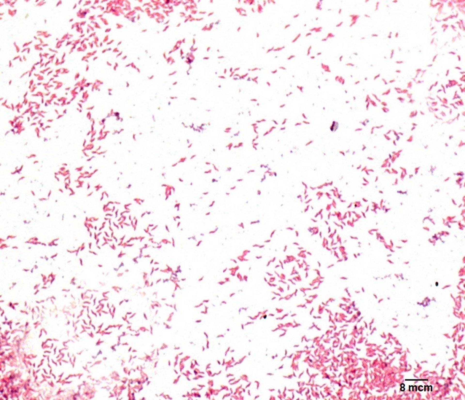
Micrograph showing Campylobacter jejuni in a Gram-stained smear of intestinal cells from a dog in experimentally induced enteritis.
Courtesy of Ms. Paula Yant.
Fresh fecal samples should be collected and transported to the laboratory, preferably on the same day and within at most 2 days, for processing. If transport to the laboratory is delayed, maintaining transport media and storage conditions at 4°C (39.2°F) produces the best results. Campylobacter spp are very sensitive to environmental conditions, including dehydration, atmospheric oxygen, sunlight, and increased temperature.
Campylobacter spp can be quickly outgrown by contaminating microbes during prolonged transport to the laboratory, and isolation of pure colonies for downstream testing can be difficult. Filtration using 0.45-mcm filters can help, because Campylobacter organisms will pass through.
Enrichment is required for clinical sampling unless material can be transported to the laboratory immediately. When samples are collected using swabs, the use of commercially available transport tubes containing growth medium, such as Amies, is recommended. The medium can be plain agar or charcoal based. Several media have been described for the transport of fecal specimens, including Cary-Blair, modified Cary-Blair, modified Stuart medium, Campy thioglycolate medium, alkaline peptone water, and semisolid motility test medium. Other media are recommended for isolating those Campylobacter spp specifically associated with reproductive losses (16, 17).
Pearls & Pitfalls
|
PCR assay–based methods of identifying Campylobacter spp are often preferred for their increased sensitivity, especially if the sample has been somewhat mishandled. Real-time PCR assay is more sensitive than culture for identifying C jejuni, and DNA isolated directly from fecal samples can be used (16). However, a positive test is not sufficient evidence to determine causation and must be considered in conjunction with clinical signs.
Differentiation of subspecies of Campylobacter can be necessary for identification of important pathogens. Conventional biochemical techniques can be used to make this determination; at present, however, identification relies on molecular tests and/or matrix-associated laser desorption/ionization–time-of-flight (MALDI-TOF) methods.
Treatment and Control of Enteric Campylobacteriosis
Supportive care
Antimicrobials according to susceptibility testing
Treatment of campylobacteriosis is typically supportive, with antimicrobial therapy reserved for patients that are immunocompromised or have severe clinical signs. If antimicrobial therapy is indicated for the treatment of dogs with campylobacteriosis, erythromycin and tylosin are frequent choices. Enrofloxacin can also be selected, although its use should be avoided in puppies due to the risk of cartilage damage. Also, it should be noted that Campylobacter jejuni might not necessarily be the cause of disease in a dog with diarrhea, even if PCR testing for the organism yields a positive result.
Clindamycin, gentamicin, tetracyclines, erythromycin, cephalosporins, and fluoroquinolones are generally effective against C jejuni, Campylobacter helveticus, and Campylobacter upsaliensis.
Campylobacter fetus, Campylobacter hyointestinalis, Campylobacter mucosalis, and Campylobacter sputorum are usually resistant to the fluoroquinolones yet susceptible to cephalosporins.
Campylobacter coli are now showing resistance to fluoroquinolones and cephalosporins. Susceptibilities to penicillins and trimethoprim vary across Campylobacter spp. Resistance to the fluoroquinolones, tetracyclines, kanamycin, and some other antimicrobials has been documented among Campylobacter spp, mediated by both chromosomal and plasmid mechanisms.
Antimicrobial susceptibility testing helps guide selection of an antimicrobial agent that is likely to be clinically effective, thus improving patient care and supporting antimicrobial stewardship efforts (18, 19).
Zoonotic Risk of Enteric Campylobacteriosis
C jejuni is one of the leading causes of acute bacterial enteritis in humans throughout the world (20), and it generally results in self-limiting diarrhea (often bloody), fever, abdominal discomfort, malaise, and in some cases vomiting. However, infections can be invasive and lead to severe manifestations, including Guillain-Barré syndrome (an acute immune-mediated disorder of the peripheral nervous system), reactive arthritis, and irritable bowel syndrome (21). Antimicrobial resistance among Campylobacter isolates further magnifies the risk to public health.
Campylobacter transmission can occur through foodborne exposure (especially undercooked chicken, unpasteurized milk and other dairy products, undercooked beef, and produce), waterborne exposure, or direct contact with feces of infected animals.
Dogs are a potential source of zoonotic transmission via direct contact (22). A prolonged multistate outbreak of drug-resistant C jejuni in the US was traced to direct contact with pet store puppies (23).
Prevention of campylobacteriosis should include strict hand hygiene, safe kitchen practices (avoiding cross-contamination, properly cooking poultry and other meat, and not consuming unpasteurized dairy products), not feeding raw meat-based diets to pets, and avoiding contact with feces.
Some animals remain colonized and become persistent Campylobacter shedders despite treatment with antimicrobials. If the goal of treatment is to decrease the risk of zoonotic transmission to a susceptible household member, antimicrobial treatment alone might be inadequate. Control involves treatment, removal of the patient to a clean environment, and prospective fecal testing to ascertain shedding status; nevertheless, low infective doses and the ubiquitous distribution of the organism pose substantial challenges in the prevention of infection.
Key Points
Campylobacter spp colonize the intestine; infections are subclinical in some species (eg, chickens and cattle), while other species (eg, dogs) can develop clinical disease.
Gastroenteritis is the most common clinical syndrome in animals and humans affected by Campylobacter.
Dogs and other animals can serve as a source of zoonotic transmission, regardless of whether clinical signs are present.
Foodborne illness via ingestion of Campylobacter-contaminated food is a notable public health concern, and antimicrobial resistance is common.
For More Information
References
Pascoe B, Futcher G, Pensar J, et al. Machine learning to attribute the source of Campylobacter infections in the United States: A retrospective analysis of national surveillance data. J Infect. 2024;89(5):106265. doi:10.1016/j.jinf.2024.106265
Brown C, Martin V, Chitwood S. An outbreak of enterocolitis due to Campylobacter spp in a beagle colony. J Vet Diagn Invest. 1999;11(4):374–376. doi:10.1177/104063879901100416
Leahy AM, Cummings KJ, Rodriguez-Rivera LD, Hamer SA, Lawhon SD. Faecal Campylobacter shedding among dogs in animal shelters across Texas. Zoonoses Public Health. 2017;64(8):623–627. doi:10.1111/zph.12356
Cummings KJ, Carney PC, Goggs R, et al. Fecal Campylobacter jejuni shedding in healthy dogs and dogs with acute diarrhea in central New York. Am J Vet Res. 2026:1-7. doi:10.2460/ajvr.26.01.0032
Odendaal MW, de Cramer KG, van der Walt ML, Botha AD, Pieterson PM. First isolation of Campylobacter jejuni from the vaginal discharge of three bitches after abortion in South Africa. Onderstepoort J Vet Res. 1994;61(2):193-195. https://pubmed.ncbi.nlm.nih.gov/7596570/
Bojanić K, Midwinter AC, Marshall JC, Rogers LE, Biggs PJ, Acke E. Isolation of Campylobacter spp from client-owned dogs and cats and retail raw meat pet food in the Manawatu, New Zealand. Zoonoses Public Health. 2017;64(6):438–449. doi:10.1111/zph.12323
Del Collo LP, Karns JS, Biswas D, et al. Prevalence, antimicrobial resistance, and molecular characterization of Campylobacter spp. in bulk tank milk and milk filters from US dairies. J Dairy Sci. 2017;100(5):3470-3479. doi:10.3168/jds.2016-12084
Englen MD, Hill AE, Dargatz DA, Ladely SR, Fedorka-Cray PJ. Prevalence and antimicrobial resistance of Campylobacter in US dairy cattle. J Appl Microbiol. 2007;102(6):1570-1577. doi:10.1111/j.1365-2672.2006.03189.x
Gensler CA, Aworh MK, Atlaw N, et al. Prevalence and characterization of Campylobacter species isolated from US swine: 2021 NAHMS enteric study. J Food Prot. 2025;88(8):100567. doi:10.1016/j.jfp.2025.100567
Rollo SN, Norby B, Bartlett PC, et al. Prevalence and patterns of antimicrobial resistance in Campylobacter spp isolated from pigs reared under antimicrobial-free and conventional production methods in eight states in the Midwestern United States. J Am Vet Med Assoc. 2010;236(2):201-210. doi:10.2460/javma.236.2.201
Rath A, Rautenschlein S, Rzeznitzeck J, et al. Impact of Campylobacter spp on the integrity of the porcine gut. Animals (Basel). 2021;11(9):2742. doi:10.3390/ani11092742
Bui XT, Wolff A, Madsen M, Bang DD. Fate and survival of Campylobacter coli in swine manure at various temperatures. Front Microbiol. 2011;2:262. doi:10.3389/fmicb.2011.00262
Sithole V, Amoako DG, Abia ALK, Perrett K, Bester LA, Essack SY. Occurrence, antimicrobial resistance, and molecular characterization of Campylobacter spp in intensive pig production in South Africa. Pathogens. 2021;10(4):439. doi:10.3390/pathogens10040439
Crawshaw T. A review of the novel thermophilic Campylobacter, Campylobacter hepaticus, a pathogen of poultry. Transbound Emerg Dis. 2019;66(4):1481-1492. doi:10.1111/tbed.13229
Shang Y, Ren F, Song Z, et al. Insights into Campylobacter jejuni colonization and enteritis using a novel infant rabbit model. Sci Rep. 2016;6:28737. doi:10.1038/srep28737
Lawhon SD, Cummings KJ, Wu J, et al. Comparison of PCR and culture for detection of Campylobacter jejuni in canine feces. J Vet Diagn Invest. 2026. Online ahead of print. doi:10.1177/10406387261434179
Tuominen K, Hansson I, Söderlund R, Bertilsson S, Belaghi R, Tamminen L-M. Comparison of culture-based methods for detecting Campylobacter jejuni and Campylobacter hyointestinalis in dairy cattle feces. Microbiol Spectr. 2025;13(10):e0147525. doi:10.1128/spectrum.01475-25
Shen Z, Wang Y, Zhang Q, Shen J. Antimicrobial resistance in Campylobacter spp. Microbiol Spectr. 2018;6(2):10.1128. doi:10.1128/microbiolspec.ARBA-0013-2017
Tang Y, Fang L, Xu C, Zhang Q. Antibiotic resistance trends and mechanisms in the foodborne pathogen, Campylobacter. Anim Health Res Rev. 2017;18(2):87-98. doi:10.1017/S1466252317000135
Scallan Walter EJ, Cui Z, Tierney R, et al. Foodborne illness acquired in the United States—major pathogens, 2019. Emerg Infect Dis. 2025;31(4):669–677. doi:10.3201/eid3104.240913
Batz MB, Henke E, Kowalcyk B. Long-term consequences of foodborne infections. Infect Dis Clin North Am. 2013;27(3):599–616. doi:10.1016/j.idc.2013.05.003
Campagnolo ER, Philipp LM, Long JM, Hanshaw NL. Pet-associated campylobacteriosis: a persisting public health concern. Zoonoses Public Health. 2018;65(3):304–311. doi:10.1111/zph.12389
Francois Watkins LK, Laughlin ME, Joseph LA, et al. Ongoing outbreak of extensively drug-resistant Campylobacter jejuni infections associated with US pet store puppies, 2016–2020. JAMA Netw Open. 2021;4(9):e212520. doi:10.1001/jamanetworkopen.2021.25203