Giardiasis is an intestinal infection with the protozoan flagellate parasite Giardia spp. Most infections of veterinary health importance are by G duodenalis, a species complex of different genotypes that tend to show host specificity; production animals, companion animals, and wildlife can all be infected. In general, the clinical signs of giardiasis are abdominal discomfort and diarrhea, often with steatorrhea. Diagnosis is usually by demonstration of cysts or antigens in fecal samples. Treatment regimens are variable and depend on specific indications. Hygiene measures are an essential component of control. Humans can also be infected. However, zoonotic transmission seems to occur relatively rarely, likely because of host specificity of different genotypes/assemblages.
Giardiasis is an intestinal infection with the protozoan parasite Giardia spp.Giardia infection occurs worldwide in most domestic and wild mammals and many birds. It is also very common in humans. In some animals — particularly ruminants, but also wild mammals — Giardia infection commonly occurs without overt or apparent clinical signs.
Three major groups of Giardia have been described based on the morphology of the vegetative stage (trophozoite): G muris from rodents, G agilis from amphibians, and a third group from various warm-blooded animals, which includes at least four species, including G ardeae and G psittaci from birds, G microti from muskrats and voles, and G duodenalis (also known as G intestinalis and G lamblia).
G duodenalis comprises a complex of eight different assemblages, some of which have been divided into subassemblages. Separate species names have also been proposed for some of the assemblages and subassemblages. Together, the assemblages and subassemblages of G. duodenalishave a wide mammalian host range.
In general, when veterinarians and physicians refer to Giardia or giardiasis, they are referring to G duodenalis. Among the eight assemblages of G duodenalis, some (A and B) have a relatively wide host range and can infect humans and a range of animals, whereas others are more host specific: assemblages C and D infect dogs, assemblage E infects livestock (pigs, cattle, sheep, goats), assemblage F infects cats, assemblage G infects rodents, and assemblage H infects pinnipeds.
Pearls & Pitfalls
|
Some G duodenalis assemblages have been subdivided into genotypes with different genetic characteristics and host predilections:
Assemblage A1 predominantly infects domestic animals but is common in wildlife and also infects humans.
Assemblage A2 predominantly infects humans but also infects domestic animals and occurs rarely in wildlife.
Assemblage A3 predominantly infects wildlife and occurs occasionally in domestic animals but seldom infects humans.
The division of G duodenalis into assemblages and genotypes is important for understanding the epidemiology and the possibility of between-species transmission, particularly zoonotic transmission, such as from domestic animals to their owners.
Surveys conducted in different parts of the world have reported point prevalence estimates of Giardia infection of around 10–30% for dogs and somewhat lower for cats, with higher rates of infection in younger animals (1). However, there are wide variations between studies.
Dogs shedding Giardia cysts often show no clinical signs, and the relevance of continually treating dogs that seem to be permanently infected but apparently healthy remains a subject of debate. Various other small pet animals (such as rabbits and guinea pigs) have also been shown to be susceptible to Giardia infection (2).
Among rodents, chinchillas are particularly notable for a high occurrence of infection with clinical signs. The predominance of assemblage B infections at some commonly used typing genes is identical to the assemblage B infections reported in humans. Thus, it appears that, although zoonotic transmission of Giardia to humans is generally rare, it can be more likely to occur from infections in chinchillas. Apparent transmission of assemblage A from cervids has also been reported (3).
In small ruminants (sheep and goats), data tend to indicate an occurrence of around 20–25%, ranging from < 10% to > 40%, with similar data for cattle (4).
On farms where Giardia infection has been diagnosed in ruminant stock, a cumulative occurrence of close to 100% can be expected. Very young ruminants (neonates < 2 weeks old) are less likely to be exposed to giardia oocysts and are therefore less likely to become infected; in general, however, once exposed to oocysts, animals < 6 months old tend to be more susceptible to infection with clinical signs of disease. Regardless, even infections with very high cyst excretion are infrequently associated with clinical signs (5). Nevertheless, diarrhea, weight loss, ill thrift, and even death have also been associated with Giardia infection in some animals.
In pigs, the occurrence of Giardia tends to be lower, at around 15%. However, it is age-related, with low infection rates (around 5%) recorded from piglets and sows and higher rates reported from starter pigs and fatteners (15–30%) (6). Many infected pigs seem to be clinically normal.
In horses, Giardia occurrence rates of < 10% (from around 0.5% to 20%) seem usual, with infection more likely in foals (7). However, clinical giardiasis in horses is rarely reported.
Cycle and Transmission of Giardiasis
The life cycle of Giardia is relatively simple (see ). The flagellate vegetative stage (trophozoite) of Giardia inhabits the lumen of the small intestine, where they can attach to the brush border of epithelial cells via the ventral disk (a specialized rigid cytoskeletal organelle on the ventral side of the trophozoite), absorb nutrients, and multiply by binary fission.


The life cycle of Giardia duodenalis in a dog.
European Scientific Counsel Companion Animal Parasites. ESCCAP Guideline 06: Control of Intestinal Protozoa in Dogs and Cats. 3rd ed. ESCCAP; 2025. 82qb4jhx_1477_ESCCAP_GL6_Update_v3_1p.pdf
Giardia has no intracellular stages. The trophozoites usually live in the proximal portion of the small intestine and migrate down the intestine during the course of infection, encysting in the small or large intestine. The prepatent period is generally 3–10 days.
Giardia cysts are passed in the feces and are immediately infectious. Cyst shedding can be continual over several days to weeks. However, it is often intermittent, especially in the chronic phase of infection, resulting in a gradual increase in environmental infection pressure. Cysts are very resistant, surviving for several weeks, or even months, in an appropriate (cool, damp, and humid) environment. Overcrowding and poor hygiene favor transmission.
Although trophozoites can be passed in the feces, especially during bouts of severe diarrhea, they do not survive for long in the environment, and if ingested, they are not able to infect a new host.
Giardia transmission occurs via the fecal-oral route, either by direct contact with an infected host that is shedding infective cysts or through a contaminated environment. Characteristics that facilitate infection include the high-volume and prolonged shedding of environmentally robust cysts by infected animals and the low infectious dose.
Pathogenesis of Giardiasis
Giardia infections cause an increase in intestinal epithelial permeability, increased numbers of intraepithelial lymphocytes, and activation of T lymphocytes. Trophozoite toxins and T-cell activation initiate a diffuse shortening of brush-border microvilli and decreased activity of the small-intestinal brush-border enzymes, especially lipase, some proteases, and disaccharidases. The diffuse shortening of the microvilli leads to a decrease in overall absorptive area in the small intestine and thus an impaired intake of water, electrolytes, and nutrients (8).
In addition, the proteins secreted by Giardia trophozoites contribute to degrading the intestinal mucous barriers and disrupting the intestinal intracellular junctions. The combined effect of decreased resorption, brush-border enzyme deficiencies, and a lack of integrity between cells of the intestine leads to dysregulation of the absorption and barrier functions of the intestinal epithelium, resulting in malabsorptive diarrhea.
The decreased activity of lipase and the increased production of mucin by goblet cells might explain the steatorrhea and mucous diarrhea that has frequently been described with clinical giardiasis. Giardia infection has, in some circumstances, been associated with a lower likelihood of viral and bacterial infection, possibly via immunological pathways or by physically covering the intestinal wall, preventing access to receptors.
Clinical Findings and Lesions of Giardiasis
Presentation of Giardia infections seems to vary both between and within animal groups. Giardia infection in cats, or more typically dogs, can be present without clinical signs and diagnosed only during routine fecal checks. However, it can also be associated with chronic diarrhea or steatorrhea, which can be continual or intermittent, particularly in puppies and kittens. Weight loss can also occur.
In clinical giardiasis in cats and dogs, feces usually are soft, poorly formed, pale, and malodorous, contain mucus, and appear fatty. Watery diarrhea is unusual, and blood is usually not present in feces. Vomiting is unusual; however, it can occur.
Pearls & Pitfalls
|
Giardiasis must be differentiated from other causes of nutrient malassimilation and intestinal malabsorption (e.g., exocrine pancreatic insufficiency). Clinical laboratory findings are usually normal.
Among small animal pets, chinchillas appear to be particularly prone to Giardia infection, especially kits. Although clinical signs of infection appear to be relatively common, infection in apparently healthy chinchillas has also been reported frequently in surveys (9).
In calves and lambs, and to a lesser extent in other production animals, giardiasis can result in diarrhea that does not respond to antimicrobial or coccidiostatic treatment. Excretion of pasty to fluid feces with mucus might indicate giardiasis, especially when the diarrhea occurs in young animals (1–6 months old).
Experimental Giardia infection of goat kids, lambs, and calves resulted in decreased feed efficiency and subsequently decreased weight gain (10). However, as with other animals, subclinical infection also occurs; very large numbers of cysts can be observed in the feces of cows and sheep with no apparent clinical signs.
Gross intestinal Giardia lesions are seldom evident, although microscopic lesions, consisting of villous atrophy and cuboidal enterocytes, can be present.
Diagnosis of Giardiasis
Detection of Giardia cysts or trophozoites in fecal samples
Detection of Giardia antigen in fecal samples
Detection of Giardia DNA in fecal samples
The two most used methods to diagnose Giardia infection in the veterinary setting are (i) identification of Giardia cysts and, considerably lessfrequently, trophozoites in fecal samples and (ii) detection of Giardia antigen in fecal samples. Molecular methods are becoming more frequently employed. However, they are less likely to be used in a veterinary clinical setting.
Giardia cysts are oval (9–15 × 7–10 mcm) and can be detected in feces concentrated by the centrifugation-flotation technique using zinc sulfate (specific gravity 1.18). Sodium chloride, sucrose, or sodium nitrate flotation media can be too hypertonic and distort the cysts. Staining cysts with iodine aids in identification (see ).

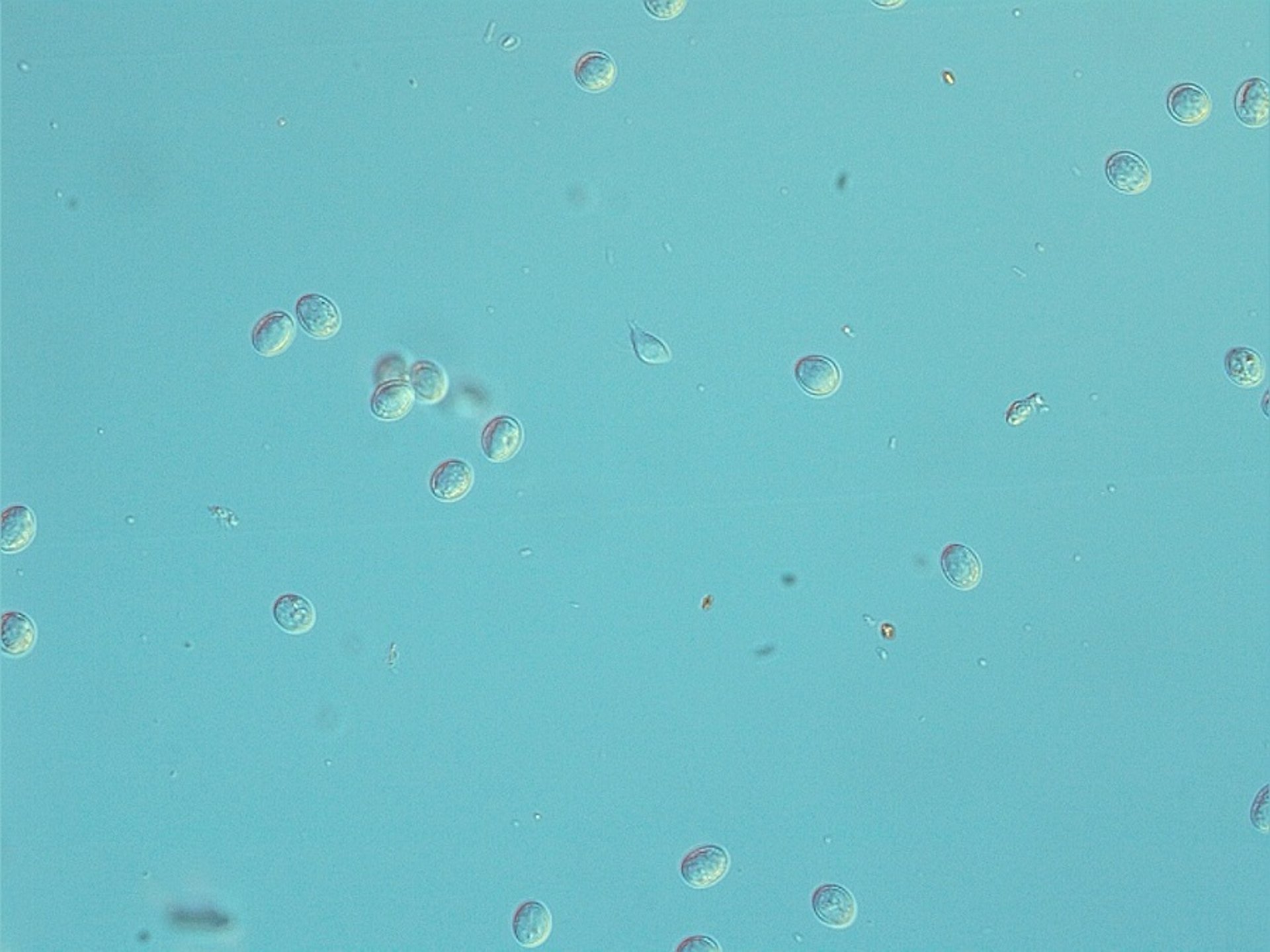
Photomicrograph of Giardia cysts in a fecal sample from a dog. Light microscopy, 1000X.
Courtesy of Dr. Lucy Robertson.
Because Giardia cysts are excreted intermittently, infections can be missed; therefore, several fecal examinations should be performed if giardiasis is suspected (eg, three samples collected over 3–5 consecutive days).
Pearls & Pitfalls
|
Because Giardia cysts rupture if dried or exposed to heat or extreme cold, empty cysts can be difficult to see and might not have the same flotation characteristics as intact cysts.
For laboratories with a fluorescent microscope, an immunofluorescent antibody staining of the cyst walls (IFAT) is a rapid and easy way to detect Giardia cysts. Several commercial IFAT kits are available, with the antibodies usually coupled to fluorescein isothiocyanate (see ).

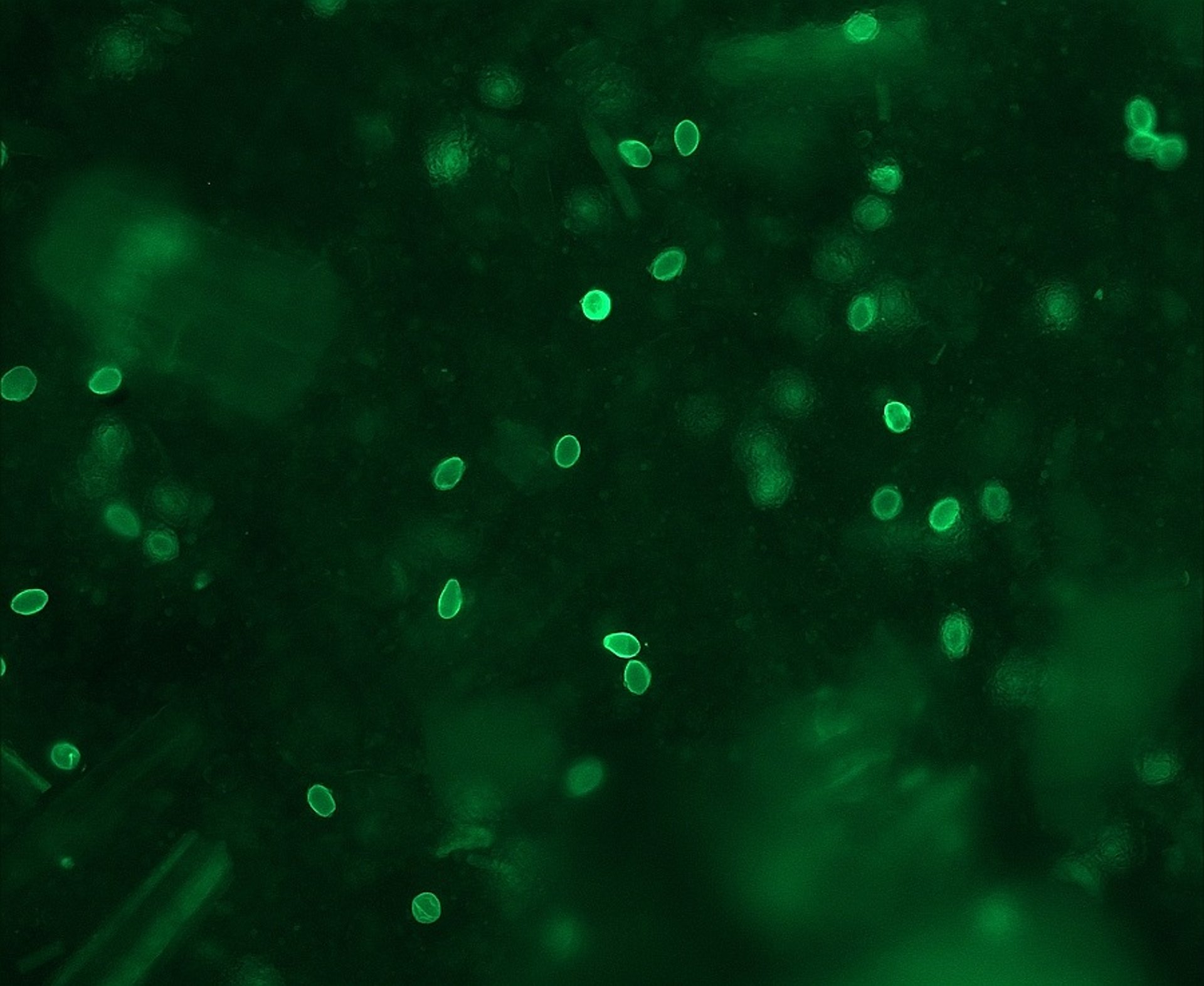
Photomicrograph of Giardia cysts from a fecal sample obtained from a cat infected during an outbreak at a cattery. The cysts are stained with fluorescein isothiocyanate bound to an antibody reactive against antigens in the cyst wall (IFAT stain).
Courtesy of Dr. Lucy Robertson.
Although cysts are the transmission stage of Giardia and are most often observed in feces, very occasionally the motile, piriform trophozoites (12–18 × 7–10 mcm) can be observed in saline smears of loose or watery feces. They should not be confused with yeast or with trichomonads, which have a single rather than double nucleus, an undulating membrane, and no concave ventral surface. The "falling leaf" swimming motion of Giardia trophozoites is also characteristic.
Giardia antigen that occurs in feces can be a useful method to diagnose Giardia infection, should infection be suspected but no cysts observed on microscopic examination. Microtiter plate-format ELISA tests to diagnose Giardia infection in animals are not commercially available. However, some veterinarians have reported success in evaluating dog samples with those designed for use in humans. In-house plate-format ELISA tests can also be used.
More commonly, and available from different commercial suppliers, are ELISA-based in-clinic tests, mostly for dog samples, that use lateral-flow technology; these are simple to use and can provide a result within minutes. The tests vary in their sensitivity and specificity, and information about these differences is proprietary. Given their relative expense and defined shelf-life, it is useful to investigate before investing in a particular test and to compare the results with other tests (such as IFAT or in-house plate-format ELISA) as a component of ongoing quality control.
Molecular methods (PCR assay) are also available for diagnosing Giardia, often as part of a panel assessing the presence of potential enteric pathogens. However, the generally high sensitivity of PCR to even low pathogen burdens may make interpretation of positive results challenging: the presence of Giardia, especially in small quantities, may be incidental or secondary to another cause of gastrointestinal disease, and in some parts of the world, PCR panels might not be readily available. Molecular tests may be most useful in cases where identifying the assemblage is relevant (eg, in outbreaks or if a human case and an infected animal are in the same household).
Protocols to identify the various assemblages have been published; however, relatively large numbers of nucleated cysts are necessary for successful DNA extraction and amplification at target genes. Unequivocal results can be elusive, particularly in dogs.
Treatment of Giardiasis
Halt clinical signs
Fenbendazole initially
No drugs approved for cats or livestock
There is some debate concerning whether animals without clinical signs that continue to shed Giardia cysts, even after treatment, should continue to be treated. Such cases need to be evaluated on an individual basis, preferably using a clinical decision-tree type format (11). Indeed, some guidelines advise against treating animals with subclinical infection; however, risk of transmission to other susceptible hosts should be considered, and strict hygiene measures are relevant.
Drug treatment regimens should probably aim to stop clinical signs rather than eliminate cyst shedding. Drug treatment approval varies between countries. There are no drugs approved for treatment of giardiasis in dogs and cats in the US, and no drug is licensed for treatment of Giardia infection in ruminants or other livestock.
Fenbendazole (50 mg/kg, PO, every 24 hours for 3–5 days) (12) is considered the first-line agent and is approved for treatment of dogs and cats in most countries in Europe. It is reported to stop shedding of Giardia cysts in dogs, with no adverse effects reported, and is safe for pregnant and lactating animals.
Metronidazole can also be considered, either alone or together with fenbendazole, should clinical signs continue. In the US, metronidazole oral suspension is FDA approved for use in dogs (25 mg/kg, PO, every 12 hours for 5 days) (13)., However, lower doses (10-25 mg/kg, PO, every 12 hours for 5-8 days) (12) are the most commonly used therapy. Although successful treatment of dogs with metronidazole has been reported, and it is licensed in most European countries for treating both dogs and cats, it has been associated with serious adverse effects of the CNS in dogs after chronic treatment or high doses.
In the US, no drugs are approved for treatment of giardiasis in cats.
Albendazole is not recommended for use in dogs or cats because of possible bone marrow suppression. A combination of praziquantel (5 mg/kg), pyrantel (14.4 mg/kg), and febantel (15 mg/kg), PO, every 24 hours for 3 days (14), has been shown to be effective for treating dogs and is licensed in most European countries and also outside the EU.
Other agents for treating giardiasis have also been tried experimentally, especially when more usual treatment regimens have been unsuccessful. These include, but are not limited to, ronidazole, tinidazole, secnidazole, azithromycin, nitazoxanide, and chloroquine. In general, published reports of such investigations indicate that the novel treatment has been successful.
Although there are no licensed treatments for Giardia in livestock, fenbendazole (15 mg/kg, PO, every 24 hours for 3 days) (15) and both fenbendazole and albendazole (5–20 mg/kg, PO, every 24 hours for 3 days) (16) have been shown experimentally to decrease cyst shedding in cattle and provide some clinical benefit (decreased diarrhea and weight gain). In a severe outbreak of giardiasis in sheep, treatment with fenbendazole at a dose of 10mg/kg, PO, every 24 hours for 3 consecutive days, was successful at clearing the infection (17). Paromomycin (25–75 mg/kg, PO, every 24 hours for 5 days) has also experimentally been found to be efficacious in cattle (18). Other treatments have been tried on livestock and have been reported to be effective (eg, secnidazole, 10 mg/kg, PO, once) (19).
That different giardiasis treatments are often tried indicates frequent frustration with cyst shedding, and sometimes clinical signs, that continues after treatment. It is unclear whether this indicates treatment failure or reinfection, although both can play a role.
Supportive or supplementary therapies for giardiasis, such as probiotic treatment, have been suggested. However, there is little reliable evidence that such therapies are effective.
Control of Giardiasis
Giardia cysts are immediately infective when passed in the feces and survive well in the environment, particularly in damp and cool conditions, in which cysts can stay infective for months. Cysts are a source of infection and reinfection for animals, particularly those in crowded conditions (eg, kennels, catteries, or intensive rearing systems for production animals). Thus, control should include removing feces as soon as possible (at least daily) and disposing of potentially infectious waste.
Infected dogs and cats should be bathed to remove Giardia cysts from the coat, and use of a shampoo containing chlorhexidine digluconatehas been suggested (11). Prompt and frequent removal of feces limits environmental contamination, as does subsequent disinfection. Cysts are inactivated by most quaternary ammonium compounds, steam, and boiling water.
Although no disinfectants are registered to kill Giardia cysts, regular frequent washing and drying of blankets, bedding, and food and water containers, along with other normal good hygiene practices, can limit transmission to other hosts and reinfection after recovery. Isolation of diarrheic pets or diagnosed carriers will also limit transmission.
To increase the efficacy of disinfectants, solutions should be left for 5–20 minutes before being rinsed off contaminated surfaces. Disinfection of grass yards or runs is impossible, and these areas should be considered contaminated for at least a month after infected dogs were last present.Cysts are susceptible to desiccation, so areas should be allowed to dry thoroughly after cleaning.
Key Points
Giardia duodenalis infects many hosts, including humans and most pets and livestock, although not all isolates are infectious to all animals.
Many Giardia infections are subclinical. Clinical infections are generally associated with chronic diarrhea with steatorrhea.
Various diagnostic options are focused on identification of either transmission stages (cysts) in feces or fecal antigen testing. PCR assay testing is infrequently used in the veterinary clinic.
Various treatment options, some of which are extralabel, aim at suppressing clinical signs rather than halting cyst shedding.
Good hygiene practices should be considered an essential component of control.
For More Information
ESCCAP: GL6 Control of Intestinal Protozoa in Dogs and Cats. Currently available in 9 languages (Dutch, English, French, German, Hungarian, Italian, Polish, Spanish, Swiss French, Swiss German, Ukrainian). English version updated in 2025.
CAPC: Giardia (Dog, updated 2025); (Cat, updated 2025).
Scorza, V. (2013). Giardiasis: an overview. Clinicians Brief.
Tysnes KR, Skancke E, Robertson LJ. Subclinical Giardia in dogs: a veterinary conundrum relevant to human infection. Trends in Parasitol. 2014;30(11):520-527. doi:10.1016/j.pt.2014.08.007
Tysnes KR, Luyckx K, Cantas L, Robertson LJ. Treatment of feline giardiasis during an outbreak of diarrhoea in a cattery: potential effects on faecal Escherichia coli resistance patterns. J Feline Med Surg. 2015;18(8):679-682.
Also see pet owner content regarding giardiasis in dogs, cats, and horses.
References
Bouzid M, Halai K, Jeffreys D, Hunter PR. The prevalence of Giardia infection in dogs and cats, a systematic review and meta-analysis of prevalence studies from stool samples.Vet Parasitol. 2015;207(3-4):181-202. doi:10.1016/j.vetpar.2014.12.011
Pantchev N, Broglia A, Paoletti B, et al. Occurrence and molecular typing of Giardia isolates in pet rabbits, chinchillas, guinea pigs and ferrets collected in Europe during 2006–2012. Vet Rec. 2014;175(1):18. doi:10.1136/vr.102236
Utaaker KS, Ytrehus B, Kifleyohannes T, Robertson LJ. 2026. From the field - a case of zoonotic transmission of Giardia duodenalis from wild reindeer?Epidemiol Infect. 2026. doi:10.1017/S0950268826101460
Li Z, Wei W, Ma Y, Hao L, Guo Y, Wang R. Prevalence and risk factors of Giardia duodenalis in sheep and goats in global: A systematic review and meta-analysis. Prev Vet Med. 2025;244:106623. doi:10.1016/j.prevetmed.2025.106623
Kifleyohannes T, Skorpen E, Rosnes Hansen K, Stuen S, Robertson LJ. Cryptosporidium and Giardia infections of lambs in Southwest Norway: a longitudinal study. Acta Vet Scand. 2025;67:40. doi:10.1186/s13028-025-00823-8
Asghari A, Ebrahimi M, Shamsi L, Sadrebazzaz A, Shams M. Global molecular prevalence of Giardia duodenalis in pigs (Sus domesticus): A systematic review and meta-analysis. Heliyon. 2023;9(2):e13243. doi:10.1016/j.heliyon.2023.e13243
Mizani A, Taherkhani P, Kia Lashaki E, Hosseini SA, Basirpour B, Dodangeh S. The global prevalence of Giardia infection in horses: A systematic review and meta-analysis. J Equine Vet Sci. 2025;150:105596. doi:10.1016/j.jevs.2025.105596
Adam RD. Giardia duodenalis: Biology and Pathogenesis. Clin Microbiol Rev. 2021;34(4):e00024-19. doi:10.1128/CMR.00024-19
Zikmundová V, Horáková V, Tůmová L, et al. Pet chinchillas (Chinchilla lanigera): Source of zoonotic Giardia intestinalis, Cryptosporidium ubiquitum and microsporidia of the genera Encephalitozoon and Enterocytozoon. Vet Parasitol. 2024;331:110275. doi: 10.1016/j.vetpar.2024.110275
Koudela B, Vitovec J. Experimental giardiasis in goat kids. Vet Parasitol. 1998;74(1):9-18. doi:10.1016/s0304-4017(97)00146-5
Tysnes KR, Skancke E, Robertson LJ. Subclinical Giardia in dogs: a veterinary conundrum relevant to human infection. Trends Parasitol. 2014;30(11):520-527. doi:10.1016/j.pt.2014.08.007
Giardia. Companion Animal Parasite Council (CAPC). Accessed March 26, 2026. https://capcvet.org/guidelines/giardia/
AYRADIA (metronidazole oral suspension) for dogs. US Product Label for Dogs. Virbac AH, Inc.; 2023. Accessed March 26, 2026. https://us.virbac.com/products/antibiotics/ayradiaTM-metronidazole-oral-suspension-for-dogs
European Scientific Counsel Companion Animal Parasites. ESCCAP Guideline 06: Control of Intestinal Protozoa in Dogs and Cats. 3rd ed. ESCCAP; 2025. 82qb4jhx_1477_ESCCAP_GL6_Update_v3_1p.pdf
Geurden T, Vercruysse J, Claerebout E. Field testing of a fenbendazole treatment combined with hygienic and management measures against a natural Giardia infection in calves. Vet Parasitol. 2006;142(3-4):367-371. doi:10.1016/j.vetpar.2006.07.019
O'Handley RM, Cockwill C, Jelinski M, McAllister TA, Olson ME. Effects of repeat fenbendazole treatment in dairy calves with giardiosis on cyst excretion, clinical signs and production. Vet Parasitol. 2000;89(3):209-218. doi:10.1016/s0304-4017(00)00200-4
Aloisio F, Filippini G, Antenucci P, et al. Severe weight loss in lambs infected with Giardia duodenalis assemblage B. Vet Parasitol. 2006;142(1-2):154-158. doi: 10.1016/j.vetpar.2006.06.023
Geurden T, Claerebout E, Dursin L. et al. The efficacy of an oral treatment with paromomomycin against an experimental infection with Giardia in calves. Vet Parasitol. 2006;135(3-4):241-247. doi:10.1016/j.vetpar.2005.09.006
Volpato A, Fortuoso BF, Campigotto G. et al. Secnidazole for control of giardiasis in dairy calves. Exp Parasitol. 2018;189:16-18. doi:10.1016/j.exppara.2018.04.008