


Many of the dental disorders of dogs are similar to those found in people. Proper dental care, including preventive methods like tooth brushing, can help keep your dog’s teeth and gums healthy.
Dental Terms
What Most People Call It | What Your Veterinarian Might Call It |
|
|
Gum Disease
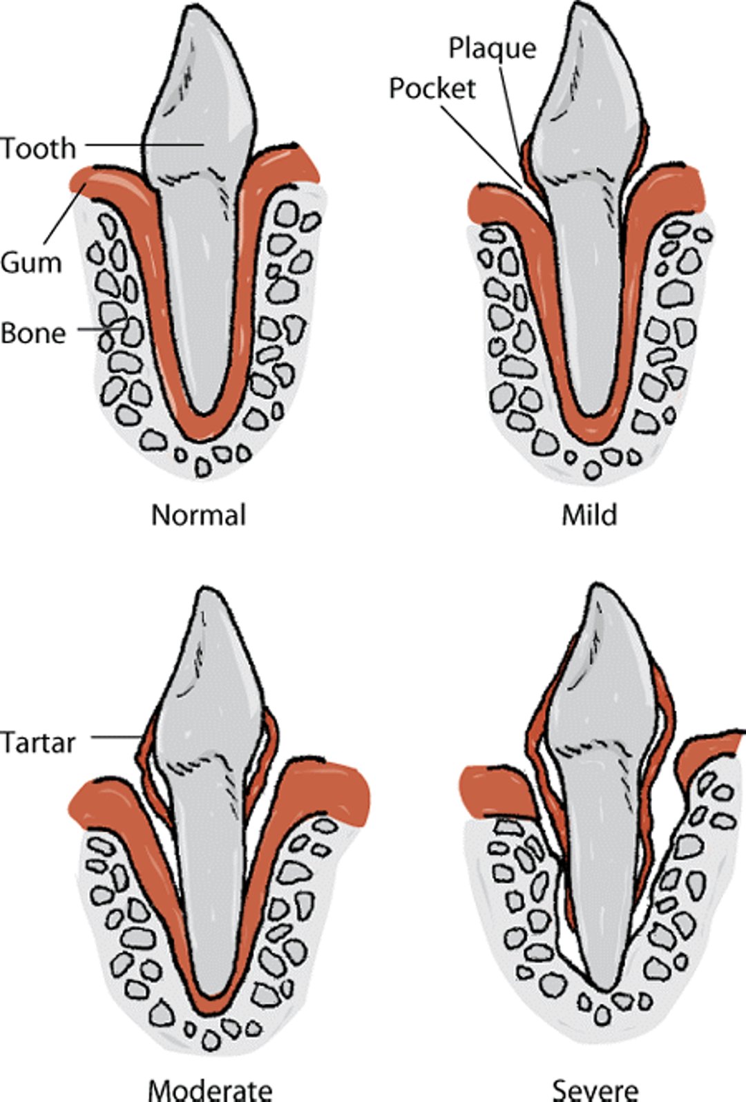
Bacterial infection of the tissue surrounding the teeth causes inflammation of the gums, the ligaments that anchor the teeth, and the surrounding bone. If gum (periodontal) disease goes untreated, teeth can be lost due to the loss of their supporting tissues. This is the major reason for tooth loss in dogs.
Gum disease is caused by accumulation of bacteria (plaque) at the gum line due in part to a lack of proper oral hygiene. Other contributing factors may include breed, genetics, age, and diet. As the number of bacteria below the gumline increases, bacterial waste products, such as hydrogen sulfide, ammonia, acids, and other compounds, accumulate and damage tissues. The dog’s own response to this infection (inflammation) also causes tissue breakdown and loss of the tooth’s supporting tissues. There are 2 forms of gum disease: gingivitis and periodontitis.
Gingivitis
In gingivitis, the gums become inflamed because of bacterial plaque, but the ligaments and bone are not yet affected. The gums change in color from coral-pink to red or purple, and the edge of the gum swells. The gums tend to bleed on contact. Bad breath is common. Gingivitis can be reversed with proper tooth cleaning but, if untreated, may lead to periodontitis (see below).
Progression of periodontal disease in dogs

Gingivitis usually can be treated by thorough professional cleaning of the teeth while the dog is under anesthesia. This should include cleaning below the gum line. If gingivitis does not improve, the dog should be examined again in case more extensive cleaning is required. When cleanings are completed, your veterinarian may apply a sealant to the teeth to prevent bacterial buildup and improve healing. Dogs that do not respond to treatment should be evaluated for other disease, such as immune system problems and diabetes. Gingivitis will reoccur if the teeth are not kept clean and free of plaque. Therefore, at-home oral hygiene methods, such as brushing, as well as regular cleanings by your veterinarian are important.
Periodontitis
In periodontitis, the tissue damage is more severe and includes the gums, ligaments, and bone. It usually is seen after years of development of plaque, tartar, and gingivitis. It is irreversible and results in permanent loss of tooth support. Small-breed dogs usually have more problems with periodontitis than large-breed dogs. Dogs that have a regular diet of hard kibble develop fewer problems due to the mechanical cleaning effect on the teeth as the food is chewed. Back teeth are affected more often than front teeth. The upper teeth are affected more severely than the lower teeth, and the cheek surfaces of the teeth have more disease than the surfaces near the tongue. Gingivitis is often first noticed at about 2 years of age but improves if treated. Periodontitis usually begins at 4 to 6 years of age and, if untreated, progresses to tooth loss.
Periodontitis is treated with thorough professional cleaning above and below the gum line. In some cases, surgery will be needed to gain access to the root surface for cleaning. Your veterinarian can determine the extent of bone support loss by taking x-rays of the jaws. These are usually recommended as a normal part of periodontal disease diagnosis and treatment planning. Extractions are often necessary for dogs with periodontitis. Extractions allow the tissue to heal, and dogs do surprisingly well without the teeth. Finally, veterinarians will treat any factors contributing to periodontitis, such as tooth crowding or underlying diseases.
If your dog has been treated for periodontitis, you will need to continue oral hygiene care at home. Follow your veterinarian’s instructions, which might include daily toothbrushing, dietary changes, plaque prevention gel, and oral rinses.
Prevention
The most important thing to remember is that gum disease will not develop around clean teeth. At-home methods to keep your pet’s teeth clean, such as toothbrushing and diet, along with regular dental examinations, are the best ways to help prevent gum disease. Daily toothbrushing is best, but wiping the teeth with a gauze at least every 2–3 days can remove plaque in dogs that do not allow toothbrushing. Only the outside surface of the teeth needs to be brushed or wiped. Toothpastes made for people should not be used. Your veterinarian might recommend foods, toys, and treats to help clean plague off of teeth. Reliable recommendations for treats and food that can help control plague are available at the Veterinary Oral Health Council website.
Endodontic Disease
Endodontic disease occurs inside the teeth. Several different conditions fall into this category. The causes include injury, tooth fracture, enamel abnormality, and tooth decay. Teeth can be fractured from external trauma (eg, aggressive play or automobile accident) or from biting inappropriate objects (eg, real bones, hooves, antlers, hard nylon toys, rocks, fences, or cages). Treatment is either tooth extraction or a root canal procedure. Signs can include painful teeth that your pet resists having touched or tapped; a tooth with a reddish-brown, purple, or gray color; a visible fracture; a red or black hole on a crown; a swelling on the face; or a decrease in appetite. In advanced cases dental fistulas (draining tracts) occur. However, most dogs mask their pain, making diagnosis difficult. X-rays of the mouth are used to identify affected teeth and help determine the proper treatment.
Developmental Abnormalities
There are several developmental abnormalities that affect the teeth of dogs. Many of these have a genetic component. In general, abnormalities that affect a dog's comfort, health, or ability to function require treatment; those that only affect the look (esthetics) of the tooth do not.
Unerupted Teeth
Some breeds, especially small breeds (such as Maltese), are prone to having teeth that remain under the gumline (unerupted teeth). Breeds that have a shortened, flattened head (brachycephalic breeds) can have teeth that fail to erupt because they are in an incorrect position. X-rays should be taken of any areas where teeth are missing. Repeating these x-rays on a regular basis is important because an unerupted tooth can form a cyst that can destroy a large section of the jaw. Any unerupted teeth causing a problem should be extracted. Dogs that have any unerupted teeth should be spayed or neutered so that the condition is not passed to offspring.
Improper Bite
Proper growth and development of the mouth and teeth depend on a series of events that must occur in proper sequence or longterm complications will occur. Early detection and intervention is the best way to prevent more serious problems. Dental development can be divided into 3 stages, each of which can have its own set of problems that require inspection by a veterinarian. Stage 1 is from 0 to 16 weeks of age, Stage 2 is from 16 weeks to 7 months of age, and Stage 3 is from 7 months to 1½ years of age.
Stage 1: Puppies are born with relatively long upper jaws (“overbite”), which allow them to nurse. As the dog grows and begins to eat solid food, the lower jaw goes through a growth spurt. If certain of the lower baby teeth come in before the growth spurt, they can get caught behind the upper teeth and prevent the lower jaw from developing to its proper length. If your veterinarian notices this pattern in a puppy, he or she will probably recommend removing several lower baby (deciduous) teeth. If this is done, the lower jaw will have the opportunity to reach its full length, thus averting problems with the permanent teeth. Some dogs will naturally develop a significant overbite, whether or not tooth extraction is performed.
The reverse situation can also occur. In these cases, the lower jaw grows faster than usual and becomes too long for the upper jaw, producing an “underbite.” This condition can be detected as early as 8 weeks of age. Again, certain teeth from the upper jaw may become caught behind those of the lower jaw, preventing proper growth of the upper jaw. The usual treatment is to extract several upper baby teeth. Early detection and correction of such problems will produce the best longterm results.
Other congenital and developmental problems in Stage 1 that may require treatment include extra teeth (which should be extracted only if they are causing problems) or incorrect position of a baby tooth, which should be extracted if it is interfering with other teeth. If jaw growth is different between the left and right sides, teeth may be extracted from the less-developed side. This procedure provides the best opportunity for the uneven growth to correct itself.
Stage 2: The most important problem that can occur during this stage is the retention of baby teeth. Normally, shedding begins around 14 weeks of age with the loss of the upper central incisors. For the next 3 months, the baby teeth are replaced with permanent teeth. Additional permanent teeth that complete the dog’s tooth pattern also erupt during this stage. If the baby teeth are not lost at the time the corresponding permanent teeth are coming in, abnormal tooth position and bite may result. If retained baby teeth are removed by a veterinarian as soon as they are noticed; complications can usually be prevented from occurring later on.
Another developmental problem noted in Stage 2 is abnormal positioning (tilting) of lower or upper canine teeth. Depending on the specific situation and age of the dog, orthodontic treatment (that is, “braces” for your pet) can be used to align teeth in their correct positions. Tilting of the upper canines typically is seen in Shetland Sheepdogs, although it has been reported in many other small breeds. Overbite is sometimes noticed during this stage and can be treated with a special plate fitted in the mouth. In more severe cases, tooth shortening or extractions might be necessary.
Stage 3: Additional types of incorrect tooth placement can occur during this stage of your pet’s growth. Treatment, if necessary, may include orthodontic treatment or tooth extraction. Crowding of teeth is resolved by extracting one or more teeth. Likewise, teeth that are significantly rotated, as are often found in flat-faced (brachycephalic) breeds, are usually removed.
Enamel Defects
During the development of tooth enamel on both baby and permanent teeth, fevers and the deposition of chemicals within the tooth may cause permanent damage. The canine distemper virus is especially damaging because it both attacks the enamel-producing cells of the teeth and causes a fever. This results in tooth enamel that is thinner than normal. Other fever-causing diseases may result in enamel that does not develop properly and is weaker than normal. Severe malnutrition in young dogs may result in enamel defects. Enamel defects in isolated teeth are most likely the result of trauma or infection. Often, the infections in fractured baby teeth affect the enamel of the permanent teeth that come in behind them. Enamel defects may also be inherited, especially in Siberian Huskies.
Treatment of these conditions can include bonding of synthetic materials to the teeth, fluoride treatment, and frequent dental preventive care.
Trauma to the Face and Jaw
Fractured teeth and jaws often occur because of trauma (eg, falls, chewing, fighting with other animals, and automobile accidents). The jaw can also be fractured because it has been weakened by severe periodontitis or cancer. Fractured teeth should be inspected by a veterinarian to determine whether there has been damage to the tooth pulp. If fractures extend into the pulp, root canal (endodontic) treatment or tooth extraction will be needed. Restorative techniques such crowns can repair defects in tooth structure if the damage is limited to the hard tissues or the pulp has already been treated with root canal treatment. Wounds to the gums or other soft tissues should be treated by the veterinarian as well.
Fractures of the bone will need to be stabilized by the veterinarian. Stabilization may require the use of wires, pins, or other materials. As long as the correct bite position can be maintained, healing is rapid and most of the supporting material can be removed by the veterinarian in about 6 to 8 weeks. A feeding tube may be needed if the dog has difficulty eating while the injury heals.
Tooth Decay (Cavities)
Tooth decay is uncommon in dogs. When it does occur, decay usually is seen as pits on the bite surfaces of the molar teeth. If cavities do occur, they can be filled in a way similar to that used in human dentistry.
For More Information
Also see professional content regarding dental disorders.


