

The eyes of animals, including dogs' eyes, function much like yours. Animals also develop many of the same eye problems that people can have, including cataracts, glaucoma, and other problems. It is important for your dog to receive good eye care to protect its sight and allow it to interact comfortably with its environment.
The eye is an active organ that constantly adjusts the amount of light it lets in and focuses on objects near and far. It produces continuous images that are quickly relayed to the brain.
Anatomy of the eye

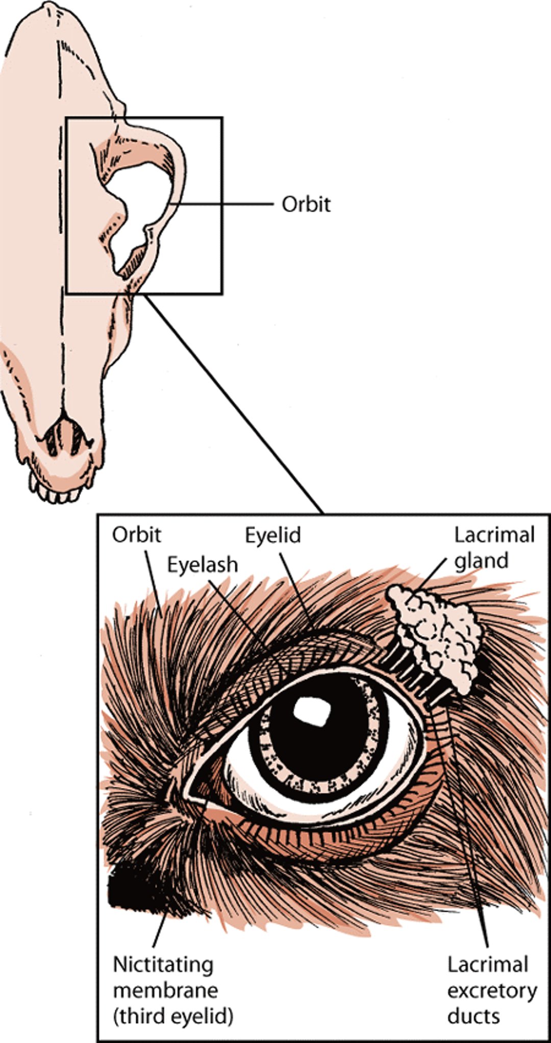
The bony cavity or socket that contains the eyeball is called the orbit. The orbit is a structure that is formed by several bones. The orbit also contains muscles, nerves, blood vessels, and the structures that produce and drain tears.
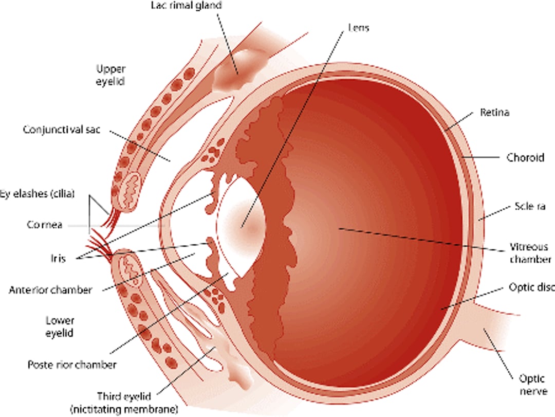
The white of the eye is called the sclera. This is the relatively tough outer layer of the eye. It is covered by a thin membrane, called the conjunctiva, located near the front of the eye. The conjunctiva runs to the edge of the cornea and covers the inside of the eyelid. The cornea is a clear dome on the front surface of the eye that lets light in. The cornea not only protects the front of the eye, but also helps focus light on the retina at the back of the eye. The iris is the circular, colored area of the eye. It controls the amount of light that enters the eye by making the pupil larger or smaller. The pupil is the black area in the middle of the eye. The pupil is controlled by the circular sphincter muscle. When the environment is dark, the pupil enlarges to let in more light; when the environment is bright, the pupil becomes smaller to let in less light.
The lens, which sits behind the iris, changes its shape to focus light onto the retina. Small muscles (ciliary muscles) contract to cause the lens to become thicker, which allows the lens to focus on nearby objects. The ciliary muscles relax to cause the lens to become thinner when it focuses on distant objects. These lens changes are limited in dogs. The retina contains the cells that sense light (photoreceptors). The most sensitive area of the retina is called the area centralis in dogs; this area contains thousands of tightly packed photoreceptors that make visual images sharp. Each photoreceptor is attached to a nerve fiber. All the nerve fibers are bundled together to form the optic nerve. The photoreceptors in the retina convert the image into electrical impulses, which are carried to the brain by the optic nerve.
The upper and lower eyelids are thin folds of skin that can cover the eye and reflexively blink to protect the eye. Blinking also helps spread tears over the surface of the eye, keeping it moist and clearing away small particles. The eyes of a dog are protected not only by the same types of eyelids that people have, but also by the nictitating membrane, which is sometimes called the third eyelid. This additional eyelid is a whitish pink color, and it is found under the lower eyelids on the inside corner of the eye (near the nose). The third eyelid extends across the eye when needed to protect the eyeball from scratches (for example, while traveling through brush) or in response to inflammation.
To function properly, eyes must be kept moist. Tears are the source of this needed moisture. Tears are comprised of water, oil, and mucus. Lacrimal glands produce the watery portion of tears. They are located at the top outer edge of each eye. Mucus glands in the conjunctiva (called goblet cells) produce mucus. Meibomian glands within the eyelids produce the oily portion. The mixture of water, oil, and mucus creates a more protective tear that is slower to evaporate. Nasolacrimal ducts allow tears to drain from each eye into the nose. Each of these ducts has openings at the edge of the upper and lower eyelids near the nose.
Structures that protect the eye, dog

Physical Examination of the Eye
Because of the importance of sight to your dog, one of the critical aspects of any examination or checkup will be an examination of your pet’s eyes. Be prepared to provide any background or medical history (such as any previous injury to the eye, history of treatments or medications used, any signs of visual problems, and vaccination history) that might help with the diagnosis of any eye problem.
The first step of the examination involves checking to be sure that the shape and outline of the eyes are normal and that there are no obvious abnormalities. Then, using light and magnification in a darkened room, the reflexes of the pupils and the front part of the eye are examined. Depending on these findings and the reasons for the checkup, additional tests may be needed. Some parts of the examination may require sedation or anesthesia.
A test, called the Schirmer tear test, may be performed to ensure that the eyes are producing enough tears to keep them moist. This is a relatively simple test in which a small paper strip is inserted under the eyelid to measure the amount of moisture produced. Another common test involves placing a small drop of fluorescein stain into each eye, which allows defects—such as scratches in the cornea of the eye—to be detected.
Pressure within the eye is measured painlessly using an instrument called a tonometer. (If eye pressure is too high, optic nerve damage can occur, leading to irreversible blindness.) A swab may also be done to culture for bacteria or fungi. The eyelids may be turned inside out to examine the underside. The nasolacrimal tear duct may be flushed to evaluate the external parts of the eye. Drops may be added to the eyes to allow the pupils to become dilated so that the veterinarian may examine the internal parts of the eye using an ophthalmoscope.
For More Information
Also see professional content regarding eye structure, function, and physical examination.


