

The Rule of 20 is a list of 20 critical parameters that should be evaluated at least daily in all critically ill animals; many of these should be assessed several times per day.
Anticipation, not reaction, is the key to successful management of critically ill animals. Animals must be effectively treated and actively monitored to detect or prevent organ compromise before organ failure occurs. This often requires aggressive and repeated fluid balance evaluation, close patient monitoring, and support throughout the course of definitive therapy.
Tissue hypoxia and organ dysfunction or failure can be a direct result of the primary disease, secondary to a complication of the primary disease, or even due to adverse events during therapy. Organs frequently affected include the heart and blood vessels, kidneys, lungs, GI tract, and liver. When the disease process is multisystemic, problems such as malnutrition and coagulopathies must be anticipated.
Optimal care requires a thorough and methodical approach to diagnostic procedures, monitoring, specific therapeutics, and supportive care.
Using the Rule of 20 ensures that the clinical status and therapeutic strategy for each animal is comprehensive and meets the patient's ongoing needs.
Like any monitoring tool, the Rule of 20 is not a static concept but a dynamic one; the specifics of each parameter will change with advancements in laboratory testing, understanding of disease pathology, and current concepts in critical care. In addition, the systems examined in the Rule of 20 are not singularities; each is impacted by and can impact other parameters, so each parameter should be assessed while considering the patient as a whole.
Applications of the Rule of 20 are always evolving. Since the Rule of 20 was first published in 2017, developments of note include the following:
monitoring of advanced coagulation assessment
alterations in management of diabetic ketoacidosis (DKA) and relatively new developments of euglycemic DKA with sodium-glucose cotransporter-2 (SGLT2) inhibitors
ultrasonographic assessment techniques
understanding of endothelial physiology
growing concerns about antimicrobial resistance
in-hospital relaxation techniques
the addition of many consensus statements
diagnosis and management of a variety of diseases
Diagnostic tools being investigated that might apply to the Rule of 20 in the future include biomarkers for predicting cardiovascular risk (eg, NT-proBNP).
Various scoring systems help to monitor critically ill patients, such as the animal trauma triage score, modified Glasgow Coma Scale (MGCS), and the Glasgow Composite Measure Pain Scale.
Fluid Balance in the Critically Ill Animal
The goal of fluid therapy is to provide adequate perfusion (intravascular volume) and hydration (interstitial volume) without overloading the interstitial space.
Peripheral perfusion can be assessed by physical parameters such as heart rate, mucous membrane color, pulse quality, and mentation, as well as by measured parameters such as blood pressure, central venous pressure, urine output, and blood lactate concentration. Hydration can be assessed by physical parameters such as mucous membrane and corneal moistness and skin turgor, and by measured values (eg, body weight, PCV, total solids).
Animals with systemic inflammatory response syndrome (SIRS) diseases may require more fluid than expected because of peripheral vasodilation and loss of endothelial integrity, making the administration of colloids with crystalloid solutions optimal. When veterinarians treat fluid deficits, intravascular deficits should be addressed rapidly first; interstitial deficits should be treated using standard calculations to correct dehydration and monitor ongoing losses. Fluids should be reassessed regularly, and typical fluid prescriptions include three components; replacement fluids, maintenance fluids, and ongoing losses. The American Animal Hospital Association (AAHA) updated their fluid therapy guidelines for dogs and cats in 2024 (1).
As with any drug, fluid choice and amount can have detrimental effects if administered inappropriately, so fluid prescription must be appropriate for the patient's needs. Administration of crystalloids carries the risk of injury as well, particularly if done in excess; endothelial injury may occur with crystalloid administration, and fluid overload can negatively impact the function of many organs.
Oncotic Pull/Albumin in the Critically Ill Animal
Albumin provides the major intravascular oncotic pull in the normal vasculature. In conditions in which massive blood loss or leakage of plasma proteins due to an exudative process has occurred, albumin is lost from the intravascular space. This loss of intravascular oncotic pressure, combined with increased capillary permeability associated with many SIRS diseases, requires treatment using synthetic colloids with a higher molecular weight than that of albumin.
Colloid oncotic pressure (COP) can be measured with colloid osmometers; however, these are not commonly available in veterinary practice. Formulas are available to calculate COP based on plasma protein concentrations; however, they are not reliable predictors of measured COP.
Normal COP in dogs is approximately 20 mm Hg. In patients with moderate to severe decreases in COP or in total proteins, natural and synthetic colloids should be administered:
Natural colloids include plasma products, concentrated or lyophilized human or canine albumin, and stroma-free Hgb.
Synthetic colloids include dextrans and hydroxyethyl starches.
Traditional understanding of transcapillary fluid shifts was described in Starling's model, focusing on intravascular and interstitial COP and hydrostatic pressures. The discovery and understanding of the endothelial glycocalyx has led to a revision of Starling's principles of transcapillary fluid shifts (2, 3).
In the endothelial glycocalyx model, the vascular system does not absorb fluid from the interstitial space; rather, fluid is returned to the vasculature through the lymphatic system. As our understanding of endothelial cell function and the impact of the endothelial glycocalyx model expands, novel therapeutic options or shifts in best practices in oncotic therapy will likely follow.
Part of the oncotic activity normally provided by albumin can be provided by synthetic colloids; however, only albumin can perform other functions, such as drug, cation, and hormone transport; free radical scavenging; and acid-base balance.
Albumin can be lost with a variety of diseases (GI, renal, or SIRS); it is also a negative acute-phase protein, so production of albumin drops during critical illness. Interstitial albumin stores may be drawn upon to replace serum albumin; however, this "autotransfusion" effect is now thought to be quite limited.
Serum albumin concentrations < 2 g/dL have been associated with complications and a poor prognosis in several disease states (4, 5); however, neither restoration of albumin concentrations nor administration of albumin compared to crystalloids has clearly improved overall rate of survival to discharge. Plasma and albumin transfusions are often administered to supplement the albumin to reach a minimum target of 2 g/dL; however, large volumes of plasma are required to reach this goal and can often be cost-prohibitive. Lyophilized canine serum albumin is available in 5-g vials, allowing for replacement of albumin with lower total volumes compared to plasma transfusions; however, cost remains a factor, and availability is inconsistent.
Human serum albumin (HSA) products have been used in critically ill dogs but may result in severe organ dysfunction because anti-HSA antibodies may be preexisting or may develop days to weeks later.
Interstitial stores of albumin also play an important role in critically ill patients with fluxes occurring from illness as well as from fluid administration; these stores must also be replenished, making albumin physiology substantially more complex than simply monitoring serum concentrations.
Glucose in the Critically Ill Animal
The goal with a critically ill animal is to maintain glucose between 80 and 120 mg/dL (approximately 4.4–6.6 mmol/L). Severe hypoglycemia can cause hypotension or neurological dysfunction ranging from weakness to stupor or seizures. The following conditions can result in hypoglycemia:
sepsis
inadequate nutrition (in young or critically ill patients)
heatstroke
severe renal or hepatic disease, including portosystemic vascular anomalies
certain types of neoplasia (insulinoma)
toxicities (xylitol, some medications)
iatrogenic insulin administration
In addition, neonates and small stature dogs have lower liver glycogen stores and minimal body fat reserves, which increases risk of hypoglycemia. Toy and miniature breeds of dogs are especially at risk, likely due to a suspected alanine deficiency that impairs normal gluconeogenesis.
Dextrose supplementation is warranted in any animal that is hypoglycemic. Solutions with a dextrose concentration > 5% are best administered through a central line.
Animals with clinical hypoglycemia despite administration of solutions with high dextrose concentrations should be assessed for insulinoma and might benefit from glucagon infusions. A difference of > 20 mg/dL (1.1 mmol/L) in blood glucose concentration and abdominal fluid glucose concentration has high sensitivity and specificity for septic peritonitis in animals that have not recently had surgery (6).
Pearls & Pitfalls
|
Insulin treatment of hyperglycemia in diabetic animals is important to offset diabetic ketoacidosis or hyperosmolar complications. CRI of regular insulin can result in the slow and controlled lowering of blood glucose concentration (to help avoid rapid changes of blood osmolality); close monitoring of blood glucose concentrations should be performed. Insulin glargine and regular insulin administered together for cats with diabetic ketoacidosis resulted in faster resolution of pH and ketonemia in one study (7).
Standard dosages of fixed-rate regular insulin administration are available (see the table ). However, there is wide variability in insulin dosing and individual glycemic control. Higher dosages of IV regular insulin CRI (> 2.2 U/kg/day) were administered to cats with diabetic ketoacidosis (DKA) in one study with no adverse effects, and higher concentrations of IV regular insulin CRI (2.2 U/kg/240 mL bag of saline solution [0.9% NaCl]) were associated with improved outcome in cats with DKA, compared with lower concentrations (1.1 U/kg/240 mL bag of saline solution) in another study (8, 9).
Insulin should be administered early (< 6 hours after onset) in the treatment of ketosis, which resolves faster with earlier treatment. Although continuous infusions have historically been the standard treatment for DKA, ongoing investigations are challenging long-standing paradigms and examining various options for insulin therapy, including intermittent glargine compared to a continuous regular infusion in cats and the use of glargine and short-acting insulin together with similar outcome in cats. However, the ideal combination of long-acting, short-acting, fixed-rate versus variable-rate insulin administration remains undetermined. In general, the trend has been to be more aggressive with insulin therapy to resolve ketosis as quickly as possible.
Sodium-glucose cotransporter 2 (SGLT2) inhibitors, which have been routinely used in human patients, are also available on the veterinary market. Although these drugs have been demonstrated to be successful in stable diabetic cats, their use in critically ill patients is not recommended.
The advent of SGLT2 inhibitors has changed the face of diabetic complications. Although less common than classical DKA, euglycemic diabetic ketoacidosis (EDKA) is a potential complication of treatment with SGLT2 inhibitors. EDKA is characterized by acidosis with relative euglycemia (blood glucose concentration < 250 mg/dL [13.9 mmol/L]).
Minimal published data exist on EDKA in veterinary patients. However, anecdotally, treatment of EDKA involves the following steps:
permanent discontinuation of the SGLT2 inhibitor
insulin infusions
similar fluid therapy to correct dehydration and electrolyte changes
investigation to determine underlying causes
Compared with the case in classical DKA, patients with EDKA are more likely to require dextrose infusions.
Tight glycemic control of increased blood glucose with insulin infusions in nondiabetic critically ill patients has improved neurological outcome in critical human surgical patients but not in human medical patients; in addition, increased incidences of hypoglycemia may occur with tight glucose control protocols. Acutely traumatized animals are prone to insulin resistance because of large amounts of circulating cortisol and epinephrine and may develop hyperglycemia severe enough to require treatment with insulin; however, the benefit of tight blood glucose control has not been clearly demonstrated in veterinary medicine.
Patients on aggressive insulin therapy should be monitored closely. Continuous glucose monitors (CGMs) that have been developed for human diabetic patients are now more practical and affordable for veterinary patients and have proven useful in monitoring patients with glucose disorders (see images of and with GCMs). These monitors have specific advantages over traditional (ear prick) glucose curves: CGMs provide more information, minimize patient discomfort, eliminate concerns of iatrogenic anemia, are well tolerated, and may detect periods of low or high blood sugar that were not previously recognized.


A Boston Terrier with continuous glucose monitor. These monitors measure interstitial glucose concentrations every 1–5 minutes for several days and can be used for a variety of conditions, including diabetic ketoacidosis, hypoglycemia, sepsis, and others.
Courtesy of Dr. Andrew Linklater.

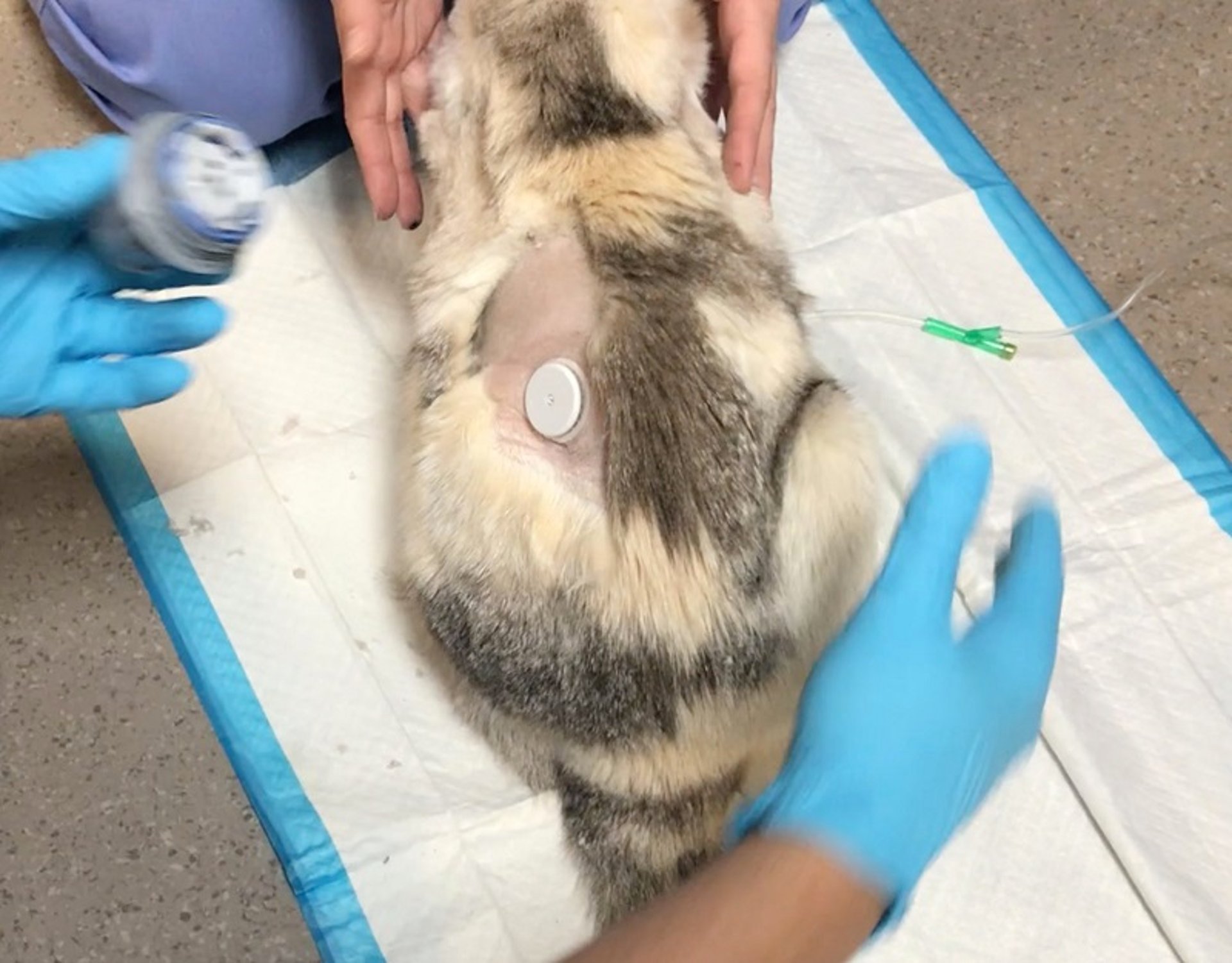
A cat with a continuous glucose monitor placed. These monitors measure interstitial glucose concentrations every 1–5 minutes for several days and can be used for a variety of conditions, including diabetic ketoacidosis, hypoglycemia, sepsis, and others.
Courtesy of Dr. Andrew Linklater.
Electrolytes in the Critically Ill Animal
Hypokalemia can be a contributing factor in weakness and ileus of critically ill animals. Affected animals commonly have decreased oral intake and/or increased GI and urinary potassium losses that require potassium supplementation in IV fluids.
Hyperkalemia can be a life-threatening complication of urinary tract rupture or obstruction, impaired potassium excretion in acute kidney injury or end-stage chronic kidney disease, reperfusion injury, or massive cellular death. Hyperkalemia commonly results in bradyarrhythmias and can be temporarily treated with calcium gluconate and insulin, concurrently with dextrose and/or sodium bicarbonate and occasionally beta-adrenergic receptor agonists.
In patients with hyperkalemia, if there is bradycardia or other substantial changes in the ECG, 10% calcium gluconate (0.5–1 mL/kg, IV slowly over 10–20 minutes) should be administered (10). The goal of calcium gluconate administration is to temporarily reset the difference between the resting and threshold potential, improving ECG changes; the other listed therapies are often administered concurrently to help correct serum potassium concentration. The underlying pathology that led to hyperkalemia must be addressed.
Other important electrolytes to monitor include sodium, ionized calcium, phosphorus, magnesium, and chloride; all can be increased or decreased in critically ill animals and can affect other body systems (eg, nervous and cardiovascular systems) as well as serum osmolality, RBCs, and acid-base balance.
Acid-Base Balance in the Critically Ill Animal
Acid-base assessment in critically ill patients is often complex. The assessment can be made in several ways: the traditional (or Henderson-Hasselbalch) approach, the strong ion approach, and the semiquantitative approach.
The traditional approach involves assessment of the pH, determination both of the metabolic and respiratory components and of whether the process is compensated or mixed, and assessment of anion gap (AG).
The most common causes of metabolic acidosis include lactic acidosis, renal disease, and DKA. Type A lactic acidosis can result from cellular hypoxia caused by hypoperfusion or hypoxia; causes of type B lactic acidosis include liver or kidney disease, excessive muscle activity (with tremors, seizures), and toxins.
Lactate concentration can be easily measured with handheld or benchtop analyzers. Resolution of hyperlactatemia from hypoperfusion with adequate fluid resuscitation is associated with improved rate of survival to discharge. Treatment of lactic acidosis involves maximizing blood flow and tissue oxygen delivery and investigating and treating underlying disease.
Rarely is the administration of sodium bicarbonate (NaHCO3) warranted for perfusion-related acidosis; however, it may be required with severe metabolic disease.
Once perfusion and oxygenation are corrected, the acid-base status is reassessed.


If severe metabolic acidosis (as occurs with ketosis or uremia) persists, cautious administration of fluids with NaHCO3 supplementation may be warranted, restoring serum concentration to > 15 mEq/L. The point at which to administer NaHCO3 remains controversial, especially given the potential adverse effects, anecdotal suggestions include when HCO3 concentration remains below approximately 10 mEq/L or pH remains below 6.9 after perfusion has been restored
The dose of NaHCO3 (in mEq) is calculated as follows:
NaHCO3dose = Vd × (Target [HCO3] − Patient [HCO3]) × BW
where Vd is an the volume of distribution of NaHCO3 (approximately 0.3 L/kg), [HCO3] is bicarbonate concentration (in mEq/L), BW is body weight (in kg), and the target NaHCO3 is 10–15 mEq/L (11, 12).
Serum bicarbonate concentrations are carefully monitored to meet patient requirements. Consequences of NaHCO3 administration can include metabolic alkalosis, hypernatremia, hypokalemia, and total and ionized hypocalcemia. Other possible complications with sodium bicarbonate administration include impairment of tissue oxygenation, hypotension, hyperosmolarity, paradoxical intracellular acidosis, hypercapnea, hypervolemia, hypocalcemia, hypokalemia, and hypomagnesemia.
The AG can be calculated when blood electrolytes are measured:
AG = [Na] + [K] – [HCO3] – [Cl]
Normal AG is 12–24 mEq/L in dogs and 13–27 in cats (12, 13). An increased AG indicates some unmeasured anion present in the blood, which can include ketones, lactate, uremic compounds, or toxins (eg, salicylates, ethylene glycol, ethanol, methanol, indomethacin, isoniazid, paraldehyde, propylene glycol).
In the semiquantitative acid-base analysis, which is an expansion of the strong ion difference evaluation, several factors are examined, each of which has an impact on acid-base status: sodium/free water, chloride, phosphate, albumin, lactate, and unmeasured anion effects. Each factor has a quantifiable effect on acid-base status evaluation of a patient and needs to be considered individually to determine the magnitude of its effects on base excess. This particular assessment might help clarify which components are contributing to acid-base balance; however, it is more cumbersome to calculate on the clinic floor.
See also videos on respiratory acidosis, respiratory alkalosis, and acid-base maps and compensatory mechanisms,
Oxygenation and Ventilation in the Critically Ill Animal
Pulmonary function can be compromised in critical illness for a variety of reasons (pneumonia, acute respiratory distress syndrome, thromboembolism, congestive heart failure, etc). Early diagnostic tests (eg, imaging, blood work, tracheal washes, urine blastomycosis antigen testing, etc) and targeted therapeutics will help limit extension of pulmonary disease.
Aspiration pneumonia is a particular challenge, because it is most commonly a "second hit" disease (secondary to another systemic illness); veterinarians must be diligent to prevent it. Therapeutics such as antiemetics and prokinetics, as well as nasogastric tubes to prevent aspiration pneumonia, should be used whenever appropriate (see images of dog with and ).


A dog with a nasogastric tube and nasal oxygen line in the same naris. This dog was the victim of a gunshot and required an emergency laparotomy and thoracotomy.
Courtesy of Dr. Andrew Linklater.
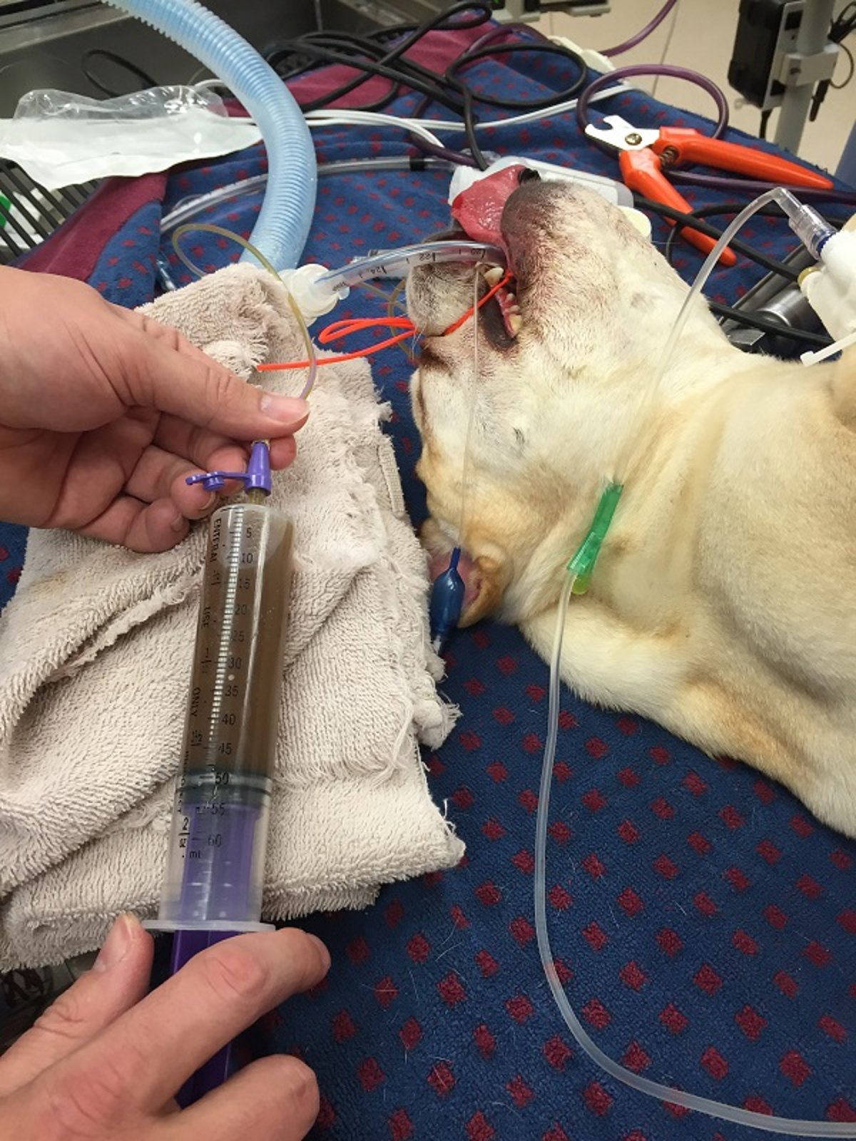
A brachycephalic dog going to surgery for an intestinal obstruction due to a foreign body. Notice the nasogastric tube placed. These tubes can be suctioned to prevent aspiration pneumonia and measure gastric residual volumes as well as to administer medications and nutrition.
Courtesy of Dr. Andrew Linklater.
Arterial blood gas measurement is the gold standard for detecting hypoxemia or hypercarbia. Pulse oximetry is a noninvasive way to determine the oxygen saturation (SpO2) of Hgb and is widely available. Supplemental oxygen and/or therapeutic ventilation may be indicated with SpO2 < 96%. Hypercarbia can be detected using end-tidal CO2 (ETCO2) monitoring through an endotracheal tube or nasal catheter and has been shown to correlate with arterial CO2 tension in animals (14, 15).
Serial monitoring is recommended in the initial management of animals with respiratory compromise to determine the adequacy of oxygen supplementation and the need for mechanical ventilation.
Several methods of oxygen supplementation exist, including flow-by oxygen, cage-based oxygen, nasal or nasotracheal oxygen, high-flow oxygen, and ventilation, each with its own advantages and disadvantages. (See images of dogs and cats receiving oxygen via , an , and described in the Primary Survey topic.)
If hypoxemia is unresponsive to oxygen supplementation (PaO2 < 60 mm Hg or SpO2 < 90%), if hypercarbia (hypoventilation) is present (PaCO2 > 60 mm Hg), or if respiratory effort (work of breathing) is substantially increased, manual or mechanical ventilation is necessary. Ventilation should not be delayed until respiratory failure or arrest.
Pearls & Pitfalls
|
The prognosis for animals that require ventilation is variable; those with hypoxemia from congestive heart failure (62% [16]) or hypoventilation from tick paralysis (64% [17]), cervical spinal disease (71% [18]), or envenomation (91% [19]) have a better prognosis than those that require ventilation for hypoxemia due to primary pulmonary (22% [20]) disease such as pneumonia (20% [21]), contusions (30% [16]), or lower motor neuron disease (21% [22]). Invasive (arterial blood gas) and noninvasive (ETCO2/SpO2) monitoring should be performed during mechanical ventilation to determine the need to adjust the ventilator settings.
Neurological Status in the Critically Ill Animal
Routine neurological monitoring of any patient is important, and in patients with neurological disease, serial examinations should be performed. Using an objective score, such as a modified Glasgow Coma Scale (MGCS), will help provide more objective data between clinicians. MGCS scores have been associated with outcome in dogs and cats with traumatic brain injury (23). A decline in an animal’s MGCS or level of consciousness warrants investigation to exclude metabolic causes, such as hypoglycemia, hyperglycemia, hepatic encephalopathy, acidosis, electrolyte or osmotic derangements, or sudden development of hypertension, hypotension, or shock.
An increase in intracranial pressure can result from intracranial hemorrhage, fluid overload (cerebral edema), primary brain/meningeal disease, and/or ischemia. The drugs the animal is receiving should be carefully evaluated for adverse effects that can lead to altered mentation or level of consciousness.
Cerebral edema may be responsive to medical management with osmotic agents such as mannitol therapy, occasionally used in conjunction with furosemide; 7% hypertonic saline may be an alternative for patients sustaining trauma. Steroids may be indicated in certain inflammatory diseases (eg, meningitis, neoplasia), and antimicrobials in infectious disease (eg, toxoplasmosis). Craniotomy may be needed in animals not responsive to medical management.
It is essential to maintain normal cerebral blood flow to ensure adequate oxygen and nutrient delivery to the brain; cerebral blood flow is largely dependent upon cerebral perfusion pressure (CPP), and CPP is the net pressure gradient that drives blood flow to the brain. CPP is calculated as follows:
CPP = MAP – ICP
where MAP is mean arterial blood pressure (in mm Hg) and ICP is intracranial pressure (in mm Hg).
ICP is usually directly measured via intracranial pressure transduction; however, ICP is not routinely measured or CPP calculated in cats and dogs. Nevertheless, given the importance of maintaining CPP, understanding the core concepts behind blood flow to the brain helps optimize medical management in those with severe brain disease.
Elevating the head 15° and avoiding procedures that might increase venous pressure and, subsequently, intracranial pressure are essential. Maintaining normal oxygenation/ventilation, blood pressure, glucose concentration, and serum electrolyte and water balance is essential for animals with brain disease. Neurological status may be evaluated on a regular basis using a scoring system such as the MGCS, which can provide an objective assessment and help identify when intervention is necessary; lower MGCS scores are associated with a poorer prognosis (24).
Spinal injury that is severe enough to cause paralysis (particularly with lack of deep pain sensation) or inability to ventilate most often warrants immediate advanced imaging and surgical intervention. Loss of deep pain is associated with a poor return to function.
Blood Pressure in the Critically Ill Animal
Blood pressure should be monitored via direct or indirect methods. The minimum goal is to maintain organ perfusion by maintaining a mean arterial blood pressure > 60 mm Hg(systolic > 90 mm Hg); however, normal blood pressure is considered an ideal goal. In hypotensive animals with adequate cardiac function, treatment consists of intravascular volume infusion, oxygen administration, and pain control.
Hypotension unresponsive to intravascular volume replacement can be due to one or more of a variety of causes:
hypoglycemia
acidosis
alkalosis
electrolyte disorders (eg, potassium, calcium, magnesium)
brainstem pathology
cardiac arrhythmias
metabolic toxins (eg, hepatic, renal)
ongoing fluid loss
relative or absolute hypoadrenocorticism (eg, cortisol deficiency)
cardiac or pericardial disease
excessive vasodilation
excessive vasoconstriction
Patient assessment for these causes should be performed and immediately addressed. (Also see Assessment of Resuscitation Efforts in Animals.)
The need for cardiovascular support with positive inotropes or vasopressors is considered when the causes listed above are ruled out.
An experienced ultrasonographer might be able to assess ventricular contractility and/or capacitance (caudal vena cava) vessel size to provide an estimate of preload and/or a patient's potential response to fluid therapy.
Once intravascular volume (central venous pressure > 8 cm H2O, although this is measured less commonly) and cardiac function are assessed as adequate, vasopressor therapy with IV CRI of norepinephrine (0.05–2 mcg/kg/min), epinephrine (0.02–0.2 mcg/kg/min), dopamine (5–20 mcg/kg/min), or another pressor (vasopressin or ephedrine) agent is instituted (25, 26). The choice of which vasopressor to use has not been well established in veterinary patients. Norepinephrine is often considered a first-line choice, followed by epinephrine or vasopressin, and then followed by ephedrine or dopamine. Vasopressin is usually limited because of its cost, and epinephrine is specifically indicated with anaphylaxis.
A CRI begins at the lower end of the dosage range and is increased incrementally until blood pressure goals are reached. Objective measurements of global perfusion can also include lactate monitoring; animals with a substantially increased lactate concentration can have a poorer prognosis. Studies have demonstrated that serial lactate monitoring is more useful than a single measurement (27).
Central venous oxygen saturation (ScvO2) is another objective measurement of global perfusion. ScvO2 is measured from the vena cava or right atrium (versus SvO2, which is measured from the pulmonary artery). Normal ScvO2 is 65–75%, whereas lower ScvO2 may indicate increased oxygen extraction (28).
Hypertension can lead to catastrophic problems if severe. The American College of Veterinary Internal Medicine classifies risk of target-organ damage from hypertensioninto four categories based on systolic blood pressure:
I: < 150 mm Hg = minimal risk
II: 150–159 mm Hg = mild risk
III: 160–179 mm Hg = moderate risk
IV: ≥ 180 mm Hg = severe risk
Hypertension can lead to retinal detachment or to neurological derangements from intracranial hemorrhage and can exacerbate proteinuria in animals with chronic kidney disease.
Moderate to severe hypertension can be treated with oral antihypertensive agents such as calcium channel blockers (eg, amlodipine), angiotensin II receptor blockers (ARBs; eg, telmisartan) in nonazotemic animals, direct arterial dilators (eg, hydralazine), or systemic injectable antihypertensive agents such as nitroprusside administered as a continuous infusion, titrated to effect. Angiotensin-converting enzyme (ACE) inhibitors (eg, benazepril) are commonly chosen as first-line therapy in dogs. However, they are generally not effective as sole therapy for managing systemic hypertension in cats and may be inadequate as monotherapy in dogs with severe hypertension. Benazepril has been used in cats that require a second antihypertensive agent (29, 30).
Blood pressure must be monitored continuously to assess response to therapy. Chronic hypertension that is rapidly decreased can result in decreased renal perfusion; the goal should be to decrease blood pressure by no more than 25% in the acute setting. See also ACVIM consensus guidelines for the management of hypertension in dogs and cats.
Heart Rate, Rhythm, and Contractility in the Critically Ill Animal
The electrical and mechanical systems of the heart should be evaluated separately. ECG assessment is necessary to identify arrhythmias and to institute specific antiarrhythmic therapy.
Arrhythmias can occur for a variety of reasons, such as SIRS diseases, splenic disease, organ torsion (eg, gastric dilation and volvulus), and electrolyte abnormalities (eg, hyperkalemia); the underlying condition must be identified and treated concurrently. Oxygen, fluids, and analgesia are considered first-line agents to treat underlying conditions and rule out common causes of sinus tachycardia.
An arrhythmia requires treatment when it results in decreased cardiac output and clinical signs of shock. Some ventricular rhythms (such as ventricular premature contractions and accelerated idioventricular arrhythmias) may not necessarily require immediate therapy if cardiac output is not considerably decreased.
Pearls & Pitfalls
|
Indications for treatment of a ventricular rhythm include the following:
tachycardia (rates > 160 bpm in dogs or 240 bpm in cats)
clinical signs of poor perfusion (low blood pressure, poor pulse quality, etc)
multiform arrhythmias
R-on-T phenomenon or torsades de pointes (see )


Ventricular tachycardia from a dog with gastric dilation and volvulus. Note that the three beats on the right demonstrate R-on-T phenomenon (where there is no baseline between the beats).
Courtesy of Dr. Andrew Linklater.
Other tachyarrhythmias may respond to class I, II, III, or IV antiarrhythmics; bradyarrhythmias can be challenging to treat medically. If a bradyarrhythmia does not respond to beta agonists or parasympatholytics (eg, atropine), pacemaker placement may be necessary. Note that some patients have persistent bradycardia due to high vagal tone (disease affecting the eyes, neck, chest, or abdomen); when that is occurring, the underlying disease must be treated.
An echocardiogram can be performed to evaluate cardiac contractility in SIRS diseases and to detect underlying cardiac diseases such as dilated cardiomyopathy. If cardiac contractility is decreased, dobutamine (dogs: 5–20 mcg/kg/min or cats: 2.5–5 mcg/kg/min, IV CRI) is given to provide inotropic support if there is evidence of poor cardiac output (26, 31).
Studies have demonstrated that dogs with mitral valve disease and dilated cardiomyopathy have a poorer prognosis if their cardiac troponin 1 (cTnI) and/or NT-proBNP is increased. However, these tests are not available in all hospitals and do not necessarily direct therapy or diagnose or differentiate cardiac diseases (32).
Temperature in the Critically Ill Animal
Body temperature is considered part of the initial database and should be measured regularly in every critically ill animal. A variety of diseases can result in increased or decreased body temperature.
Rectal temperature is considered the (noninvasive) clinical standard for temperature measurement and correlates well with core body temperature measured through a central catheter. Esophageal temperature may be used in anesthetized patients; axillary and auricular methods of obtaining a temperature are considered less accurate but can be used to monitor trends in patients that will not allow rectal temperature to be obtained.
Temperature-sensing microchips are becoming more common, and they correlate well with rectal temperature in normothermic and febrile cats but might not be as accurate in dogs (33).
Increased temperatures can be observed with environmental exposure (eg, heatstroke), increased activity (eg, exercise, excitement), and infectious, inflammatory, or neoplastic diseases. Severe increases of temperature (> 40.8°C [105.5°F]), particularly when prolonged, can lead to severe metabolic disease, such as hemorrhagic diathesis, disseminated intravascular coagulation, and SIRS diseases, which can lead to multiorgan dysfunction.
Effective means of cooling hyperthermic animals include fluid therapy, using wet towels with fans, and placing alcohol on paw pads and axillary and inguinal regions. Animals should not be immersed in cold water, because this causes peripheral vasoconstriction and decreases core heat dissipation.
Pearls & Pitfalls
|
Fever of unknown origin warrants a systemic evaluation.
Hypothermia is most commonly associated with anesthesia in small animals; however, severe systemic disease (particularly in cats) and environmental exposure might be contributing factors. Mild hypothermia can be a common sequela of severe cardiovascular disease and is a prognostic marker in cats with hindlimb thromboembolism. Temperature is a vital parameter to monitor and treat in cats with clinical signs of shock, and active warming is an essential component of therapy.
Therapeutic hypothermia might have some neuro-sparing effects in animals with traumatic brain injury or in postresuscitation (CPR) care; however, further investigation is needed. In animals with induced hypothermia, blood flow to most organs can be substantially decreased, and coagulation can be affected.
Altered body temperature is part of the definition of SIRS-type diseases; other parameters include an increased or decreased heart rate, increased or decreased WBC count, and an increased respiratory rate.
Coagulation in the Critically Ill Animal
Disseminated intravascular coagulation (DIC) can develop in any animal that has undergone a period of relative vascular stasis such as occurs during shock; severe tissue or capillary damage as occurs with trauma; exposure of capillary endothelial cells to circulating inflammatory mediators as occurs during sepsis or SIRS; or moderate to severe alterations in body temperature.
In the early stages of DIC, there may be few or no clinical signs. However, as DIC progresses, its effects are obvious and catastrophic. The goal is to detect DIC in the early stages and to slow or prevent its progression:
Early DIC is characterized by a hypercoagulable stage in which serum antithrombin concentrations are decreased and the coagulation cascade is activated by any of the precipitating causes.
Activation of the coagulation cascade throughout the body rapidly depletes the clotting factors and the blood platelet count as platelets are incorporated into the clots. At this stage, the prothrombin time (PT) and partial thromboplastin time may be decreased; however, this is a challenging stage to identify and diagnose.
This rapidly progresses to a hypocoagulable stage as the coagulation factors are consumed.
In this late stage, the PT and activated partial thromboplastin time (aPTT) or activated clotting time (ACT) are prolonged, and fibrinogen degradation products are increased.
DIC occurs on a continuum between these stages and can be very challenging to diagnose early in the course of disease: thrombocytopenia is common, but D-dimer assays may not be commonly available. Viscoelastic testing (thromboelastography) can help identify patients at risk for thromboembolism or those in early stages of DIC. Although viscoelastic testing is becoming more available, it must be run in-house and is often limited to specialty centers; specific points of intervention have not been determined.
Treatment of DIC focuses on treating the underlying disease and removing the stimulus for continued activation of the coagulation cascade. In the early hypercoagulable stages, treatment focuses on maximizing the function of antithrombin, which is the most abundant natural inhibitor of the serine proteases of the coagulation cascade.
Antithrombin activity is typically measured as part of a DIC panel and reported as a percentage relative to a species-specific reference pool, which is defined as 100%.When antithrombin activity is adequate, heparin can be administered (50–100 U/kg, SC, every 8 hours) to patients at risk of thrombosis; doses are individualized to obtain an aPTT 1.5–2 times normal (34).
In human medicine, if antithrombin activity is < 70%, it has been suggested that plasma transfusions should also be given to increase antithrombin activity to ≥ 80%. Note that adequate antithrombin levels in veterinary patients have not been determined and that these data are from human medical literature (35).
In animals with diseases known to predispose to DIC, coagulation parameters and platelet counts should be monitored. Thromboelastography provides another means of global assessment of the clotting cascade and can be a useful tool with suspected hypo- or hypercoagulable states; hypercoagulable states are challenging to diagnose, and thromboelastography is one of the few methods that can provide an accurate assessment.
Thrombosis occurs without DIC when there are alterations in the Virchow triad of endothelial injury, blood stasis, and hypercoagulable state. Abnormalities in one or more of these components may be present with the following:
vascular anomalies
atrial enlargement (cats)
severe systemic illness (SIRS, immune-mediated hemolytic anemia)
trauma
neoplasia
renal disease
protein-losing renal and GI disease
as a primary disease in Greyhounds
The most common severe manifestations of hypercoagulability are aortic and pulmonary thromboemboli. Pulmonary thromboemboli should be suspected when hypoxemia (PaO2 < 80 mm Hg) is present with minimal lung changes on thoracic radiographs. Anticoagulation therapy and oxygen support should be implemented, and oxygenation and ventilation monitored.
Pearls & Pitfalls
|
Arterial thromboembolism can occur in cats with underlying heart disease. Antithrombotics are warranted in these cases; options include aspirin and/or clopidogrel, heparin (low molecular weight or unfractionated), rivaroxaban or apixaban, and warfarin. Some of these drugs require close monitoring of clotting times to achieve therapeutic goals. This disease can be painful, and opioid medications are often warranted, as is monitoring for reperfusion injury. The CURATIVE guidelines outline the use of antithrombotics in veterinary critical care (36).
Disease states that result in relative hypocoagulability can include the following:
fulminant liver failure
heatstroke
severe thombocytopenia
dilutional hypocoagulability from fluid and colloid administration
congenital defects in the coagulation cascade, such as von Willebrand disease, hemophilia A or B, or platelet defects in specific breeds (Boxers)
Therapy should be specific to the inciting cause; plasma products such as fresh frozen plasma (10–20 mL/kg) are often necessary to correct life-threatening coagulopathies (37).
Hyperfibrinolysis (rapid clot breakdown) is a condition characterized by excessive and accelerated breakdown of blood clots, which can lead to a hypocoagulable state and increased bleeding. In certain breeds (Greyhounds) and certain disease states (trauma), excessive bleeding can occur after clot formation; hyperfibrinolysis is also reported to occur in a variety of other disease processes. This syndrome can be identified or diagnosed with viscoelastic testing and is treated with either tranexamic acid or aminocaproic acid.
Red Blood Cell and Hemoglobin Concentration in the Critically Ill Animal
Because hemoglobin carries most of the oxygen in the blood, maintaining adequate Hgb concentrations is essential to maintaining adequate oxygen delivery. When anemia is associated with clinical signs of tachycardia, increased respiratory rate, altered mentation, severe lethargy/weakness, and hypotension, then packed RBCs, whole blood, or hemoglobin-based oxygen carriers should be administered to bring the PCV to a minimum of 20% or the Hgb concentration to a minimum of 7 g/dL. In some cases of hemolytic or chronic anemia, the PCV can be maintained at a lower percentage before transfusion is required if there are no corresponding clinical signs.
In animals that require multiple blood sampling (such as diabetic patients) or in very small animals, blood sampling should be minimized to prevent iatrogenic blood loss; this can be performed using CGMs in diabetic patients. Optimal Hgb target concentrations have not been determined in veterinary patients across multiple disease processes; however, conservative transfusion management in humans (Hgb goal of 7 g/dL or PCV of 20%) has been associated with improved rate of survival to discharge compared with more liberal transfusion goals (Hgb of 10 g/dL or PCV of 30%) (38).
In 2021 the Association of Veterinary Hematology and Transfusion Medicine released a consensus statement regarding transfusion reactions in dogs and cats (39).
Very few severe transfusion reactions are reported in transfusion-naive recipient dogs; therefore, dog erythrocyte antigen 1 (DEA 1) mismatched blood may be given as a first transfusion in dogs. Otherwise, it is best to administer DEA 1 type–specific blood.
Crossmatching may not be necessary for naive recipients either; however, substantial knowledge gaps exist in this area. In one study, mean hematocrit after transfusion was higher in dogs with a crossmatch performed (40), and crossmatch incompatibilities have been reported in 15% of naive cats (41) and 17% of naive dogs (40). In dogs that have received a transfusion ≥ 4 days previously, a crossmatch is recommended. Only type-specific blood should be administered to cats because life-threatening transfusion reactions can occur.
In dogs and cats with acute cavitary (pleural or peritoneal) hemorrhage, blood may be salvaged from the cavity with aspiration (via centesis or exploratory surgery when indicated) and an autologous blood transfusion administered through a blood filter. Xenotransfusions, primarily canine blood administered to cats, have been performed in some veterinary patients. Although this has resulted in positive outcomes, transfusion reactions can be high, antibodies will develop within 2–4 days, and allogenic (same-species) transfusion remains the preferred choice.
Rarely, disease states result in altered Hgb (such as methemoglobinemia) or altered oxygen-carrying capacity (such as carboxyhemoglobinemia), often recognized by altered mucous membrane color (muddy or brick-red, respectively). Despite normal measured Hgb concentrations, oxygen is not being delivered to tissues in affected animals, and oxygen supplementation is necessary, along with treatment of the underlying disease.
An alternative means to increase oxygen-carrying capacity of the blood is a commercial hemoglobin-based oxygen carrier. However, no commercial products are available. Monitoring PCV does not provide an adequate assessment of oxygen delivery after use of hemoglobin-based oxygen carriers.
Erythrocytosis can occur as either relative (from hypovolemia) or absolute (normovolemia). Animals with relative or absolute erythrocytosis (a hematocrit or PCV above the reference range), other than sight hounds and animals at high altitudes, can have poor microvascular flow (due to the altered blood rheology) and hypertension (which impairs microvascular delivery of oxygen to the tissues and can result in neurological signs). Treatment is directed to improve this microvascular flow and oxygen delivery to the tissues.
Some patients develop transient erythrocytosis secondary to splenic contraction, which most often occurs with hemorrhagic shock.
Hypovolemic erythrocytosis usually occurs as a result of severe dehydration, such as with acute hemorrhagic diarrhea syndrome, heatstroke, etc. Treatment usually involves aggressive IV fluid resuscitation and rehydration.
Absolute polycythemia can occur as a result of hypoxia, cardiac disease, renal and endocrine disease secondary to erythropoietin-secreting tumors, polycythemia vera, and more. Treatment in these cases includes treating the underlying disease, phlebotomy with fluid or plasma readministration, and hydroxyurea (20–25 mg/kg [dog] or 10–15 mg/kg [cat], PO, every 12 hours, then tapered to lowest effective frequency) (42).
Renal Function in the Critically Ill Animal
In animals that have had a hypotensive episode, are receiving potentially nephrotoxic medications, or have primary renal compromise, renal function should be evaluated daily.
Urinalysis is ideally performed before fluid administration to assess renal function. Normal urine output is 1–2 mL/kg/h and can be closely monitored with an indwelling urinary catheter. Animals in polyuric renal failure are most often managed medically; however, animals in oliguric (< 0.8 mL/kg/h), anuric (< 0.03 mL/kg/h), or relative oliguric (less than expected) renal failure can require peritoneal dialysis or hemodialysis to maintain fluid, acid-base, and electrolyte balance. If monitoring urine output with a catheter is not possible, then estimating urine output by measuring absorbent pads or litter boxes is necessary. Body weight should also be recorded regularly.
Serial measurement of serum BUN, creatinine, electrolytes, and phosphorus concentrations will detect changes and help guide therapy. Measurement of symmetric dimethylarginine (SDMA) concentration may help detect renal dysfunction before creatinine concentration exceeds the upper limit of the reference interval; however, changes in SDMA concentration will often mirror changes in creatinine concentration.
Serial urinalyses to detect glucosuria, proteinuria, or renal tubular casts help evaluate acute tubular injury before the damage progresses to overt renal failure and azotemia.
Additional diagnostic tests include urine culture and susceptibility testing, urine protein:creatinine ratio, and specific testing for renal-specific disease (eg, ethylene glycol, leptospirosis). Animals may also be monitored using a scoring system to provide additional objective monitoring: the International Renal Interest Society has a staging system to monitor dogs and cats with chronic renal disease based on serum creatinine concentration, blood pressure, and proteinuria.
Infection Identification and Immune Status in the Critically Ill Animal
Strict aseptic technique should be observed when treating animals with severe neutropenia (< 1,000 neutrophils/mcL or lower), including those receiving immunosuppressive drugs. These animals should be isolated from other animals and handled by a single person who adheres to appropriate barrier nursing techniques (washes hands, wears gloves and gown before handling the animal, etc). All veterinary staff should be encouraged to wash hands between patients, wear examination gloves routinely, treat wounds in a clean manner, and administer IV injections only after swabbing an IV port with alcohol.
Patients with particularly infectious disease processes (parvoviral enteritis, infectious respiratory tract infections, etc) should ideally be housed in a separate ward, with barrier nursing techniques used for individual patients, including gowns, gloves, masks, and eye and foot protection. Educating hospital staff on appropriate patient-handling techniques may help limit development of nosocomial infections, which develop ≥ 48 hours after hospital admission.
Ultimately, antimicrobial selection should be based on the results of culture and susceptibility testing; however, empirical treatment, based on the site of infection and suspected type of bacteria, is necessary pending these results. Empirical therapy may be based on common organisms found at the affected site and/or Gram stain and cytological evaluation, which should be performed immediately. Repeated culture and susceptibility testing may be necessary in animals not responding to therapy as expected or if prolonged antimicrobial therapy is anticipated.
An antimicrobial protocol should be established for veterinary hospitals to minimize the number of antimicrobials administered empirically on a routine basis to decrease the development of resistant organisms in the hospital environment and to improve their susceptibility patterns. Handwashing and use of antimicrobial stewardship guidelines (limiting use of specific classes of antimicrobials) have been demonstrated to limit development of antimicrobial resistance, decrease antimicrobial costs, and decrease surgical site infections in humans; however, there are limited veterinary data (43, 44).
Antimicrobial resistance is a growing concern in both human and veterinary species, particularly with the development and increased recognition of methicillin-resistant Staphylococcus pseudintermedius in veterinary species. Targeted surveillance, rational antimicrobial use, and public awareness are essential to preserving antimicrobial effectiveness (45, 46).
Facility-specific monitoring of culture and susceptibility results for evidence of nosocomial infections and bacterial resistance patterns can help identify and control sources of infection and limit development of resistance. If multiple antimicrobials are started, the antimicrobial spectrum should be narrowed and the antimicrobial choice adjusted as soon as the organism's susceptibility pattern is identified (de-escalation).
Recurrent infections should be investigated for an underlying pattern of resistance, nidus of infection, or immune-compromising disease.
Empirical therapy, even for a septic patient, should not continue for more than 3 days, and antimicrobials should be de-escalated to the lowest-tier single agent as soon as culture results are available.
The advantages and disadvantages of each antimicrobial agent administered should be closely considered. Cost, penetration into various tissues (site of infection), volume of distribution, adverse effects, and disease process present should all be considered.
Options for first-line, broad-spectrum coverage include the following:
First-generation cephalosporins (eg, cefazolin at 20–35 mg/kg, IV slowly, IM, or SC, every 6–8 hours) provide good gram-positive and gram-negative coverage. They may be paired with metronidazole (10–15 mg/kg, slow IV, every 12 hours) to help cover anaerobic infections (47).
Aminopenicillins with a beta-lactamase inhibitor (eg, ampicillin/sulbactam, 10–40 mg/kg, slow IV, every 8 hours) provide good gram-negative, gram-positive, and anaerobic coverage (47).
For gram-negative infections, fluoroquinolones (eg, enrofloxacin in dogs: 5–20 mg/kg, IM or slow IV, every 24 hours; in cats: 5 mg/kg, IM or PO, every 24 hours) (34).
Newer generations and classes of antimicrobials, such as carbapenems (eg, imipenem), third-generation cephalosporins (eg, ceftazidime), and vancomycin, should be reserved for use in animals with bacterial infections demonstrated to be resistant to other antimicrobials.
WBC counts performed on a semiregular basis (every 2–3 days) can indicate an appropriate response to infection/inflammation, resolution of disease, or patient deterioration.
Various molecular markers or biomarkers have been investigated in SIRS-type diseases (including high-mobility group box chromosomal protein 1, C-reactive protein [CRP], plasma IL-1-beta and IL-6) to help understand and stratify disease, including outcome. However, other than CRP, many of these are not routinely available or not available in a timely manner to practitioners.
GI Motility and Mucosal Integrity in the Critically Ill Animal
Critically ill animals, even those without a primary GI disease, are prone to gastric atony, ileus, and gastric ulceration. Regular auscultation for bowel sounds can provide insight on a physical examination; ultrasonography may also be used to assess GI motility.
Metoclopramide (0.5 mg/kg, every 8 hours, IV, IM, or PO, or 0.4 mg/kg/h, CRI, followed by 0.3 mg/kg/h and increased up to 1 mg/kg/h) is useful because of its central antiemetic effects and its ability to increase progressive gastric and intestinal motility. Other motility modifiers to consider include cisapride (dogs: 0.1–0.5 mg/kg, IV or PO, every 8 hours; cats: 2.5–5 mg/cat, PO, every 8–12 hours [up to 1 mg/kg every 8 hours]), erythromycin (0.5–1 mg/kg, IV or PO, every 8–12 hours) (34).Motility modifiers should be avoided if gastric or intestinal obstruction, hemorrhage, or perforation is suspected or has been confirmed.
Pearls & Pitfalls
|
Placement of a nasogastric tube facilitates regular (every 2–8 hours) removal of accumulated gas and fluid from the stomach, which minimizes nausea, gastric distention, and vomiting, thereby decreasing the possibility of aspiration of refluxed gastric contents. The nasogastric tube also can be used to introduce small amounts of a glucose and electrolyte solution or a liquid diet to provide nutrition directly to enterocytes, which helps prevent gastric ulceration and intestinal mucosal compromise with secondary bacterial translocation.
Antiemetics are used in animals that continue to vomit frequently despite placement of a nasogastric tube, thus improving patient comfort and decreasing the incidence of aspiration, vasovagal syncope, and bradycardia that can accompany the vomiting reflex. The following antiemetics are used in small animals:
Metoclopramide blocks the dopaminergic receptors in the chemoreceptor trigger zone (CRTZ) and central vomiting center and acts peripherally by promoting gastric emptying (34).
Ondansetron (0.5–1 mg/kg, IV or PO, every 8–12 hours) and dolasetron (0.6–1 mg/kg, IV or PO, every 24 hours) are potent antiemetics that block serotonin receptors and act at the CRTZ and the central vomiting center (48, 49).
Maropitant (1 mg/kg, SC or IV, every 24 hours) is a neurokinin-1 (NK-1) receptor antagonist that blocks vomiting at the CRTZ, vomiting center, and peripheral receptors (34).
Chlorpromazine (dogs: 0.5 mg/kg, IM or SC, every 6–8 hours; cats, 0.2–0.5 mg/kg, IM or SC, every 6–8 hours) can be used in patients with normal blood pressure (34).
A combination of antiemetics that have different mechanisms of action might be required to arrest refractory emesis in severe illness. If a patient requires multiple antiemetics, GI obstruction must be ruled out.
GI ulceration often accompanies critical diseases such as hypotension and hypergastrinemia associated with liver and other diseases, including drug toxicities, neurological disease, and respiratory disorders requiring ventilation. Histaminergic H2-receptor antagonists such as famotidine (dogs, 0.5–1 mg/kg, PO, IV or SC, every 12–24 hours; cats: 0.2–0.25 mg/kg, IM, SC, PO, or IV, every 12–24 hours) and proton pump inhibitors such as omeprazole (0.5–1 mg/kg, PO, every 12 hours) and pantoprazole (0.5–1 mg/kg, slow IV, every 24 hours) are commonly administered (34). Note that changing the pH of the stomach can result in altered microbial flora.
Agents such as sucralfate (0.25–1 g/animal, PO, every 8–12 hours) and barium bind to esophageal and gastric erosions and ulcers but are more cumbersome to administer due to the increased frequency and must be given at least 1 hour before or after other medications. Misoprostol (2–5 mcg/kg, PO, every 12 hours) may help prevent NSAID-induced ulceration (34).
Diarrhea is also very common in the ICU, occurring with a variety of underlying disease processes. Often, diarrhea resolves with appropriate dietary changes and supportive care with fluids for dehydration and electrolyte changes.
Controversy exists regarding nonspecific treatment options for diarrhea. Metronidazole has not consistently demonstrated improved outcome compared to ; no antimicrobials or probiotics in several studies; a variety of probiotics are available and have demonstrated some improved outcome (40, 50). Other agents, such as carvacrol, attapulgite, and loperamide (dogs: 0.12 mg/kg, PO, every 8–12 hours; cats: 0.08–0.16 mg/kg, PO, every 12 hours), have shown only anecdotal evidence of improved outcome; loperamide may cause GI cramping (34).
Specific therapy for diarrhea may include anthelmintics (eg, fenbendazole, 50 mg/kg, PO, every 24 hours) if parasites are present (34), parvovirus monoclonal antibody for parvoviral infections (51), and antimicrobials for specific bacterial infections (Salmonella, Campylobacter, etc).
Fecal microbiota transplantation (50–100 g/20 kg of body weight, diluted 1:1 with saline solution [0.9% NaCl] and administered rectally) has demonstrated faster improvement of fecal consistency in some studies (52, 53, 54).
Drug Dosages and Metabolism in the Critically Ill Animal
An active medications list should be kept with each animal's medical record and carefully reviewed daily for potential drug interactions, drug dosages, and possible adverse effects.
Each drug administered enterally or parenterally should be assessed for its compatibility with other fluids and drugs.
If renal or hepatic function is compromised, or if protein (albumin)-binding capacity is decreased, some drug dosages should be decreased to account for altered metabolism, elimination, or protein binding.
The daily review also should ensure that the dosage has been calculated correctly and that it is appropriate for the animal’s current weight and body condition score.
The sudden onset of any new clinical signs should be investigated in light of the medications and their potential adverse effects.
Nutrition in the Critically Ill Animal
When nutritional needs are not met, animals rapidly develop a negative energy balance, which can result in GI dysfunction, organ dysfunction, hepatic lipidosis (cats), poor wound healing, and even death. Direct enteral nutrition is always preferred because it will improve the normal GI barrier, function, and motility; most animals tolerate trickle flow feeding techniques through a temporary feeding tube.
Short-term options include syringe or forced feeding; however, this can lead to food aversion and is not comfortable for most animals. Easy to place and well tolerated, short-term feeding tubes that allow trickle feeding include nasogastric and nasoesophageal tubes. Nasogastric tubes also allow gastric suctioning to monitor GI function and might help to limit continued vomiting and risk of aspiration pneumonia; nasojejunal tubes can be challenging to place.
Pearls & Pitfalls
|
Long-term feeding tubes include esophagostomy, pharyngostomy, gastrostomy, and jejunostomy tubes. Each of these tubes is well tolerated by most animals, and all require anesthesia to place; esophagostomy is a minor surgical procedure, and gastrostomy tubes can be placed with endoscopic assistance.
Feeding by trickle flow is initiated with small volumes of a dilute veterinary liquid diet solution. If an animal has been starved for an extended period of time, nutrition should start at 25–33% of the daily caloric requirement and increase by 25–33% of daily caloric requirements per day to avoid refeeding syndrome, which can result in hyperglycemia, hypokalemia, hypophosphatemia, and hypomagnesemia (55).
Before each bolus feeding and every 6 hours during a CRI, the gastric feeding tube should be suctioned to determine residual gastric volume. After suctioning or administering a liquid diet, the person administering the food should flush the tube with saline solution (0.9% NaCl) or water.
If this initial feeding is not tolerated, the patient should be reevaluated for underlying disease and have prokinetics added. Diet volume should be decreased or the diet diluted with water or an electrolyte solution. As the animal recovers and is able to tolerate at least 50% resting energy requirements, bolus feeding can be introduced by gradually decreasing meal frequency and increasing volumes.
Appetite stimulants, such as the serotonin antagonist cyproheptadine, the serotonin agonist mirtazapine, and the ghrelin agonist capromorelin, are commonly used but with varying success; transdermal mirtazapine is available for cats. Oral benzodiazepines can cause hepatotoxicity in cats and are not good alternatives; injectable benzodiazepines or propofol may be used as a short-term solution for animals with rapidly resolving disease. The use of appetite stimulants provides inconsistent food intake and is not recommended as the primary way to administer nutrition in critically ill animals.
Pearls & Pitfalls
|
When nutritional needs cannot be met by enteral feeding, parenteral feeding is used. Partial parenteral nutrition, consisting of amino acid and carbohydrate solutions, can be infused through a peripheral vein, providing part of the animal's caloric requirements in a readily metabolizable form. Total parenteral nutrition (including the lipid component) must be delivered through a central venous catheter because high osmolarity of the solutions can cause phlebitis and RBC lysis. In animals with prolonged anorexia, vitamin supplementation might also be necessary.
Pain Control in the Critically Ill Animal
Pain activates the stress hormone systems of the body and contributes to increased morbidity and death.
Clinical signs of pain are quite variable in animals and include the following:
decreased normal behavior (appetite, ambulation, grooming, etc)
development of abnormal behaviors (vocalizing, inappropriate urination, altered posture, agitation or aggression, etc)
reaction to touch or abnormal body posture
altered objective physical parameters (increased heart rate, pale mucous membranes, dilated pupils, etc), which can mimic clinical signs of shock
Multidimensional and facial expression–based scoring systems have been validated for pain assessment in cats (56, 57).
Animals that do not show obvious clinical signs of pain but are known to have a painful condition should receive analgesics as part of their treatment. (Also see Systemic Pharmacotherapeutics of the Nervous System.) Preemptive administration of analgesics is recommended, when possible.
Pain should be assessed using a validated pain assessment tool and monitored on a regular basis during the course of hospitalization to ensure adequate analgesia. A scoring system such as the Composite Glasgow Pain Scale is a useful monitoring tool to help determine need and response to analgesia.
Analgesia in critically ill animals can safely be provided by opioids titrated to effect. See the table .
Opioids Used in Emergency Practice
Drug | Dosage | Comments |
|---|---|---|
Morphine | Dogs: 0.05–0.4 mg/kg, IV, q 1–4 h; 0.2–1 mg/kg, IM, SC, or slow IV, q 2–6 h; 0.1 mg/kg diluted with 0.9% saline solution to 0.3 mL/kg administered epidurally at 0.23 mL/kg, q 8–24 h Cats: 0.2–0.5 mg/kg, IV, IM, or SC, q 2–4 h | Incremental IV bolus technique: dogs—increments of 0.1 mg/kg until analgesia appears adequate; cats—increments of 0.02 mg/kg In dogs, this can be followed by CRI at 0.1 mg/kg/h that can be increased incrementally if needed up to 1 mg/kg/h. Rapid IV injections may cause histamine release. |
Oxymorphone/hydromorphone | Dogs: 0.015–0.03 mg/kg/h, IV CRI; 0.05–0.2 mg/kg, IV, IM, or SC, q 2–6 h Cats: 0.015–0.03 mg/kg/h, IV CRI; 0.05–0.1 mg/kg, IV, IM, or SC, q 2–6 h | Minimal cardiovascular effects; may cause panting or emesis |
Fentanyl | Dogs: 2–10 mcg/kg, IV, q 30–60 min or as a loading dose for a CRI; 2–20 mcg/kg/h, IV CRI Cats: 2–5 mcg/kg, IV loading dose; 2–5 mcg/kg/h, IV CRI | With short half-life, fentanyl is best administered as a CRI. Higher doses may cause bradycardia. |
Fentanyl transdermal patch | 12.5 mcg patch for dogs < 5 kg body wt; 25 mcg patch for dogs 5–10 kg; 50 mcg patch for dogs 10–20 kg; 75 mcg patch for dogs 20–30 kg; 100 mcg patch for dogs > 30 kg | The patches cannot be cut. More than one patch may be used in larger animals. Injectable repository fentanyl solutions may be an alternative if available. |
Butorphanol | Dogs: 0.1–0.4 mg/kg, IM, IV, or SC, q 1–4 h Cats: 0.1–0.4 mg/kg, IM, IV, or SC, q 1–4 h | Has a ceiling effect; short duration of effect (1–2 h) in most dogs Can be given as a CRI with the dose divided over 4 hours |
Buprenorphine | Dogs (0.3 mg/mL formulation): 0.005–0.02 mg/kg, IM or IV, q 6–12 h Cats (0.3 mg/mL formulation): 0.005–0.03 mg/kg, IM, IV, or sublingual, q 6–12 h | May be more difficult to reverse Sublingual absorption reported to be excellent in cats |
Methadone | 0.1–0.5 mg/kg, IV, IM, or SC, q 2–6 h; dogs, up to 1 mg/kg | Less vomiting than morphine, less panting than hydromorphone |
Dosages are from Pigott A. Nutritional status. In: Kirby R, Linklater A, eds. Monitoring and Intervention for the Critically Ill Small Animal: The Rule of 20. Wiley Blackwell; 2017 and Papich MG. Papich Handbook of Veterinary Drugs. 5th ed. Saunders; 2021. | ||
Opioids provide potent analgesia (given IV, IM, or SC) with minimal cardiovascular adverse effects, and their actions are reversible with antagonists (eg, naloxone).
Long-acting opioids are best avoided in unstable animals.
IV administration of morphine has been reported to cause hypotension due to histamine release (58); however, this adverse effect does not seem to be of clinical importance if the drug is given over 5–10 minutes or as a CRI. Other medications, such as hydromorphone, oxymorphone, and fentanyl, can be given without this risk.
CRI provides constant analgesia and is often more convenient and less painful than intermittent IM or SC injections.
In cats, injectable buprenorphine is absorbed systemically after sublingual administration.
Neuroleptanalgesia can be provided by combination of an opioid with a sedative (eg, benzodiazepine) or tranquilizer (eg, acepromazine) in animals without contraindications to these medications.
For long-term control of pain, transdermal fentanyl patches or repository fentanyl injections are used but require up to 12 hours to reach therapeutic blood concentrations; analgesia must be provided by injection until adequate blood concentrations have been reached.
Additional formulations exist of some medications, such as long-acting injectable buprenorphine (0.24 mg/kg, SC, every 24 hours for up to 3 days) and buprenorphine transdermal solution (2.7–6.7 mg/kg topically with analgesia provided for 4 days) (59, 60). These drugs are not often chosen for critically ill patients because the dose cannot be tapered.
If pain is not adequately controlled with opioids alone, then ketamine, an N-methyl-D-aspartate (NMDA) receptor antagonist, can be administered with opioids (0.5–1 mg/kg, IV, load followed by 0.1–1 mg/kg/h as a continuous IV infusion) (61). Ketamine may have variable effects on the cardiovascular system, making patient selection crucial, and it should not be used as a sole agent for pain relief.
Dexmedetomidine, administered as an IV CRI or on the buccal mucosa, may provide additional analgesia but has substantial sedative and cardiovascular effects. Lidocaine, a local anesthetic, can be used as an adjunct for systemic pain relief when delivered as a CRI and combined with ketamine and/or an opioid.
Maropitant has shown promise as a potential adjunctive analgesic agent and was found to decrease anesthetic requirements during noxious stimuli in dogs (62).
Local pain relief can be provided using local infiltrative blocks (lidocaine, bupivicaine, or liposome-encapsulated bupivicaine) or other local nerve or regional blocks. Intermittent infusions of bupivicaine administered through thoracotomy tubes or abdominal catheters can provide pleural and peritoneal analgesia. Epidural injections by needle or infusion soaker catheters can provide pain relief from pelvic, limb, and abdominal injuries or disease. Many local blocks are combined with additional medications, including dexmedetomidine, epinephrine, and opioids, which may help prolong or add to the blocks' effects.
NSAIDs are not as commonly used in critically ill animals because of their effects on the GI tract, kidney, and liver; however, they might be appropriate in animals with high fevers or orthopedic injury that are not systemically ill.
Other oral classes of medication that are well tolerated for mild to moderate pain include tramadol, amantadine, and gabapentin.
Acetaminophen might have some value in dogs; however, its effect as an analgesic in this species is not well known. Acetaminophen should not be used in cats because they lack the liver enzyme (glucuronyl transferase) to metabolize it, so it is toxic to them.
Pearls & Pitfalls
|
Adjuvant methods of pain relief include placing ice packs on regions of swelling, acupuncture, laser therapy, therapeutic pulsed electromagnetic field devices, massage, and other adjuvant analgesic techniques.
Wound Care and Bandages in the Critically Ill Animal
It is essential to have appropriate wound care in the critically ill patient. This applies to not only traumatic wounds but also surgical wounds and iatrogenic wounds created for necessary therapy, such as IV catheter sites, drains, stoma sites for various tubes, etc. Each of these sites should be properly prepared, covered, protected, and monitored.
Bandages provide a variety of therapeutic benefits by preventing contamination and/or further infection, applying light compression (to minimize seroma formation and edema), preventing self-inflicted injury, and minimizing movement (decreasing pain).
Bandages must be changed whenever they become soiled or wet, with appropriate bandage materials applied directly to the wound. Initial traumatic or infected wounds many require bandage changes several times per day. All open wounds should be bandaged on arrival to prevent further contamination and nosocomial infection until wound assessment and surgical debridement can be performed. Areas of skin swelling or bruising should be marked to determine progression or resolution of the pathology.
The severity of wounds has been associated with increased risk for infection and in some cases (burns) associated with outcome. Several patient factors can also impact the ability of wounds to heal, including age, underlying disease processes, foreign material, infection, blood supply, and severity of initial injury.
Veterinary Nursing Care and TLC in the Critically Ill Animal
Providing nursing care to critically ill animals requires a skilled, knowledgeable, attentive, and highly trained nursing staff. Nursing care must be tailored to specific conditions.
A well-trained nursing staff can recognize deterioration or alterations in an animal often before the attending clinician because of the substantial amount of hands-on time they spend with patients. Veterinary nursing staff play a vital role in the veterinary profession and are effective patient advocates, often recommending potentially beneficial therapy that may have been initially omitted by a veterinarian.
Recumbent animals should be turned from one side to the other every 4 hours or maintained in variations of sternal recumbency to prevent decubital ulcers and atelectasis. Physical therapy 3–4 times a day is important to maintain range of motion, muscle tone, and blood flow; PT can be provided through massage, passive range of motion, encouraged activity, etc.
Activity may also improve GI motility, help minimize pain, improve joint mobility, and provide a time when animals can urinate and defecate outside of their kennel. Various mobility devices (harnesses) and patient hoists are available to help mobilize patients without taxing staff excessively.
Catheters should be labeled and marked with the date of placement, and catheter sites should be inspected on a routine basis for clinical signs of infection, displacement, or development of a new fever. When catheters are removed, the entrance site should be inspected for signs of inflammation/infection.
Urine and fecal soiling should be immediately cleaned. Recumbent animals require regular inspection and cleaning to prevent urine scalding of the skin; tail wraps minimize contamination from diarrhea. Urinary catheters and rectal catheters (for liquid feces) are available for recumbent patients to minimize soiling.
Owner visits should be encouraged, depending on the demeanor of the patient. Animals should be handled and spoken to kindly to minimize stress and anxiety. Having familiar items such as toys or blankets from home is helpful for some pets.
When the animal’s condition permits, consolidating several treatments at one time, as well as turning down the lights at night (or specific diurnal light settings), allows the animal some time to rest and sleep undisturbed with minimal noise.
Visual and auditory relaxation techniques might be helpful but are likely species-specific: reggae, soft rock, and classical music have been noted to have specific physiological benefits in dogs, and instrumental music (with or without a purring background) has been found to be beneficial for cats (63, 64).
Key Points
For any critically ill or injured patient, regular patient reevaluation is required to detect problems in patients before they become more difficult to manage.
Using the Rule of 20 as a guide for monitoring critically ill patients will help identify many problems so the clinician and nursing staff can make corrections during care.
Many parameters of the Rule of 20 can interact with each other and should also be evaluated in light of the patient as a whole.
The Rule of 20 is a constantly developing concept; changes and updates will continue to occur as our understanding of physiology increases and more scientific evidence is published.
For More Information
Kirby R, Linklater A, eds. Monitoring and Intervention for the Critically Ill Small Animal: The Rule of 20. Wiley Blackwell; 2017.
Pardo M, Spencer E, Oduanayo A, et al. 2024 AAHA fluid therapy guidelines for dogs and cats. J Am Anim Hosp Assoc. 2024;60(4):131-163.
Drobatz KJ, Hopper K, Rozanski E, Silverstein DC, eds. Textbook of Small Animal Emergency Medicine. Wiley Blackwell; 2018.
Burkitt Creedon JM, Davis H, eds. Advanced Monitoring and Procedures for Small Animal Emergency and Critical Care. 2nd ed. Wiley Blackwell; 2023.
Silverstein DC, Hopper K, eds. Small Animal Critical Care Medicine. 3rd ed. Saunders; 2023.
American College of Veterinary Internal Medicine consensus statements, 2000–2024. Wiley Online Library.
Bell S. A veterinary nurse's guide to the Modified Glasgow Coma Scale. The Veterinary Nurse. July 2, 2024. Accessed March 19, 2025.
Lopez Quintana A. Treatment of Encephalic Trauma. World Small Animal Veterinary Association Congress Proceedings; September 2016; Cartagena, Colombia.
Also see pet owner content regarding emergency monitoring for dogs and cats.
References
Pardo M, Spencer E, Oduanayo A, et al. 2024 AAHA fluid therapy guidelines for dogs and cats. J Am Anim Hosp Assoc. 2024;60(4):131-163. doi:10.5326/JAAHA-MS-7444
Levick JR. Revision of the Starling principle: new views of tissue fluid balance. J Physiol. 2004 ;557(pt 3):704. doi:10.1113/jphysiol.2004.066118
Woodcock TE, Woodcock TM. Revised Starling equation and the glycocalyx model of transvascular fluid exchange: an improved paradigm for prescribing intravenous fluid therapy. Br J Anaesth. 2012;108(3):384-394. doi:10.1093/bja/aer515
Vigano F, Perissinotto L, Bosco VRF. Administration of 5% human serum albumin in critically ill small animal patients with hypoalbuminemia: 418 dogs and 170 cats (1994-2008). J Vet Emerg Crit Care (San Antonio). 2010;20(2):237-243. doi:10.1111/j.1476-4431.2010.00526.x
Terradas Crespo E, Marting LG, Davidow EB. Retrospective evaluation of indications, transfusion protocols, and acute transfusion reaction associated with eh administration of lyophilized canine albumin: 53 cases (2009-2020). J Vet Emerg Crit Care (San Antonio). 2023;33(5):567-576. doi:10.1111/vec.13316
Bonczynski JJ, Ludwig LL, Barton LJ, Loar A, Peterson ME. Comparison of peritoneal fluid and peripheral blood pH, bicarbonate, glucose and lactate concentration as a diagnostic tool for septic peritonitis in dogs and cats. Vet Surg. 2003;32(2):161-166. doi:10.1053/jvet.2003.50005
Gallagher BR, Mahony OM, Rozanski EA, Buob S, Freeman LM. A pilot study comparing a protocol using intermittent administration of glargine and regular insulin to a continuous rate infusion of regular insulin in cats with naturally occurring diabetic ketoacidosis. J Vet Emerg Crit Care (San Antonio). 2015;25(2):234-239. doi:10.1111/vec.12269
Claus MA, Silverstein DC, Shofer FS, Mellema MS. Comparison of regular insulin infusion doses in critically ill diabetic cats: 29 cases (1999-2007). J Vet Emerg Crit Care (San Antonio). 2010;20(5):509-517. doi:10.1111/j.1476-4431.2010.00567.x
Cooper RL, Drobatz KJ, Lennon EM, Hess RS. Retrospective evaluation of risk factors and outcome predictors in cats with diabetic ketoacidosis (1997-2007): 93 cases. J Vet Emerg Crit Care (San Antonio). 2015;25(2):263-272. doi:10.1111/vec.12298
Hoehne SN, Mellema M. Potassium disorders. In: Drobatz KJ, Hopper K, Rozanski E, Silverstein DC, eds.Textbook of Small Animal Emergency Medicine. Vol 2. Wiley Blackwell; 2019:705.
Waldrop JE. Administration of sodium bicarbonate. In: Drobatz KJ, Hopper K, Rozanski E, Silverstein DC, eds. Textbook of Small Animal Emergency Medicine. Vol 2. Wiley Blackwell; 2019:1140-1146.
DiBartola SP. Introduction to acid-base disorders. In: DiBartola SP, ed. Fluid, Electrolyte and Acid-Base Disorders. 3rd ed. Elsevier Saunders; 2006:229-251.
Gross RL, Dellinger RP. Arterial blood gas interpretation. In: Fink MP, Abraham E, Vincent J-L, Kochanek PM. Textbook of Critical Care. 5th ed. Elsevier Saunders; 2005:463-470.
Pang D, Hethey J, Caulkett NA, Duke T. Partial pressure of end-tidal CO2 sampled via intranasal catheter as a substitute for partial pressure of arterial CO in dogs. J Vet Emerg Crit Care (San Antonio). 2007;17(2):143-148. doi:10.1111/j.1476-4431.2007.00213.x
Kelmer E, Scanson LC, Reed A, Love LC. Agreement between values for arterial and end-tidal partial pressures of carbon dioxide in spontaneously breathing, critically ill dogs. J Am Vet Med Assoc. 2009;235(11):1314-1318. doi:10.2460/javma.235.11.1314
Powell L. Mechanical ventilation in the emergency room. In: Drobatz KJ, Hopper K, Rozanski E, Silverstein DC, eds. Textbook of Small Animal Emergency Medicine. Vol 2. Wiley Blackwell; 2019:1216-1218.
Webster RA, Mills PC, Morton JM. Indications, durations and outcomes of mechanical ventilation in dogs and cats with tick paralysis caused by Ixodes holocyclus: 61 cases (2008-2011). Aust Vet J. 2013;91(6):233-239. doi:10.1111/avj.12061
Beal MW, Paglia DT, Griffin GM, Hughes D, King LG. Ventilatory failure, ventilator management and outcome in dogs with cervical spinal disorders: 14 cases (1991-1999). J Am Vet Med Assoc. 2001;218(10):1598-1602. doi:10.2460/javma.2001.218.1598
Ong HM, Kelers K, Hughes D, Boller M. Retrospective evaluation of cats with elapid snake envenomation associated with neurotoxicity requiring mechanical ventilation: 12 cases (2005-2014). J Vet Emerg Crit Care (San Antonio). 2017;27(5):579-585. doi:10.1111/vec.12632
Cagle LA, Hopper K, Epstein SA. Indications and outcome associated with positive pressure ventilation in dogs and cats: 127 cases. J Vet Emerg Crit Care (San Antonio). 2022;32(3):365-375. doi:10.1111/vec.13176
Hopper K. Mechanical ventilation: core concepts. In: Silverstein DC, Hopper K, eds. Small Animal Critical Care Medicine. 3rd ed. Saunders; 2023:185-192.
Rutter CR, Rozanski EA, Sharp CR, Powell LL, Kent M. Outcome and medical management in dogs with lower motor neuron disease undergoing mechanical ventilation: 14 cases (2003-2009). J Vet Emerg Crit Care (San Antonio). 2011;21(5):531-541. doi:10.1111/j.1476-4431.2011.00669.x
Cameron S, Weltman JG, Fletcher DJ. The prognostic value of admission point-of-care testing and modified Glasgow Coma Scale score in dogs and cats with traumatic brain injuries (2007-2010): 212 cases. J Vet Emerg Crit Care (San Antonio). 2022;32(1):75-82. doi:10.1111/vec.13108
Davros AM, Gregory CW, Cockrell DM, Hall KE. Comparison of clinical outcomes in cases of blunt, penetrating and combination trauma in dogs: a VetCOT registry study. J Vet Emerg Crit Care (San Antonio). 2023;33(1):74-80. doi:10.1111/vec.13253
Pascoe PJ, Ilkiw JE, Pypendop BH. Effects of increasing infusion rates of dopamine, dobutamine, epinephrine, and phenylephrine in healthy anesthetized cats. Am J Vet Res. 2006;67(9):1491-1499. doi:10.2460/ajvr.67.9.1491
Hart S, Silverstein DC. Catecholamines. In: Silverstein DC, Hopper K, eds. Small Animal Critical Care Medicine. 3rd ed. Saunders; 2022:855-860.
Zollo AM, Ayoob AL, Prittie, JE, Jepson RD, Lamb KE, Fox PR. Utility of admission lactate concentration, lactate variable and shock index in outcome assessment in dogs diagnosed with shock. J Vet Emer Crit Care. 2019;29(5):505-513. doi:10.1111/vec.12868
Waddell LS. Hemodynamic monitoring. In: Silverstein DC, Hopper K, eds. Small Animal Critical Care Medicine. 3rd ed. Saunders; 2022:1030-1036.
Brown SA, Brown CA, Jacobs G, Stiles J, Hendi RS, Wilson S. Effects of the angiotensin converting enzyme inhibitor benazepril in cats with induced renal insufficiency. Am J Vet Res. 2001;62(3):375-383. doi:10.2460/ajvr.2001.62.375
Elliott J, Fletcher M, Souttar K, et al. Effect of concomitant amlodipine and benazepril therapy in the management of feline hypertension (abstract). J Vet Intern Med. 2004;18:788.
Wolf J, Gerlack N, Weber K, Klima A, Wess G. Lowered N-terminal pro-B-type natriuretic peptide levels in response to treatment predict survival in dogs with symptomatic mitral valve disease. J Vet Cardiol. 2012;14(3):399-408. doi:10.1016/j.jvc.2012.05.005
Fonfara S, Loureiro J, Swift S, James R, Cripps P, Dukes-McEwan J. Cardiac troponin I as a marker for severity and prognosis of cardiac disease in dogs. Vet J. 2010;184(3):334-339. doi:10.1016/j.tvjl.2009.04.004
Goig M, Godino J, Tejedor MT, Burgio F. Correlation of temperature-sensing microchip and rectal temperature measurements in cats. Front Vet Sci. 2024:10:1319722. doi:10.3389/fvets.2023.1319722
Papich MG. Papich Handbook of Veterinary Drugs. 5th ed. Saunders; 2021.
Lee S, Kim J, Ji M, Moon H, Lee W. Clinical outcomes of antithrombin III supplementation in an overt disseminated intravascular coagulation: a longitudinal single-institutional experience and retrospective analysis. Ann Palliat Med. 2024;13(3):477-495. doi:10.21037/apm-23-535
Goggs R, Bacek L, Bianco D, Koenigshof A, Li RHL. Consensus on the rational use of antithrombotics in veterinary critical care (CURATIVE): domain 2: defining rational therapeutic usage. J Vet Emerg Crit Care (San Antonio). 2019;29(1):49-59. doi:10.1111/vec.12791
Musulin SE. Transfusion medicine. In Silverstein DC, Hopper K, eds. Small Animal Critical Care Medicine. 3rd ed. Saunders; 2022:409-421.
Carless PA, Henry DA, Carson JL, Hebert PP, McClelland B, Ker K. Transfusion thresholds and other strategies for guiding allogenic red blood cell transfusion. Cochrane Database Syst Rev. 2010;(10):CD002042. doi:10.1002/14651858.CD002042.pub2
Davidow EB, Blois SL, Goy-Thollot I, et al. Association of Veterinary Hematology and Transfusion Medicine (AVHTM) transfusion reaction small animal consensus statement (TRACS). Part 1: definitions and clinical signs. J Vet Emerg Crit Care (San Antonio). 2021;31(2):141-166. doi:10.1111/vec.13044
Odunayo A, Garraway K, Rohrback BW, Rainey A, Stokes J. Incidence of incompatible crossmatch results in dogs admitted to a veterinary teaching hospital with no history of prior red blood cell transfusion. J Am Vet Med Assoc. 2017;250(3):303-308. doi:10.2460/javma.250.3.303
McClosky ME, Brown DC, Weinstein NM, et al. Prevalence of naturally occurring non-AB blood type incompantibilties in cats and influence of crossmatch on transfusion outcomes. J Vet Intern Med. 2018;32(6):1934-1942. doi:10.1111/jvim.15334
Vail DM. Hematopoietic tumors. In: Ettinger SJ, Feldman EC, eds. Textbook of Veterinary Internal Medicine Expert Consult. 7th ed. Saunders; 2010:2159.
He Y, Jiang H, Wu G, et al. Clinical pharmacist-led Antimicrobial Stewardship under volume-based procurement: a retrospective quasi-experimental study on rational use and resistance control. Antimicrob Steward Healthc Epidemiol. 2025;5(1):e244. doi:10.1017/ash.2025.10155
Mousavi PS, Ali AA, Jawhar DS, et al. Impact of an integrated stewardship intervention at a secondary care hospital in Ras Al Khaimah, United Arab Emirates: findings from September 2018-March 2019. Antimicrob Resist Infect Control. 2025;14(1):145. doi:10.1186/s13756-025-01654-y
Horodyska I, Kasperska P, Michalski K, Bubak J, Herman I, Miszczak M. Natural microbiota of dogs and cats as as source and vector of resistance genes—clinical significance. Int J Mol Sci. 2025;26(16):7717. doi:10.3390/ijms26167717
Viegas FM, Santana JA, Silva BA, et al. Occurrence and characterization of methicillin-resistant Staphylococcus spp. in diseased dogs in Brazil. PLoS One. 2022;17(6):e0269422. doi:10.1371/journal.pone.0269422
Sykes JE, Papich MG. Antibacterial drugs. In: Sykes JE, ed. Greene’s Infectious Diseases of the Dog and Cat. 5th ed. Elsevier; 2023:103-126.
Sedlacek H, Ramsey D, Boucher J, Eagleson J, Conder G, Clemence R. Comparative efficacy of maropitant and selected drugs in preventing emesis induced by centrally or peripherally acting emetogens in dogs. J Vet Pharmacol Ther. 2008;31(6):533-537. doi:10.1111/j.1365-2885.2008.00991.x
Quimby JM, Lake RC, Hansen RJ, Lunghofer PJ, Gustafson DL. Oral, subcutaneous, and intravenous pharmacokinetics of ondansetron in healthy cats. J Vet Pharmacol Ther. 2014;37(4):348-353. doi:10.1111/jvp.12094
Ellis C, Odunayo A, Tolbert MK. The use of metronidazole in acute diarrhea in dogs: a narrative review. Topics in Comp Anim Med. 2023;56-57:100824. doi:10.1016/j.tcam.2023.100824
Larson L, Miller L, Margiasso M, et al. Early administration of canine parvovirus monoclonal antibody prevented mortality after experimental challenge. J Am Vet Med Assoc. 2024;262(4):506-512. doi:10.2460/javma.23.09.0541
Gal A, Barko PC, Biggs PJ, et al. One dog's waste is another dog's wealth: A pilot study of fecal microbiotia transplantation in dogs with acute hemorrhagic diarrhea syndrome. PLoS One. 2021;16(4):e0250344. doi:10.1371/journal.pone.0250344
Pereira GQ, Gomes LA, Santos IS, Alfieri AF, Weese JS, Costa MC. Fecal microbiota transplantation in puppies with canine parvovirus infection. J Vet Intern Med. 2018;32(2):707-711. doi10.1111/jvim.15072
Chaitman J, Ziese AL, Pilla R, et al. Fecal microbial and metabolic profiles in dogs with acute diarrhea receiving either fecal microbiota transplantation or oral metronidazole. Front Vet Sci. 2020:7:192. doi:10.3389/fvets.2020.00192
Tonozzi C. Nutritional status. In: Kirby R, Linklater A, eds. Monitoring and Intervention for the Critically Ill Small Animal: The Rule of 20. Wiley Blackwell; 2017:285-301.
Belli M, de Oliveira AR, de Lima MT, Trindade PHE, Steagall PV, Luna SPL. Clinical validation of the short and long UNESP-Botucatu scales for feline pain assessment. PeerJ. 2021;9:e11225. doi:10.7717/peerj.11225
Steagall PV, Monteiro BP, Trindade PHE, Bukhari SSUH, Luna SPL. Understanding the Feline Grimace Scale: a study of dimensional structure, importance of each action unit and variables affecting assessment. Vet J. 2025;314:106448. doi:10.1016/j.tvjl.2025.106448
Rosow CE, Moss J, Philbin DM, Savarese JJ. Histamine release during morhpine and fentanyl anesthesia. Anesthesiology. 1982;56(2):93-96. doi:10.1097/00000542-198202000-00003
Simbadol. Package insert. Zoetis; 2022.
Zorbium. Package insert. Elanco US; 2022.
Lamont LA. Adjunctive analgesic therapy in veterinary medicine. Vet Clin North Am Small Animal Pract. 2008;38(6):1187-1203. doi:10.1016/j.cvsm.2008.06.002
Cubeddu F, Masala G, Sotgiu G, Mollica A, Versace S, Careddu GM. Cardiorespiratory effects and desflurane requirement in dogs undergoing ovariectomy after administration maropitant or methadone. Animals (Basel). 2023;13(14):2388. doi:10.3390/ani13142388
Bowman A, Dowell FJ, Evans NP. The effect of different genres of music on the stress level of kennelled dogs. Physiol Behav. 2017;171:207-215. doi:10.1016/j.physbeh.2017.01.024
Paz JE, da Costa FV, Nunes LN, Monteiro ER, Jung J. Evaluation of music therapy to reduce stress in hospitalized cats. J Feline Med Surg. 2022;24(10):1046-1052. doi:10.1177/1098612X211066484


