

Advancement flaps can be used to move skin and relieve tension. The simplest type of advancement flap involves sliding skin to cover an adjacent defect. These flaps are elevated without regard to their vascular supply.
Flap survival depends on the subdermal vascular plexus from the flap base and revascularization from the recipient bed. With subdermal plexus flaps, the vascular supply is affected by the width of the flap base. A high length-to-width ratio decreases the likelihood of flap survival, because blood supply will not reach the distal end of the flap. Any flap placed in tension carries a high risk of failure.

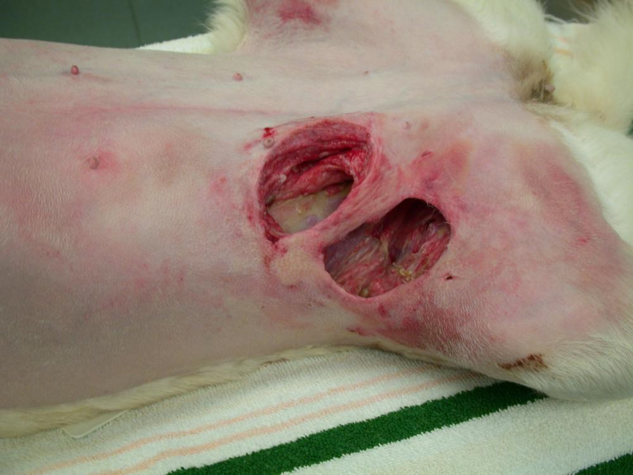
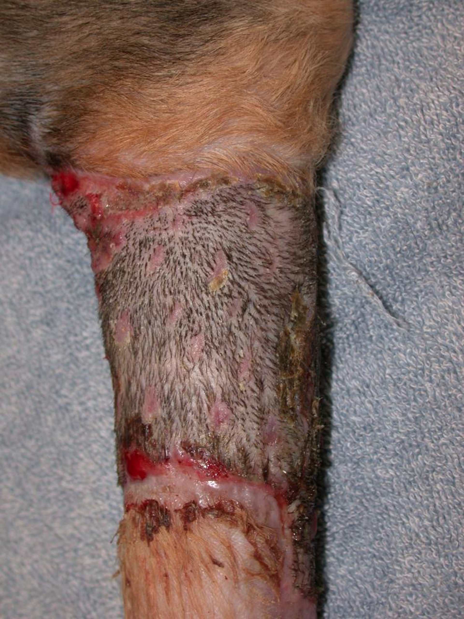
Preoperative view of wound to be covered with a superficial epigastric axial pattern flap.
Courtesy of Dr. Kevin Winkler.
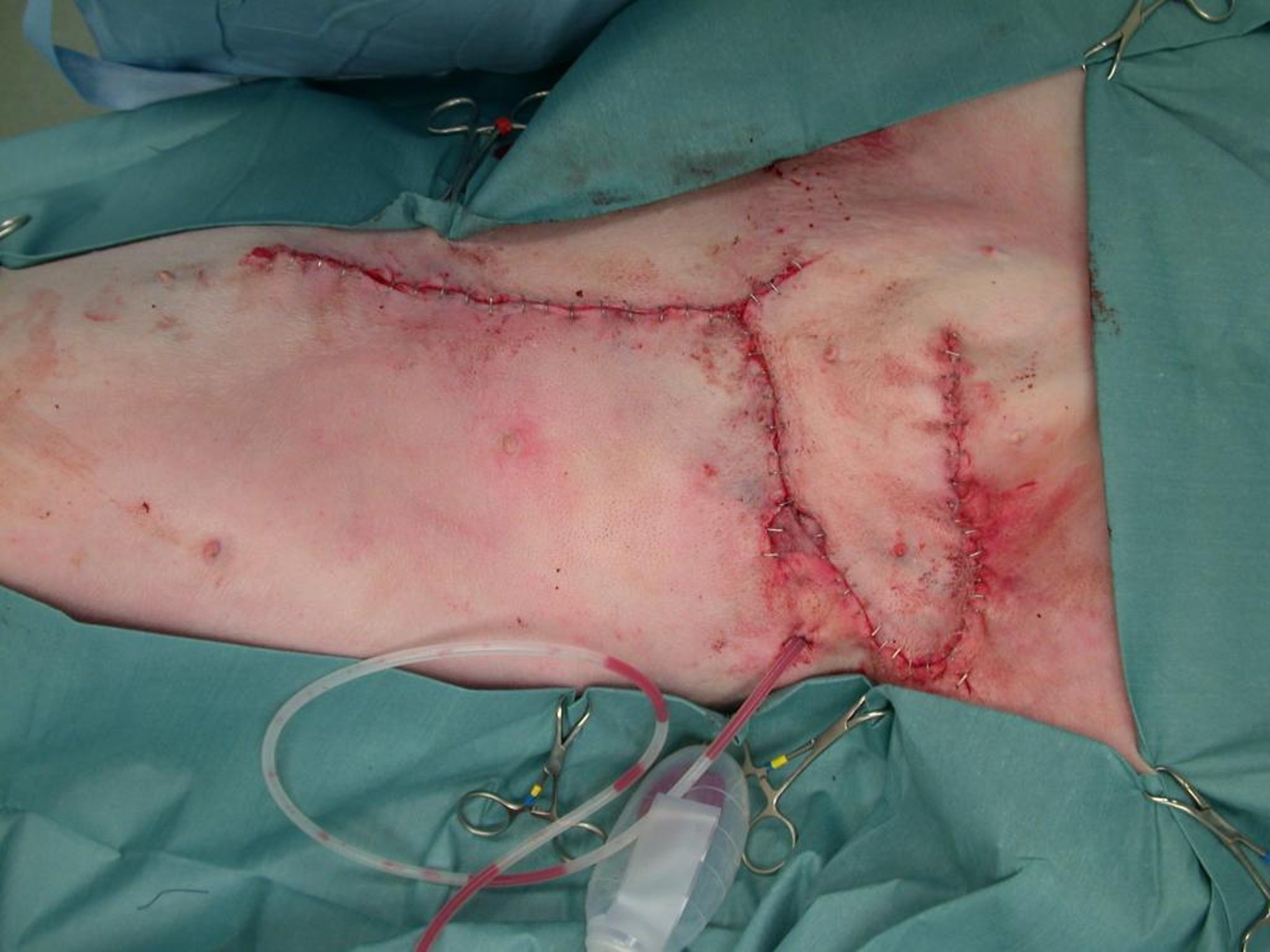
Postoperative view of a superficial epigastric axial pattern flap.
Courtesy of Dr. Kevin Winkler.
The basic advancement flap technique is known as the single pedicle advancement flap. Two slightly divergent incisions are made perpendicular to the defect. The tissue is undermined, advanced, and sutured to close the original defect. For larger wounds, two single pedicle flaps are safer than one large flap. Two advancement flaps are combined to form the H plasty.
There are several other well-described flap techniques, including the bipedicle advancement flap and the V-Y advancement flap. In each of these techniques, the coverage depends on stretching the skin over the defect. For this reason, the use of these techniques may be limited by vascular supply or the regional anatomy, such as the area around the eyelids.
Flaps designed to incorporate a direct cutaneous artery are known as axial pattern flaps (arterial pedicle grafts). The flaps can be used to cover a large area of tissue and carry along a new blood supply to ensure flap survival. Muscle-based pattern flaps can also be used to reconstruct a body wall defect in addition to covering a loss of skin.
The surviving area of axial pattern flaps is 50% greater than a corresponding subdermal plexus flap and therefore allows coverage of a larger area. Because axial pattern flaps are based on arteries, they must have consistent landmarks and do not cover all regions of the body.
The best-described of the axial pattern flaps is based on the caudal superficial epigastric artery. Based in the caudal aspect of the abdominal wall, this flap can extend cranially to include all but the most cranial thoracic mammae (ie, this flap can extend cranially to include the caudal thoracic mammary gland through the inguinal mammary gland). Other commonly used flaps include the thoracodorsal, omocervical, and deep circumflex iliac artery flaps.
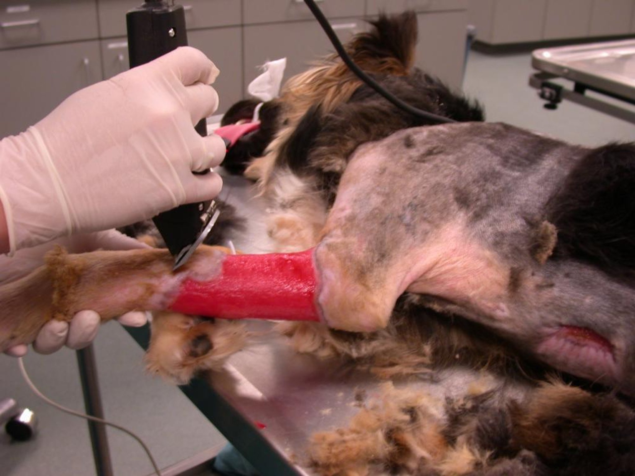
Initial wound to be grafted with a free skin mesh graft. Note the healthy granulation bed.
Courtesy of Dr. Kevin Winkler.

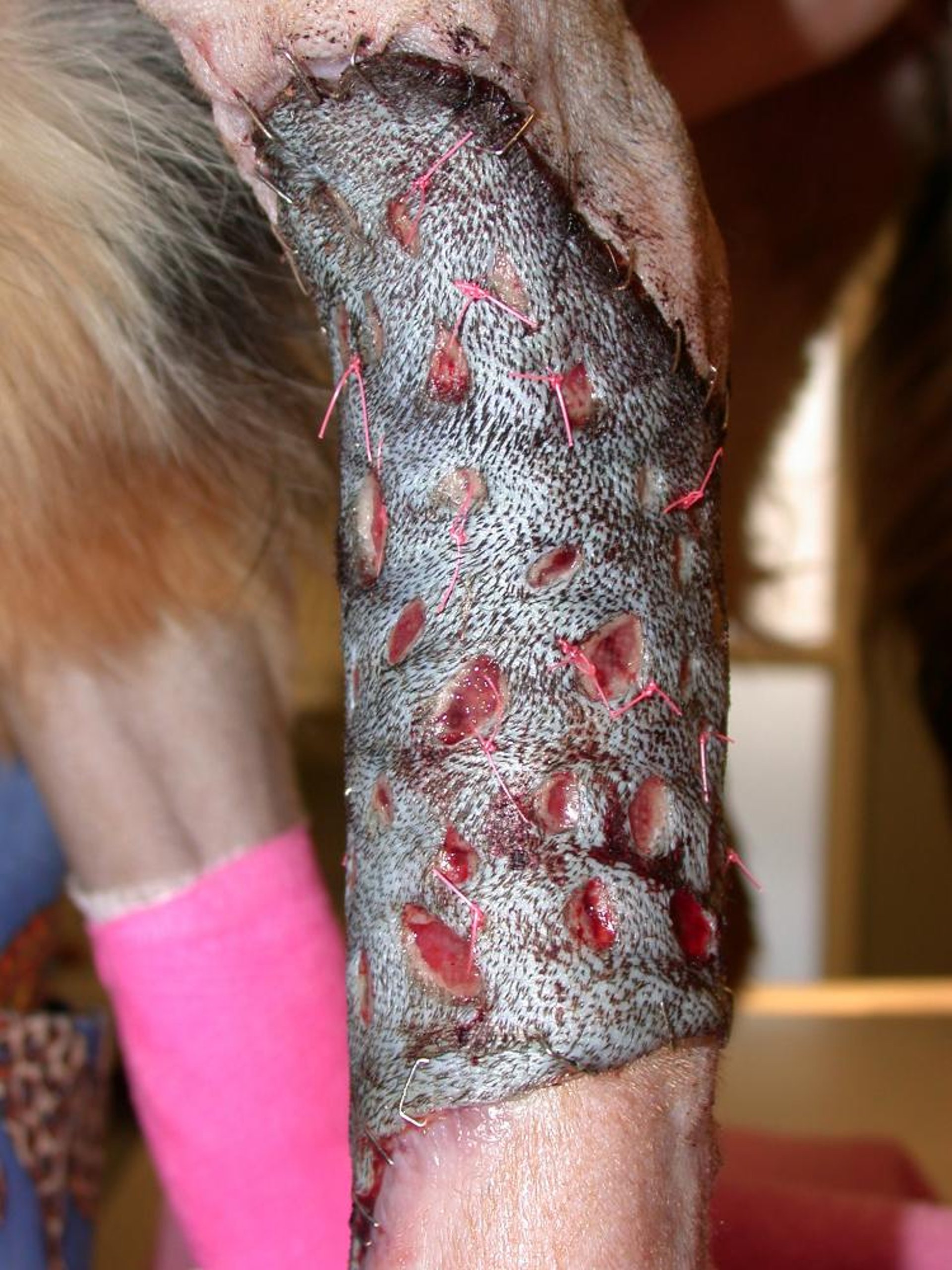
Appearance of a free skin mesh graft 24 hours after surgery.
Courtesy of Dr. Kevin Winkler.
Appearance of a free skin mesh graft 3 weeks after surgery.
Courtesy of Dr. Kevin Winkler.
Free skin grafts are used for cases with massive tissue loss such as large burns or degloving injuries. The grafts are best used as a split mesh. This allows drainage and helps prevent seroma formation.
Skin grafts will not remain viable if laid over squamous epithelium, denuded bone, cartilage, or tendon. The grafts must have a healthy, vascularized bed.
Initially, nutrition for the flap is maintained because capillary action pulls serum into the dilated capillaries of the skin graft, creating graft edema. Anastomosis with recipient bed vessels (inosculation) begins within 48–72 hours of surgery. The edema may worsen immediately after inosculation, because arterial supply is established before venous return. The edema should resolve as normal blood flow returns to the flap by day 4–6 after surgery.
All skin flaps and grafts require a clean, healthy recipient bed for survival. This is especially important for subdermal plexus flaps and free tissue transfers, because they do not contain a direct cutaneous arterial supply. The recipient bed must be free of debris, infection, and necrotic tissue.
Although flaps may have well-described anatomical markers, determining their viability may not be as easy. The simplest, but least accurate, methods to assess a flap’s viability are subjective measures, including the assessment of color, warmth, sensation, and bleeding.
Purple color cannot be used as a predictor of viability. Contused, purple skin is often viable; however, progression from deep purple to black indicates necrosis.
Skin temperature may be affected by the state of vasodilation and is therefore not an accurate method to assess viability. Viable flaps may bleed from a cut surface, as may nonviable flaps that still have some arterial function but poor or no venous return. After a flap is moved, it may develop edema for the first few days until venous vascularization is completed.
Newer techniques and medications being developed for wound care include platelet-rich plasma, extracorporeal shock wave therapy, stem cell therapy, and a variety of growth factors. Their efficacy may be controversial.
Key Points
Advancement flaps move skin without regard for their vascular supply
Axial pattern flaps rotate skin with an associated arterial supply
Free skin grafts can cover large areas where a skin flap may not be an option.
For More Information
Also see pet health content regarding surgical techniques in wound management.


