

Adequate housing, a good diet, and routine parasite control will help to minimize disease in pet reptiles, as with other animals.
Heart and Blood Vessel Disorders of Reptiles
Septicemia, caused by bacteria in the blood, is a common cause of death in reptiles. The disease affects the whole body and may result from trauma, an abscess, an infestation of parasites, or environmental stress. Death may be sudden or occur after longterm signs of illness. Common signs are trouble breathing, lack of energy, convulsions, and loss of muscle control. Reptiles with septicemia may develop small, purplish red spots on the belly skin; chelonians may have reddened plastrons. Keeping a reptile’s environment clean and well maintained can reduce the risk of septicemia. Affected reptiles should be isolated, seen by a veterinarian as soon as possible, and treated with antibiotics.
Digestive Disorders of Reptiles
Reptiles’ digestive systems can be affected by viral, bacterial, protozoal, and parasitic infections.
Adenoviruses
Adenoviruses may cause fatal liver or digestive tract diseases in certain snakes (Gaboon vipers, ball pythons, boa constrictors, rosy boas, and rat snakes), lizards (Jackson’s chameleons, savannah monitors, and bearded dragons), and crocodilians. In bearded dragons, the adenovirus appears to be transmitted by fecal-oral contamination (feces coming into contact with the mouth). Signs of infection are more common in younger dragons but can also affect adults, usually to a lesser extent. The signs are vague and include lack of energy, weakness, weight loss, diarrhea, and sudden death. The illness rate is high in young bearded dragons, but supportive care (fluid administration and assisted feeding) and antibiotics to treat secondary bacterial infections can increase survival.
The signs of adenovirus infection in bearded dragons are similar to those caused by coccidia and nutritional disorders. The diagnosis of adenovirus infection can be confirmed with a polymerase chain reaction (PCR) test of the blood or a liver biopsy. Treatment is supportive with anti-inflammatory drugs and assisted feeding.
Lizards that have recovered from the infection should be quarantined for at least 3 months. Many infected lizards fail to thrive and become sick with other illnesses. The length of time a lizard can infect other lizards after recovering is unknown, so you should not plan to sell or trade a previously infected animal.
Infectious Stomatitis
Infectious stomatitis, infection and inflammation of the tissue lining the mouth, is seen in snakes, lizards, and turtles. Early signs include tiny, purplish red spots in the mouth. Diseased tissue develops along the rows of teeth as the condition worsens. In severe cases, the infection can extend into the upper and lower jaw bones. Bacteria that are commonly found in the mouth are the most frequent causes of stomatitis. Respiratory or gastrointestinal infection may develop if stomatitis is not treated promptly. Treatment involves surgical removal of dead tissue from the mouth, cleaning with an antiseptic solution, antibiotics , and supportive care. More extensive surgery may be required in severe cases. Vitamin supplementation, especially with vitamins A and C, has been suggested but may not change the course of disease.
Intestinal Parasites
Pet reptiles that are stressed in captivity and housed in small enclosures increase their susceptibility to heavy infestations of parasites with direct life cycles—that is, parasites that require only 1 host species (the reptile) to complete their life cycle. Every effort must be taken to rid reptiles of parasite burdens and to rid the environment of intermediate host species (like insects).
Your veterinarian can test your reptile’s feces for gastrointestinal parasites. Treatment should be attempted when evidence of an infestation of parasites is found. Drugs called anthelmintics are usually needed to eliminate internal parasites. Many different anthelmintics (dewormers) are available. Your veterinarian can prescribe the one considered most effective for the particular parasite involved.
Roundworms of the stomach (stomach worms) are seen in lizards and can cause stomach ulcers in severe infestations. Numerous snakes are infected by a hookworm that lives in the upper gastrointestinal tract and causes wounds at sites of attachment. Large swellings caused by the inflammatory response to this hookworm can cause intestinal obstruction.
Parasites known as ascarids (roundworms) also frequently infect reptiles. Severe tissue damage and death may occur in infected snakes. Snakes infected with ascarids may regurgitate partially digested food or adult worms and may have no appetite. Infection may cause inflammatory swellings in the gastrointestinal tract. These swellings may abscess and perforate the intestine.
Many other species of roundworms may be found on examination of a reptile's feces. Prey animal parasites (like mouse pinworms) that do not cause disease in reptiles may also be seen in a reptile's feces after the reptile consumes an infected animal.
Protozoal Diseases
Protozoa are single-celled organisms; some cause disease in animals. Entamoeba invadens is the most serious disease-causing protozoan of reptiles. Signs of infection are loss of appetite and weight, vomiting, mucus-containing or bloody diarrhea, and death. The disease may spread quickly in large snake collections. Plant-eating reptiles appear to be less susceptible than meat-eating ones. Several kinds of reptiles that seldom become affected or die can serve as carriers; these include garter snakes, northern black racers, and box turtles. Most turtles are resistant, although giant tortoises are susceptible. Other resistant species include eastern king snakes, cobras (possibly as an adaptation that allows them to eat other snakes), and crocodiles. Susceptible snakes include most boas, colubrids (a family that includes king snakes, garter snakes, and racers), elapids (a family of venomous snakes including coral snakes and mambas), crotalids (the pit viper family, including copperheads and rattlesnakes), and other vipers. This protozoan is transmitted by ingestion of the cyst form of the parasite, which is passed in the feces of infected snakes and persists for long periods in the environment. Diagnosis is made by examining feces or tissue samples for the protozoan.
An antiprotozoal drug is usually prescribed for treatment. To help prevent transmission among reptiles, turtles and snakes should not be housed together. The potential for this disease to be passed on to humans should not be taken lightly, and strict sanitation and hygiene measures should be observed.
Cryptosporidiosis is infection caused by protozoa of various Cryptosporidium species. It can cause regurgitation, marked weight loss, and longterm weakness. In snakes, the organism affects the lining of the digestive tract, causing thickening of the stomach lining and loss of normal digestive motion of the stomach. A veterinarian may be able to feel a mass in the stomach area. X-rays or an examination and biopsy of the inside of the gastrointestinal tract using an endoscope may reveal thickening of the stomach lining. Many lizards, including Old World chameleons and savannah monitors, are affected primarily in the intestine. Cryptosporidiosis is diagnosed with tests of the feces or regurgitated food or by biopsy of the stomach. Several treatments have been suggested, but most do not work consistently. Intensive supportive care will often stabilize and help prolong the life of the affected reptile.
Cryptosporidiosis can be transferred from animals to humans. However, Cryptosporidium species typically found in reptiles appear to rarely infect humans.
Hormonal Disorders of Reptiles
Although hormonal (endocrine) disorders do not appear to be common in reptiles, diabetes mellitus (a disorder of carbohydrate metabolism) has been reported in turtles and tortoises. Signs of diabetes in these reptiles include glucose (a type of sugar) in the urine and abnormally high levels of glucose in the blood. Affected animals may have an increased appetite but lose weight. Treatment is difficult and may involve insulin therapy, diet modification, and addressing concurrent illnesses.
Eye and Ear Disorders of Reptiles
Infections of the eyes are possible in all reptiles. Ear infections are most likely to occur in turtles.
Eye Abscesses and Conjunctivitis
Snakes can develop abscesses below the spectacle (eyecap), the clear protective covering over the eye. Other reptiles can develop conjunctivitis, inflammation of the membranes around the eye. The severity of conjunctivitis ranges from mild to severe (involving all the tissues around the eye and the eyeball itself). It may result from the spread of infectious stomatitis from the mouth. Conjunctivitis in turtles and lizards without spectacles can be treated with topical eye ointment. Snakes and lizards with spectacles need surgery to drain the abscess and flush the eye with an antibiotic solution. Some affected reptiles, especially turtles, may need supplemental vitamin A.
Ear Infections
Ear infections often occur in turtles, most often box turtles and aquatic turtles. Swelling may be seen at the eardrum, and diseased tissue may be present. Many bacteria species cause ear infections. Animals with middle or inner ear infections often require surgery that punctures the eardrum, flushes the middle ear, and removes infected tissue. Ear infections can also be caused by vitamin A deficiency, so injections and dietary supplementation of vitamin A may be beneficial.
Bone and Muscle Disorders of Reptiles
Abnormal beak growth in a turtle

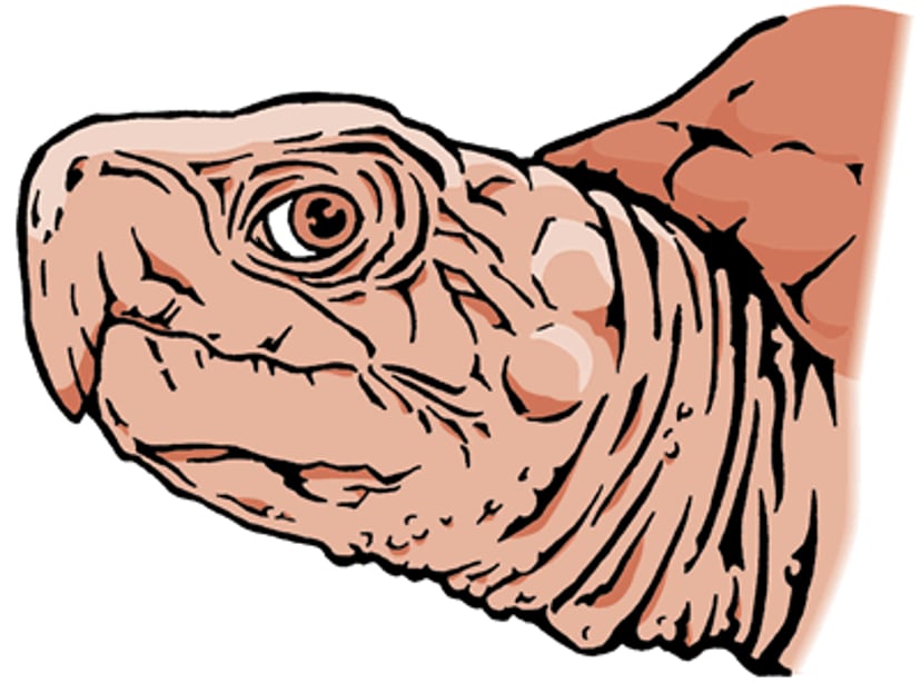
Abnormal beak growth occurs in turtles and tortoises. This condition, which interferes with feeding, is often associated with poor nutrition, a deficiency of calcium, or both. Calcium or vitamin D3 deficiency may cause the skull to become distorted as it develops. The distortion interferes with normal positioning of the upper and lower beaks when the jaw is closed and can affect wear of the beaks. Excessive levels of protein in the diet may contribute to this condition. Treatment consists of trimming or grinding the beak into a more normal shape. The condition usually returns because of the underlying abnormal position of the jaws, and long-term, repeated beak trimming is typically required. Abrasive foods that allow for some natural beak shaping during feeding should be provided to turtles and tortoises in captivity.
Metabolic Bone Diseases
Secondary nutritional hyperparathyroidism is the most common bone disease seen in pet reptiles. It is caused by a poor diet with the wrong ratio of calcium to phosphorus and a lack of vitamin D3 or poor husbandry (lack of ultraviolet B light, inadequate temperature control in the cage). Affected reptiles are generally rapidly growing plant- and insect-eating lizards, turtles, or tortoises. Signs include poor appetite, weakness, an inability to walk normally, swollen or distorted jaw or leg bones, fractures of the spine or legs, cloacal prolapse (in which the lining of the cloaca protrudes through the vent opening to outside the body), and muscle spasms. Diagnosis requires x-rays to document generalized loss of bone from the skeleton and blood tests that show low levels of vitamin D and an imbalance of calcium and phosphorus. Treatment of critical cases requires fluid therapy, nutritional support, and calcium injections if blood levels are too low. Correction of the diet and husbandry (including provision of UV light to ensure adequate vitamin D is being made in the skin) are the most important parts of successful treatment.
Secondary renal hyperparathyroidism is a kidney disorder that occurs in adult reptiles. It is associated with high levels of phosphorus and low levels of calcium in the blood, abnormal calcification (hardening) of muscles and organs, and loss of calcium from bones. Diagnosis can be suspected based on the animal's history, x-rays, and blood tests, although a definitive diagnosis requires demonstration of poor kidney function or kidney damage seen on a biopsy of the kidney. Treatment options are limited because of the difficulty in correcting kidney damage and typically involve supportive care with medications and fluids.
Brain, Spinal Cord, and Nerve Disorders of Reptiles
Stargazing is a sign of some nervous system disorders in snakes and other reptiles. Stargazing describes a twisting of the neck that makes the animal appear to look upward (toward the stars). Other signs of nervous system disorders include mental dullness, abnormal posture, seizures, and inability to move normally. One of the most common causes of stargazing in boa constrictors and pythons is inclusion body disease, which is caused by a viral infection. There is no treatment for this disease, which is always fatal.
Stargazing can also be caused by exposure to excessive heat, head injuries, toxins, and infections caused by bacteria or other organisms. Bacterial meningitis or encephalitis can result from bacterial infections that move into the bloodstream and penetrate nervous system tissue. The outcome of neurologic disease varies depending on the cause, but the outlook is generally poor. Your veterinarian will perform tests to try to determine the underlying cause of the neurologic signs and to prescribe appropriate treatment. Viral diseases are typically treated with supportive care. Antibiotics are usually necessary in cases of bacterial infection.
Nutritional Disorders of Reptiles
Nutritional disorders can be caused by dietary imbalance in various nutrients, including protein, vitamins, and minerals, or by disorders that prevent proper metabolism of nutrients.
Malnutrition and Dehydration
Reptiles with poor appetites may require assisted feeding. This is a process that is best directed by your veterinarian because feeding a malnourished reptile with severe dehydration can lead to additional health problems. Initial feedings should replace fluids and electrolytes. Signs of dehydration include loose skin or sunken eyes.
A dehydrated reptile can sometimes be encouraged to drink by allowing it to bathe in shallow water within an enclosure kept within the preferred temperature range for that species. Dehydrated turtles and tortoises may also be able to absorb water through the cloaca while bathing. However, dehydrated reptiles may need fluids administered by a veterinarian through injection or a stomach tube.
A malnourished reptile might have protruding bones and a gaunt appearance, but signs of malnutrition and dehydration may not be easy to see. Some malnourished animals require assisted feeding, which should be done only under the direction of a veterinarian to reduce the possible risks.
If your reptile is not eating well, environmental factors such as temperature, light, and humidity should be set at the proper levels for that type of reptile. Reptiles cannot process fluids and nutrients properly if environmental conditions are not optimal.
Gout
Gout is seen in all orders of reptiles. It occurs when the level of uric acid in the blood is too high. Two forms of gout have been reported. Visceral gout affects the organs, and articular gout affects the joints. X-rays may show mineralized deposits from uric acid in the affected organs and joints. Visceral gout can be due to too much protein in the diet (primary visceral gout) or to other causes such as dehydration or kidney damage (secondary visceral gout). Gout can be very painful, causing discomfort to the point that some reptiles refuse to move, eat, or drink.
Primary visceral gout is treated by correcting the diet. Secondary visceral gout is treated by trying to correct the underlying problem. Drugs that have traditionally been used to treat gout in humans may be effective in reptiles if the diagnosis is made early. However, the outlook for recovery is poor in advanced cases. Medical treatment usually must be long-term because signs of gout often recur if treatment is discontinued. Euthanasia should be considered in reptiles that appear to be in pain and have no appetite.
Lung and Airway Disorders of Reptiles
Respiratory infections, including pneumonia, are common in reptiles and can be caused by parasites of the respiratory system or the whole body, unfavorable environmental temperatures, unsanitary conditions, other diseases, malnutrition, and vitamin A deficiency. Open-mouth breathing, discharge from the nose, and difficulty breathing are frequent signs. Septicemia (widespread infection in the bloodstream) may develop in severe or prolonged cases. Treatment consists of improving environmental factors (such as cleanliness and temperature) and starting antibiotic therapy. A veterinarian will be able to advise you on the proper antibiotic treatment. Reptiles with respiratory infections should be kept at the middle to upper end of their preferred temperature range. Increased temperatures help stimulate the immune system and also help thin out respiratory tract secretions, making them easier for reptiles to expel. Turtles with pneumonia often have an underlying vitamin A deficiency. Turtles treated for pneumonia may not improve completely until they have been supplemented with vitamin A.
Paramyxovirus
Paramyxovirus infections are more common in vipers than other snakes but have also been reported in nonvenomous snakes. This highly contagious virus most often causes respiratory system problems. Secondary bacterial infections are common, and affected animals may have discharge from the nose, open-mouth breathing, dried pus in the mouth, and labored breathing. Neurologic signs, such as tremors and abnormal posture, are sometimes seen. The virus appears to be passed from one animal to another through respiratory tract secretions.
A snake with a respiratory infection that does not respond to typical treatments (including antibiotics) may have a paramyxovirus infection. Blood tests can be used to screen animals for infection and prevent carriers from entering noninfected collections. There is no specific treatment, but supportive care and antibiotics may be helpful. Affected snakes should be isolated and strict hygiene practices used.
Reproductive Disorders in Reptiles
Dystocia (Egg Retention)
Sterilization is rarely performed in reptiles, and therefore reproductive disease remains a common problem. In egg-laying reptiles, eggs (with variable levels of shell mineralization) may be retained within the reproductive tract (egg retention or egg binding), whereas in species that bear live young, unfertilized eggs or fetuses may be seen within the reproductive tract. In some cases, abnormal ovarian follicles may also be present. Dystocia is generally not a sudden event as in mammals or birds, and reptiles may retain eggs or fetuses for weeks or even months after the normal timing of laying/birth. If it is not known when the reptile was bred, it may be hard to tell the difference between a normal pregnancy and dystocia in otherwise clinically healthy reptiles. Metabolic disease or infection can worsen the issue. In general, your veterinarian will be able to make the diagnosis after examining your reptile and performing x-rays and ultrasound. Blood tests may also help identify infections or metabolic diseases, such as hypercalcemia (elevated blood levels of calcium; see nutrient requirements).
Unless there is evidence of short-term disease, medical management may be tried, although it frequently fails. Improvements in husbandry (especially provision of solitude and a suitable substrate), corrections of any metabolic diseases, and hormonal treatments to induce labor may be helpful. In most cases, surgery is necessary after medical stabilization and typically requires removal of the reproductive organs.
Vent Prolapse
Several organs, including the cloaca, colon, oviduct (part of the female reproductive tract), hemipenes/phallus (like the penis in male mammals), and (if present in some species) bladder, may push abnormally through the vent of reptiles and get stuck outside the body. Common causes include dystocia, breeding trauma, inflammation of the cloaca, infections, metabolic disease, bladder stones, kidney disease, cancer, or any space-occupying mass within the abdomen that causes straining to defecate. It is important for your veterinarian to identify the prolapsed organ, because some (eg, phallus in turtles/hemipenes in snakes and lizards, required only for breeding and not for urination) can be amputated, whereas others (eg, cloaca, colon, bladder) cannot be. Your veterinarian will gently clean the prolapsed organ and attempt to replace it back into its normal position inside the body. However, it is also important to determine the cause to prevent recurrence.
Prolapses of the hemipenes and phallus can be amputated surgically; this will make the animal infertile. If the prolapsed tissue is viable and can be replaced back into its normal position inside the body, surgery can be performed to hold it in its usual place. If the tissue is dead, then careful surgery is required to remove the affected tissues and reconnect the living sections at each end.
Skin Disorders of Reptiles
Reptiles are prone to a variety of skin diseases and disorders. Good sanitation practices, such as cleaning the enclosure regularly, providing fresh water, and removing uneaten food, can help prevent infection and parasite infestation.
Abscesses
Abscesses are pus-filled sores, often accompanied by inflammation, that are usually caused by bacterial infection. They are seen in all orders of reptiles. Abscesses are often caused by bite wounds, other injuries, or poor environmental conditions. Abscesses under the skin appear as small lumps or swellings. Other conditions that may look like abscesses are parasitic infections, tumors, and hematomas (swellings filled with blood). A number of species of bacteria, often more than one kind at a time, can be present in abscesses in reptiles. Small, localized abscesses should be surgically removed to avoid recurrence, which happens frequently. Larger abscesses should be surgically opened and drained. Appropriate antibiotics may be needed.
Dermatophytosis
Dermatophytosis, a fungal infection of the skin or nails, has been described in all reptiles. In most cases, an injury to the skin provides a point of entry for the fungus. Turtles and tortoises with fungal infections of the shell can be treated by removing the dead, damaged, or infected tissue and applying an antiseptic solution. Oral antifungal drugs or topical antifungal creams may be used to treat skin infections. Exposure to ultraviolet light also may be beneficial. (See also , below)
Dysecdysis
Incomplete shedding, snake

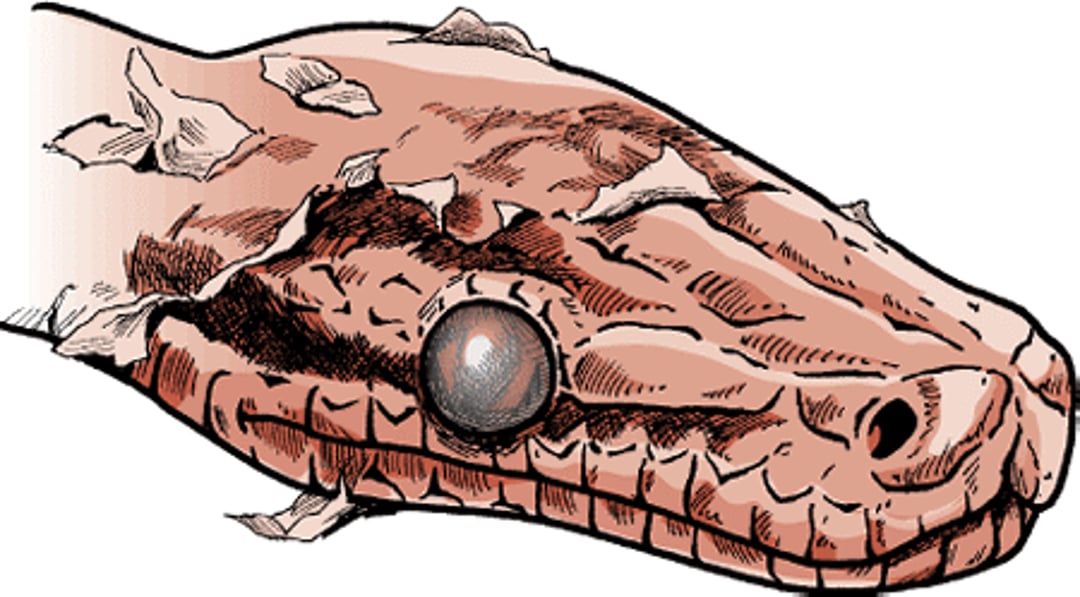
Dysecdysis refers to incomplete or abnormal shedding of the skin. Low humidity, skin parasites, nutritional deficiencies, infectious diseases, lack of suitable abrasive surfaces, and decreased thyroid function may contribute to an abnormal shed. Spectacles (eyecaps), which are the clear protective layer over the eyes, or bands of skin encircling the tail or toes, may not shed properly and may be retained. Retained spectacles are best treated by applying an eye ointment for several days until the spectacle either falls off or can be carefully and gently removed with fine forceps. Be patient; eyecaps should never be forced off because of the possibility of damaging the new one underneath.
Retained skin is best treated by soaking the reptile in warm (77°F to 85°F [25°C to 29°C]) water for several hours and then pulling on the retained skin gently with a gauze sponge. A humidity chamber also works well. This can be as simple as a 10-gallon aquarium with an undertank heater in which wet bath towels are placed. The top can be covered with a light cloth to increase humidity levels, but excessive heat must be avoided and can be relieved by allowing more ventilation. It is easier to prevent than to treat an abnormal shed, so make sure that your reptile is free of disease and parasites, eats a diet appropriate for its species, is kept at the correct humidity level, and has abrasive surfaces available to help it shed.
Skin Parasites
Parasites that live on the skin can be a problem with wild-caught and newly acquired reptiles. Infestations are best prevented by thorough examination and a period of quarantine for all new animals entering a collection.
Mites are distributed worldwide, and most species of reptiles are affected. Mites can cause reduced energy and, in heavy infestations, death by anemia (loss of red blood cells). Mites may also transmit disease-causing organisms from other infected animals. The skin of affected reptiles appears coarse, and dysecdysis may occur. Most mites are tiny (less than 1.5 millimeters long) and are often found around the eyes, skin folds on the face or neck, or any other indentation on the body.
Mites are visible to the naked eye but are hard to see in small numbers. If mites are suspected, placing the reptile on a piece of white paper and gently rubbing it may cause mites to fall off and be visible. Affected reptiles often spend a great deal of time soaking in water to drown the mites. If you examine the water dish, you might see the drowned remains of mites.
There are many methods of treatment. In all cases, cages should be cleaned thoroughly, and you should throw away substrate materials, branches, and disposable cage furniture. Newspaper bedding should be used until treatment is completed to help with frequent cleaning and to get rid of sites where mites could lay eggs. A veterinarian can tell you the type of mites your reptile has and provide advice on eliminating them from your pet and its enclosure (such as recommending a safe insecticide).
Any enclosure in which insecticide is used should be well ventilated, and any water containers should be removed while spraying insecticide and replaced when the spray has dried. Do not use any product without consulting your veterinarian; some are not safe for all reptiles.
Ticks are frequently found on reptiles and have been associated with many diseases. Heavy infestations may result in anemia (too few red blood cells) due to loss of blood. Certain ticks may cause paralysis, with muscle wasting at the site of the bite. Ticks can be removed with forceps (tweezers) or killed by insecticides recommended by your veterinarian. If you remove a tick manually, wear gloves to avoid exposure to diseases it may be carrying. Animals with ticks may require antibiotics to treat infections associated with multiple skin bite wounds and possibly transmission of disease-causing bacteria.
Leeches have been found on the legs, head, neck, and in the mouth of a variety of turtles and crocodilians. A few leeches may not be harmful, but many may cause skin damage and transmit other diseases. They may be removed by gently lifting them off with tweezers, being careful not to damage the underlying skin, or by pouring salt water on them, which may cause them to fall off.
Turtles frequently have skin maggots. Botflies create a skin wound in which to lay their eggs. The eggs hatch into bots (larvae, or maggots) that live in cyst-like structures until they are mature enough to leave the wound. The wounds often resemble a lump under the skin. These lumps have a tiny opening that may be lined with black, crusted material. Your veterinarian can remove the bot by slightly expanding the opening and carefully extracting the bot with forceps. The wound is then flushed with an antiseptic solution, and an antibiotic ointment is applied. Reptiles with multiple wounds usually need antibiotic treatment. Skin maggots may also be found in existing wounds. In these cases, the maggots must be manually removed and the animal treated with antibiotics as needed. During heavy fly season, it is recommended that turtles be housed indoors or with screens over their enclosures to offer some protection.
Scale Rot
Scale rot (ulcerative or necrotic dermatitis) is seen in snakes and lizards. Humidity and unclean environments appear to be the main factors that cause this condition. Moist, contaminated bedding allows bacteria and fungi to multiply. When coupled with exposure to feces, these organisms can cause skin sores. Secondary infection with other bacteria may result in septicemia (blood infection) and death if untreated. Reddening of the skin, areas of dead skin, skin sores, and skin discharge are common signs. Although sores are sometimes caused by skin injuries, they more often develop inside the body and then become apparent in the skin. This is the case with classic necrotic dermatitis in the ball python, which can develop even when animals are kept under ideal conditions. The disorder starts with bleeding into scales, followed by pustules (small, raised, pus-filled bumps like pimples) that eventually progress to ulcers. Treatment with antibiotics and excellent hygiene are essential.
Blister disease is an early stage of scale rot. The skin develops pustules or blisters that may heal without progressing to ulcers if treatment is started early. Low-grade heat injury may look like blister disease if it causes fluid-filled blisters.
Septicemic Cutaneous Ulcerative Disease
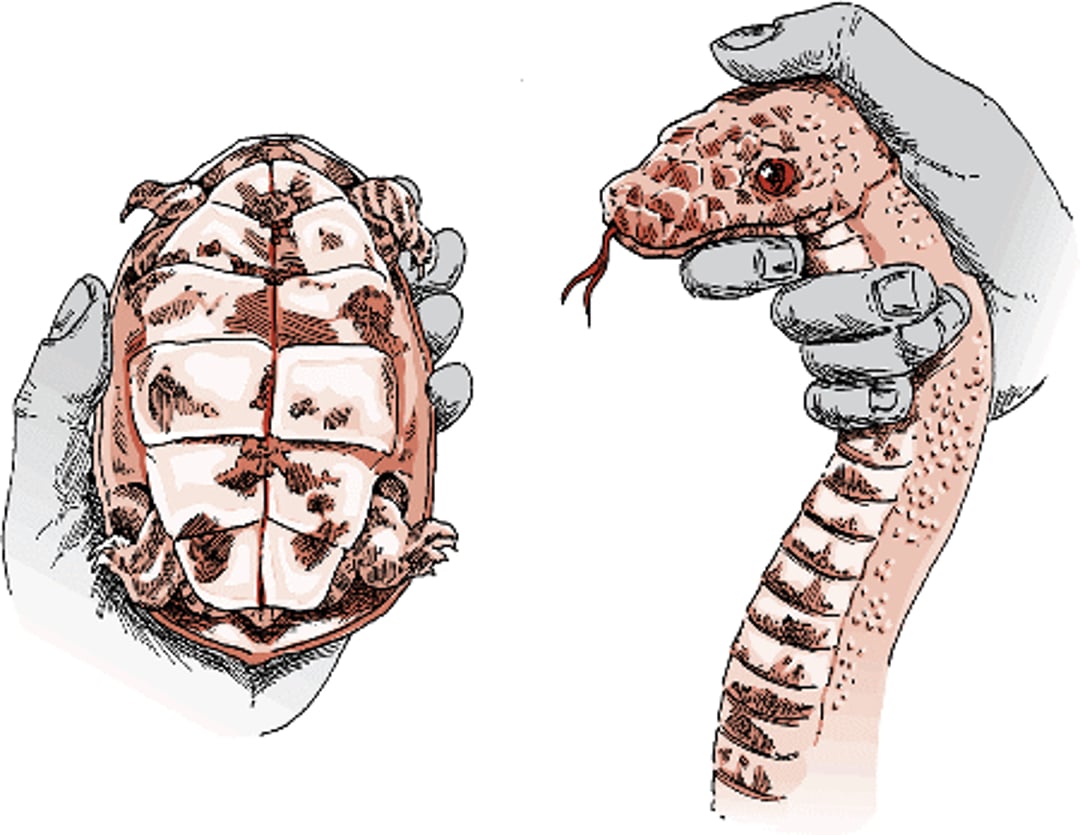
Septicemic cutaneous ulcerative disease (SCUD) is a bacterial infection of aquatic turtles. The disease causes pitting of scutes on the shell. The scutes may slough off, and an underlying pus-filled discharge may be present. Loss of appetite, lack of energy, and small, red spots on the shell (from bleeding) may occur. Liver damage is common. Treatment with an antibiotic is recommended. Good sanitation is critical to prevent SCUD.
Shell disease and scale rot

Crustacean Bacteremia
Another shell disease of turtles is caused by Beneckea chitinovora, a bacterium commonly found in crustaceans (sea creatures such as crayfish and crabs). Reddening, pitting, and ulcers of the shell are seen. Septicemia (bacteria in the blood) is uncommon. Treatment with an antibiotic and topical iodine is recommended. Feeding crayfish to turtles is often the cause of this condition and should be discouraged.
Disorders Affecting Multiple Body Systems in Reptiles
Diseases that affect more than one body system in a single animal are also known as systemic diseases.
Bacterial Infections
Bacterial diseases are common in all reptiles. Most infections occur in reptiles whose immune systems are weakened by illness or other causes. It is important to determine the type of bacteria involved and also to correct environmental and nutritional deficiencies that contribute to poor health. Antibiotic treatment will not be successful unless nutrition, environment, and sanitation are also corrected.
Coccidial Organisms
Coccidia are microscopic parasites. Several coccidial organisms have been reported to cause disease in reptiles. The severity of disease varies with the species of the coccidial organism and the type of reptile affected. These parasites can increase to tremendous numbers, especially in reptiles with suppressed immune systems. The eggs of these parasites can survive in the environment for weeks. Thorough daily cleaning is necessary to remove all feces and contaminated food and water. Insects and other food items must be removed daily because they can be a source of contamination (for instance, crickets may eat the parasitic eggs while gathering fluid from the feces). Treatment with an antimicrobial drug may take 2 to 4 weeks. Even under the best conditions, treatment will eliminate these organisms in only 50% of cases. However, reducing the number of these organisms is important to reduce the impact of the infection and lower the risk of spread to other animals.
Diseases Caused by Fungi
Excessively high humidity, low environmental temperature, existing disease, malnutrition, and other stressors may make reptiles more likely to develop fungal diseases. Little is known about the course of fungal infections that affect the whole body and develop over a long period, but maintaining good sanitation and nutrition reduces the frequency of these infections. Antifungal drugs may be used to treat fungal infections that have spread through the whole body, but reports of successful treatment in reptiles are few. For fungal infections that are limited to one area, surgical removal of the infected tissue followed by local wound treatment may be helpful. Basidiobolus species, which can cause disease in mammals, have been found in the feces of healthy reptiles.
The most frequent sites of fungal infection in reptiles are the skin and respiratory tract. Slow-healing internal sores developing on gastrointestinal tissues have been associated with some fungal infections. Fungal infections can also cause disease of the liver, kidneys, and spleen. Few signs other than weight loss may be seen before death. Animals may continue to eat until a few days before death.
Flagellates
Flagellates (a type of protozoal microorganism), especially Hexamita species, can cause urinary tract disease in turtles and tortoises and intestinal disease in snakes. An anthelmintic or antiprotozoal drug is usually prescribed for treatment.
Herpesviruses
Herpesviruses have been found in freshwater turtles, tortoises, and green sea turtles. In freshwater turtles, the virus may affect the liver. In tortoises, infection may damage tissue in the mouth and cause loss of appetite, regurgitation, and discharge from the mouth and eyes.
Treatment in tortoises includes isolating the animal, providing supportive care, and applying antiviral medication to lesions.
Inclusion Body Disease of Boid Snakes
Boa constrictors and several species of pythons are most commonly affected by inclusion body disease, which is caused by a retrovirus or arenavirus. Boas are considered to be the typical host for this virus because many are infected, and they can harbor the virus for years without signs. Inclusion body disease should be considered in every sick boa. The disease is named for the characteristic inclusion bodies, which are microscopic clumps of protein or other material that form within cells in infected animals. Signs of inclusion body disease can be related to any factor affecting the immune system and include loss of appetite or weight, secondary bacterial infections, poor wound healing, and regurgitation. As the disease progresses, some boas exhibit neurologic signs such as facial tics, abnormal tongue flicking, stargazing, twisting of the body, and seizures.
Pythons are thought to be abnormal hosts of the virus because their course of disease is more severe and neurologic signs are more extreme. Although the active disease can linger for months or more in boas, most pythons die within days or weeks of the onset of signs.
The infection appears to be spread through body fluids. Breeding, fight wounds, and contamination due to animal droppings coming in contact with the mouth (fecal-oral contract) are common ways of transferring the virus.
Inclusion body disease is not curable, and many pet owners may choose to euthanize affected snakes. However, snakes can be isolated and treated with supportive measures. It is essential that you do not sell infected specimens or their offspring; this practice has caused the disease to spread worldwide.
Infectious Cloacitis
Infection of the cloaca (the passage for both urine and feces in reptiles) can be caused by kidney stones or retention of other materials (such as retained eggs or abnormal mineral deposits) in the lower intestine, urinary tract, or reproductive passages. It is characterized by swelling and bloody discharge from the cloaca. Mineral deposits may form when vitamin or mineral imbalances are present in the diet. These deposits should be manually removed by a veterinarian and followed by dietary correction. Abscesses around the cloaca can extend to other parts of the body and can cause urinary or genital tract infections. Prompt and thorough treatment, including surgery to remove damaged tissue, local wound treatment, and appropriate antibiotics, is required. Your veterinarian may need to examine feces from your reptile to identify whether the infection is caused by parasites.
Internal Abscesses
Abscesses in internal organs may result from blood infection (septicemia). Abscesses of the female reproductive system are common and may lead to an infection of the abdomen. In this case, surgery is required because treatment with antibiotics alone is rarely successful.
Internal Parasites
Disease-causing flatworms (trematodes) infect the arteries and veins of turtles and the mouth, respiratory system, and urinary system of snakes. Veterinarians can prescribe anti-parasitic drugs to treat flatworm infections.
Tapeworms are found in all orders of reptiles but are rare in crocodilians. The complex life cycle of tapeworms and the need for intermediate hosts limit the number of cases in captive reptiles. When present, tapeworm segments (pieces of the worm’s body) may be found in the cloaca, or tapeworm eggs may be found in feces. The infective larvae of some tapeworms may cause swellings in the tissue beneath the skin. These larval stages may be removed surgically. Your veterinarian can prescribe appropriate anti-parasitic drugs to treat tapeworm infection.
Roundworms (nematodes) are found in all orders of reptiles. Examples of nematodes are pinworms and hookworms. These parasites can inhabit the intestinal tract, respiratory tract, or body wall of reptiles. They can sometimes be found in the feces. Infections often are mild but may lead to more serious diseases, such as pneumonia. In severe cases, death may result.
Some roundworm larvae may be able to penetrate the skin. Good sanitation practices to remove parasites from the environment help reduce parasite burdens in captive reptiles. Your veterinarian can prescribe appropriate anti-parasitic drugs to treat roundworm infection.
Skin sores caused by the spirurid worm (Dracunculus species) may occur in reptiles. Numerous species of spirurids infect the body cavity and blood vessels. These worms are less common in captive-bred reptiles or in reptiles that have been in long-term captivity. To treat a spirurid infection, your veterinarian may recommend increasing the reptile’s habitat temperature and giving appropriate anti-parasitic drugs.
Tongue worms (pentastomes) are found in a variety of reptiles. These worms may or may not cause signs of disease. Infections are occasionally associated with signs of pneumonia, but these parasites can inhabit any tissue, and signs will vary according to the path of their migration through tissues. Tongue worms were first noticed in venomous tropical snakes and have also been seen in other reptiles. Treatment with the usual drugs (anthelmintics) often fails to eliminate these worms. In some cases, it may be possible for your veterinarian to use an endoscope to locate and mechanically remove all the adult worms. Recognition of these infestations is important because tongue worms are thought to present a risk of infection to humans.
Mycobacterial Infections
Mycobacterial infections are often associated with chronic wasting (a gradual loss of body condition). These infections generally affect the lungs of turtles and tortoises. Lizards, snakes, and crocodilians often develop small growths on their internal organs. The drugs required to fight these infections cause liver damage in reptiles, and their long-term use is unlikely to be safe.
Cancers and Tumors of Reptiles
Tumors are much more common in reptiles than previously thought. In addition to spontaneously developing cancerous (neoplastic) diseases, tumors have been associated with parasites and certain viruses. Tumors in reptiles are usually easily identified through various tests performed by a veterinarian. Once tumors are identified, treatment protocols similar to those used in other animals could be adapted. Your veterinarian will be able to recommend the appropriate treatment.
Retroviruses
Retroviruses found in Russell's vipers, corn snakes, and California king snakes may sometimes be associated with malignant tumors. Treatment generally involves surgical removal of tumors.
Papillomas
European green lizards appear to pass viral particles from one lizard to another through bite wounds. The resulting papillomas (small growths) are approximately 1/16 to ¾ inches (2 to 20 millimeters) in diameter and may be single or multiple. Although there are no signs in the initial phase, affected lizards may lose energy and appetite and die. Single masses can be surgically removed, but regrowth is common. Isolating affected lizards may be the only way to prevent spread.
A papilloma-type virus also appears to affect Bolivian side-neck turtles and appears as white, oval skin sores on the head. Shell sores are also seen, primarily on the lower portion of the shell. Treatment is supportive care; affected animals should be isolated from other turtles.
For More Information
Also see professional content regarding diseases of reptiles.


