

Congenital abnormalities of the cardiovascular system are defects that are present at birth. They can occur as a result of genetic defects, environmental conditions, infections, poisoning, medication taken by the mother, or poor maternal nutrition. In some cases, it is a combination of these factors that causes the defect. For several defects, an inherited basis is suspected.
In cats, the frequency of congenital heart disease has been estimated to be less than 1% of the population. Among the few cats that do have congenital heart disease, common defects include atrial and ventricular septal defects, atrioventricular valve dysplasia, endocardial fibroelastosis, patent ductus arteriosus, aortic stenosis, and tetralogy of Fallot.
Detecting Congenital Heart Defects
It is important to detect a congenital heart defect as early as possible. Certain defects can be corrected with surgery, and treatment should be performed before the defect leads to congestive heart failure or irreversible heart damage. If the defect is discovered in a recently purchased cat, you may be able to return it. Pets with congenital heart defects are likely to die prematurely, causing emotional distress. Early detection also prevents continuing genetic defects into breeding lines.
The evaluation of most cats with a congenital heart defect may include a physical examination, electrocardiography (recording electrical activity of the heart), x-rays, and echocardiography (ultrasonography). These steps allow diagnosis and assessment of the severity of the defect.
General Treatment and Outlook
The medical importance of congenital heart disease depends on the particular defect and its severity. Mildly affected cats may show no ill effects and live a normal life span. Defects causing significant circulatory disturbances will likely cause death in newborn (and unborn) kittens. Medical or surgical treatments are most likely to benefit cats with congenital heart defects of moderate or greater severity. Left-to-right shunting patent ductus arteriosus is one notable exception. Surgical correction is recommended for most affected animals as long as no other diseases or abnormalities are present that would pose a risk for anesthesia or surgery.
Congenital heart defects produce signs that vary depending on the type of heart failure involved. Possible signs include shortness of breath, difficulty breathing, weakness or unwillingness to move, coughing, fainting, or an accumulation of fluid in the chest or abdomen.
Innocent Murmurs
It is very important to understand that the presence of a heart murmur in a young kitten does not necessarily indicate a congenital heart defect. Many young animals have a low-grade systolic murmur (heard while the ventricles contract) that is not associated with a congenital heart defect. These murmurs usually disappear by 6 months of age. Loud systolic murmurs and diastolic murmurs (heard while the ventricles relax) do indicate cardiac disease, however, and should prompt further investigation by your veterinarian.
Common Congenital Heart Abnormalities
The defects discussed below are those that occur with some frequency in cats. However, it is important to stress that these defects are rare.
Patent Ductus Arteriosus
The ductus arteriosus is a short, broad vessel in the unborn kitten that connects the pulmonary artery with the aorta and allows most of the blood to flow directly from the right ventricle to the aorta. Before birth, oxygenated blood within the main pulmonary artery passes into the descending aorta through the ductus arteriosus, bypassing the nonfunctional lungs. At birth, inflation of the lungs with the kitten’s first breath causes the ductus to close and allows the blood to flow to the lungs.
If the ductus does not close, the blood flow is forced from chambers of the left side of the heart to those of the right side (left-to-right shunting), resulting in many problems with circulation of blood and development of abnormal heart rhythms (arrhythmias) and enlarged chambers of the heart. Over time, heart failure develops in untreated animals. Occasionally, a large patent ductus arteriosus may cause high blood pressure in the arteries of the lungs. When severe, it can cause shunting through the ductus to slow and reverse (that is, it becomes a right-to-left shunt). Blood passing through a right-to-left shunt bypasses the lungs and does not carry adequate amounts of oxygen.
Cats with left-to-right shunts do not usually have any signs before heart failure develops, but your veterinarian will hear a continuous heart murmur. Cats with right-to-left shunts usually have a history of fatigue, tiring quickly when active, and collapse. They may also have a bluish discoloration of the skin and membranes located in the back half of the body.
Surgery to close the ductus arteriosis is successful when left-to-right shunting is present. However, when right-to-left shunting occurs, surgery is not recommended, but medical therapy is provided to reduce blood pressure in the lungs and remove excessive red blood cells.
Aortic Stenosis
The aorta is the large artery that carries blood away from the heart to the body. Obstruction or narrowing (stenosis) of the aorta is an abnormality that can affect the flow of blood, especially during exercise. Emptying of the left ventricle may be obstructed at 3 locations: subvalvular, also called subaortic, consisting of a fibrous ridge of tissue within the outflow tract of the left ventricle; valvular (the valve itself); and supravalvular (obstruction past the aortic valve).
Aortic stenosis causes thickening of the muscle fibers of the left ventricle, which creates areas of poor blood flow throughout the heart. When the supply of blood to the heart muscle is low, the heart does not get enough oxygen and is unable to rid itself of carbon dioxide and other cellular waste products. This condition is a major factor in the development of life-threatening ventricular rhythm problems. Inadequate blood flow to the body can result in lethargy, fatigue, reluctance to exercise, and collapse. In other cases, cats with no history of illness may die suddenly, and the problem is found only after death.
Sometimes, the stenosis creates a murmur that can be heard with a stethoscope. If stenosis is suspected, the veterinarian may check the electrical activity of the heart using electrocardiography. In cases where a cat faints frequently or shows other signs of possible heart rhythm problems, the veterinarian may monitor heart activity over a full day. Other tests, such as x-rays or ultrasonography, may also be used.
Treatment may include medication to improve heart function, to treat arrhythmias, and to reduce signs of heart failure. Surgery to open the narrowed region may also help but is costly and may be risky for some pets.
Pulmonic Stenosis
Pulmonic stenosis causes an obstruction to the blood flow from the right ventricle to the lungs. In most cases, the obstruction is due to abnormal development of the flaps on the valves between the pulmonary artery and the right ventricle. The stenosis can also occur in the subvalvular region (within the outflow tract of the right ventricle) or in the area beyond the pulmonary valve. The right ventricle must generate increased pressure during contraction to overcome the stenosis. In moderate to severe cases, the increased pressure can lead to severe enlargement of the right ventricle, with thickening of the muscle fibers. As muscle fibers of the right ventricle thicken, the ability of the ventricle to do its job diminishes, leading to increased right atrial pressure and congestion in the veins. The increased speed of blood flow results in an enlarged artery. In severe cases, right-side congestive heart failure may be noted.
Affected cats may have a history of failure to thrive and may tire easily. Signs include right-side heart failure (including fluid accumulation in the abdomen or limbs), weakness, and a failure to thrive. A loud murmur can be heard, and a swollen and pulsating jugular vein may also be present. Electrocardiography, x-rays, and echocardiography may help diagnose this condition. Echocardiography (an ultrasound of the heart) is valuable in determining the severity of the stenosis. Surgery may help in some cases. If heart failure or arrhythmias are present, medications can help lessen signs. The outlook is poor if atrial fibrillation or right-side congestive heart failure is present. If atrial fibrillation is noted, your veterinarian may prescribe medications to control the arrhythmia.
Atrial and Ventricular Septal Defects
The septa (plural of septum) are the membranes that divide the chambers of the heart. In an unborn kitten, there is an opening in this membrane between the atria that allows shunting of blood from the right to the left side in order to bypass the nonfunctional lungs. At birth, the opening closes and shunting stops. However, increased right atrial pressure may prevent closure of this opening and allow shunting to continue. This is not a true atrial septal defect because the membranes have formed normally. A true atrial septal defect is a consistent opening of the membranes, which allows blood to shunt from the atrium with the greater pressure. Some of these defects cause few problems for the affected animal, while others lead to heart failure (for example, accumulation of fluid in the chest cavity, abdomen, and tissues).
Ventricular septal defects (openings in the membrane separating the ventricles) vary in size and effects on blood circulation. These defects may occur with other abnormalities of the heart present at birth. Treatment depends on the severity of the signs and the direction of the shunting of blood. Most ventricular septal defects are small and do not cause many, if any, signs of disease. Large defects can result in enlargement of the right ventricle, heart failure (accumulation of fluid in and around the lungs), and right-to-left shunting.
Cats with septal defects often have a heart murmur. Chest x-rays and echocardiography (ultrasonography) may be used to confirm the defect.
With both atrial and ventricular septal defects, blood typically shunts from left to right through the opening, causing an overload of the right-side chambers. The volume of shunting depends on the size of the defect and the change in pressure across the defect. Excessive blood flow through the right-side chambers results in their enlargement and thickening of their muscle fibers. In some cases, elevated blood pressure in the lungs or heart can cause the blood flow to reverse and shunt right to left. Reverse shunts impair the delivery of oxygen to the body and are associated with a guarded to poor outcome.
Tetralogy of Fallot
Tetralogy of Fallot is a defect that produces a bluish tinge to skin and membranes because there is not enough oxygen in the blood. It is caused by a combination of defects: pulmonic stenosis (see above), a typically high and large ventricular septal defect (see above), thickening of the muscle fibers of the right ventricle, and varying degrees of the aorta rotating to the right. The effect of these abnormalities depends on their severity and size. In addition to the bluish tinge of the skin and mucous membranes, signs may include fatigue, shortness of breath, stunted growth, and seizures. Red blood cells may be abnormally increased, leading to the development of blood clots and poor circulation of blood.
Affected cats often have a heart murmur. Electrocardiographs, x-rays, and echocardiography (ultrasonography) can help confirm the diagnosis. Certain medications and treatments may be prescribed to help alleviate signs. The outlook is guarded, but cats with mild to moderate shunting may survive into adulthood.
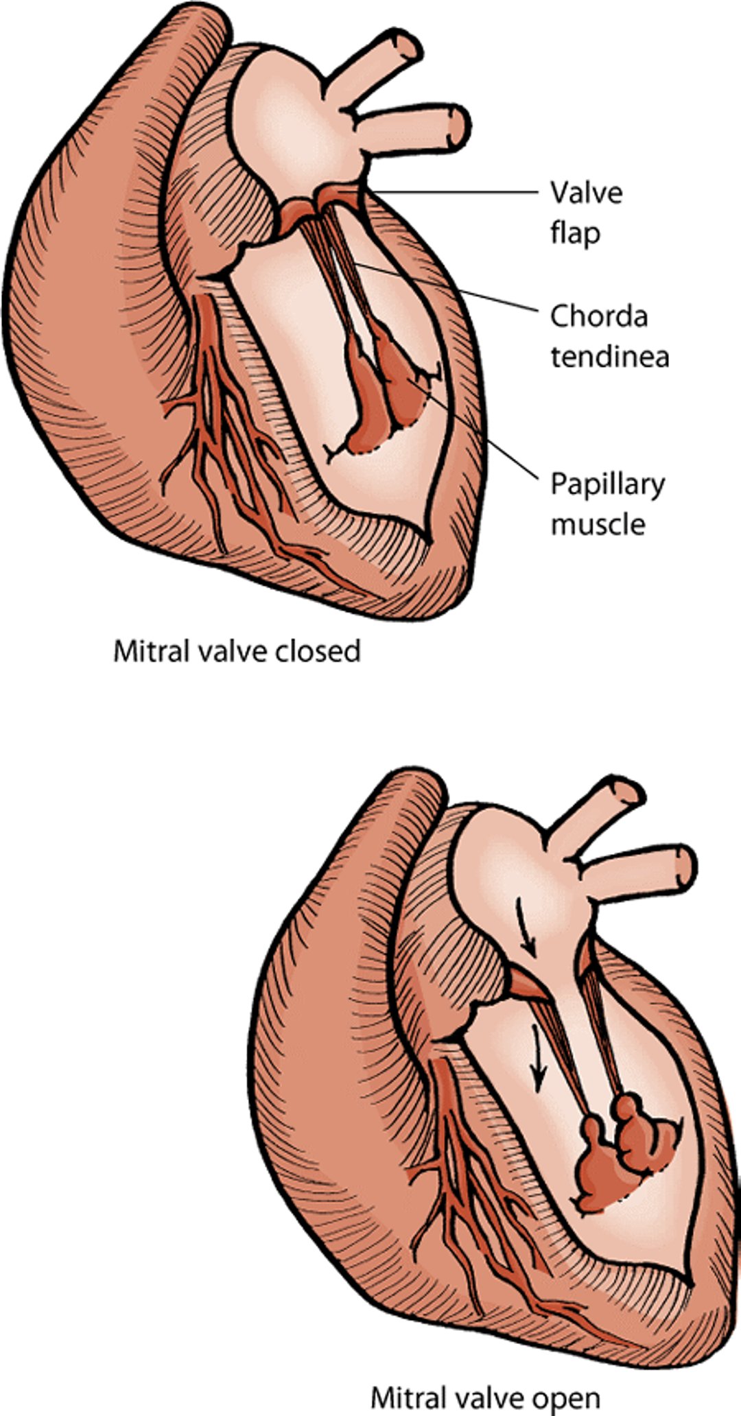
Mitral and Tricuspid Valve Dysplasia
Defects of the mitral and tricuspid valves occur more commonly in cats than in many other species. Mitral valve dysplasia refers to abnormal development or malformation of the mitral valve (which is located between the left atrium and left ventricle), allowing blood to flow back into the left atrium (regurgitation). Longterm mitral regurgitation leads to dilation of the left ventricle and atrium. Any component of the mitral valve (valve flaps, the chordae tendineae that anchor the flaps, or the small papillary muscles that attach the flaps to the ventricle) may be malformed, and often more than one component is defective.
Cats with mitral valve dysplasia have a heart murmur and may have extra heart sounds (a "gallop" rhythm). Electrocardiography, x-rays, and echocardiography (ultrasonography) can help confirm the diagnosis.
Cats with mild disease may live for years without any signs. If the defect is severe, signs of heart failure (such as difficulty breathing and reduced appetite and thirst) and arrhythmias may develop. Although these signs can be treated, the outlook is poor in most cases.
Mitral valve

Tricuspid valve dysplasia (abnormal development) is seen occasionally in newborn kittens. This prevents the tricuspid valve (located between the right atrium and the right ventricle) from performing adequately and leads to blood being regurgitated back into the right atrium. This leads to enlargement of the right ventricle and atrium. Blood flow to the lungs may be decreased, leading to fatigue and an increased rate of respiration. As the pressure in the right atrium increases, blood pools in the veins returning to the heart, causing an accumulation of fluid in the abdomen. Other defects of the heart may also be noted in affected cats.
Signs of right-sided congestive heart failure, such as accumulation of fluid in the abdomen and around the lungs, may be seen. A loud heart murmur is very noticeable. Arrhythmias, especially the sudden onset of a very high heart rate, are common and may cause death. Electrocardiography and x-rays may show enlargement of the right ventricle and atrium, while the malformed tricuspid valve and tricuspid regurgitation can be seen using echocardiography (ultrasonography).
The outlook for cats with these signs is guarded and based on the severity of disease and amount of regurgitation. Periodic draining of fluid from the abdomen may be needed. Medications may also be prescribed.
Endocardial Fibroelastosis
Endocardial fibroelastosis is an inherited defect seen most commonly in Siamese and Burmese cats. The walls of the left ventricle, the left atrium, and around the mitral valve are enlarged and thickened. Signs of heart failure, such as difficulty breathing, usually appear when affected kittens are less than 6 months old. Treatment is rarely successful and the outlook is poor.
Hernias Between the Abdomen and the Membrane Surrounding the Heart (Pericardium)
An abnormal opening between the abdomen and the membrane surrounding the heart (pericardium) is called a peritoneopericardial diaphragmatic hernia. It is the most common congenital disease of the pericardium in cats. A diaphragmatic hernia is an abnormal opening in the muscle that separates the abdomen from the chest cavity (the diaphragm) that allows abdominal organs to move up into the chest. The liver is most commonly herniated, followed by the small intestine, spleen, and stomach. Signs vary, with many patients showing no signs and the defect being discovered only after death. Chest x-rays or specialized contrast x-rays can show the intestine or liver crossing the diaphragm into the pericardium. The diagnosis can also be made using echocardiography (ultrasonography) or computed tomography (a CT scan). Patients with vomiting, trouble breathing, or other adverse conditions resulting from the hernia should have surgery to repair it.
Cor Triatriatum Sinister
Cor triatriatum sinister results from a fibrous membrane dividing the left atrium and has been reported in cats. (“Sinister” means left.) The affected atrium is divided into 2 chambers. There are commonly one or more perforations in the separating membrane, allowing communication between the 2 portions of the atrium. Affected cats can develop left-side heart failure that causes fluid accumulation in or around the lungs. Surgery can be performed to correct this disease.
For More Information
Also see professional content regarding congenital and inherited disorders of the cardiovascular system.


