

Congenital abnormalities of the cardiovascular system are defects that are present at birth. They can occur as a result of genetic defects, environmental conditions, infections, poisoning, medication taken by the mother, or poor maternal nutrition. In some cases, it is a combination of these factors that causes the defect. For several defects, an inherited basis is suspected based on breed and breeding studies. However, some studies have suggested that fewer than 1% of dogs are affected by congenital heart disease.
In addition to the congenital heart defects, many other cardiovascular disorders have been shown, or are suspected, to have a genetic basis. Diseases such as hypertrophic cardiomyopathy, dilated cardiomyopathy, and degenerative valve disease of small breeds of dogs may have a significant genetic component.
Among the few dogs that do have congenital heart disease, common defects (from most to least common) include patent ductus arteriosus, pulmonic stenosis, aortic stenosis, persistent right aortic arch, and ventricular septal defect. Less common congenital cardiac defects (occurring in less than 5% of cases) include tetralogy of Fallot, atrial septal defect, persistent left cranial vena cava, mitral valve dysplasia, tricuspid dysplasia, and cor triatriatum dexter. There are regional differences, however, in the occurrence of these defects. The most common congenital canine heart defects in the United States vary from those reported in the United Kingdom and may likely differ from those in other parts of Europe and other regions.
Detecting Congenital Heart Defects
It is important to detect a congenital heart defect as early as possible. Certain defects can be corrected with surgery, and treatment should be performed before the defect leads to congestive heart failure or irreversible heart damage. If the defect is discovered in a recently purchased dog, you may be able to return it for a refund. Pets with congenital heart defects are likely to die prematurely, causing emotional distress. Early detection also allows owners to avoid breeding dogs with genetic defects and prevent continuing genetic defects in breeding lines.
The evaluation of most animals with a congenital heart defect may include a physical examination, electrocardiography (recording electrical activity of the heart), x-rays, and echocardiography (ultrasonography). These steps allow diagnosis and assessment of the severity of the defect.
Congenital heart defects produce signs that vary depending on the type and severity of heart disease involved. Possible signs include shortness of breath or difficulty breathing, coughing, fainting, fatigue, or an accumulation of fluid in the lungs or abdomen.
General Treatment and Outlook
The medical importance of congenital heart disease depends on the particular defect and its severity. Mildly affected dogs may show no ill effects and live a normal life span. Defects causing significant circulatory disturbances will likely cause death in newborn (and unborn) puppies. Medical or surgical treatments are most likely to benefit animals with congenital heart defects of moderate to significant severity. Surgical correction, if appropriate, is indicated in most affected dogs as long as no other diseases or abnormalities are present that would pose a risk to anesthesia or surgery. If a genetic cause is likely or possible, these animals should not be bred.
Innocent Murmurs
It is very important to understand that the presence of a heart murmur in a young puppy does not necessarily indicate a congenital heart defect. Many puppies have a low-grade systolic murmur (heard while the ventricles contract) that is due to mild blood turbulence and is not associated with a congenital heart defect. These murmurs usually disappear by 6 months of age. However, the presence of signs of heart disease, loud systolic murmurs, or diastolic murmurs (heard while the ventricles relax) do indicate cardiac disease and should prompt further investigation by your veterinarian.
Common Congenital Heart Abnormalities
The defects discussed below are those that occur with the greatest frequency in dogs. However, it is important to stress that these defects are rare.
Patent Ductus Arteriosus
The ductus arteriosus is a short, broad vessel in the unborn fetus that connects the pulmonary artery with the aorta and allows most of the blood to flow directly from the right ventricle to the aorta. Before birth, oxygenated blood within the main pulmonary artery passes into the descending aorta through the ductus arteriosus, bypassing the nonfunctional lungs. At birth, inflation of the lungs upon the puppy’s first breath causes the ductus to close and allows the blood to flow to the lungs.
If the ductus does not close, the blood flow is forced from chambers of the left side of the heart to those of the right side; these defects are called left-to-right shunts. They result in overcirculation of the lungs and enlargement of the left heart chambers, which may result in arrhythmias. Over time, signs of left-sided congestive heart failure develop.
Dogs with a small ductus may reach adulthood without signs of heart failure, but most affected dogs will retain extra fluid even at a young age. Occasionally, a large patent ductus arteriosus may cause high blood pressure in the arteries of the lungs, creating an increased workload for the heart. When severe, this can cause shunting through the ductus to slow and reverse (that is, it becomes a right-to-left shunt).
In dogs with a patent ductus arteriosus with left-to-right shunting, a very noticeable, continuous, machinery-like murmur can be detected. The vibrations associated with the murmur can often be felt when touching the puppy's ribcage over the area of the base of the heart. In some newborn puppies, the ductus remains open for several days after birth. Most young puppies do not exhibit any signs. Those with a large shunt and older dogs often have signs of left-sided congestive heart failure. Arrhythmias (abnormal heart rhythms) may also be heard. Large abnormalities may show up on x‑rays. Echocardiography is valuable in excluding coexisting heart defects present since birth, as well as showing the presence of the patent ductus arteriosus.
Surgery to close the ductus in dogs with left-to-right shunting patent ductus arteriosus usually cures the problem and is almost always the best treatment option. Drugs to treat congestive heart failure may be needed before surgery can be performed. Surgery options include tying off the ductus and blocking the ductus from the inside. This latter procedure can be accomplished by using a catheter to place a device in the patent ductus arteriosus that results in blood clot formation or physical obstruction of the ductus. Your veterinarian will choose the appropriate surgical option based on the size of the ductus and the size of your dog.
In animals with a patent ductus arteriosus with right-to-left shunting, there is usually a history of fatigue, exercise intolerance, and collapse. Careful examination may reveal a slight bluish tinge to skin and membranes in the back half of the dog (that is, the limbs and trunk rather than the head and neck). Your veterinarian may note other abnormalities such as a split heart sound, a soft murmur, or an abnormal increase in red blood cells. Electrocardiography, x-rays, and echocardiography may be useful in diagnosis.
Surgically tying off the ductus should not be performed in this situation because this will cause an increase in blood pressure in the arteries of the lungs and typically death. Treatment involves control of the increase in red blood cells through periodic blood-letting and medications to decrease the blood pressure in the lungs. The longterm outlook for dogs affected with a right-to-left shunt is poor.
Pulmonic Stenosis
Pulmonic stenosis causes an obstruction to the blood flow from the right ventricle to the lungs. In most cases, the obstruction is due to abnormal development of the flaps on the valves between the pulmonary artery and the right ventricle (pulmonic valve). However, the stenosis can also occur in the cone-shaped opening, the subvalvular region (within the outflow tract of the right ventricle), or in the area beyond the pulmonary valve. The breeds at the most risk for pulmonic stenosis include English Bulldogs, Boxers, Beagles, Boykin Spaniels, terriers, and Giant Schnauzers.
The right ventricle must generate increased pressure during contraction to overcome the stenosis. In moderate to severe cases, the increased pressure can lead to severe enlargement of the right ventricle with thickening of the muscle fibers. As muscle fibers of the right ventricle thicken, the ability of the ventricle to do its job diminishes, leading to increased right atrial pressure and congestion in the veins. The increased speed of blood flow results in an enlarged artery. In severe cases, right-sided congestive failure may be noted. Other birth defects in the heart can also occur in dogs with this condition.
Dogs with pulmonic stenosis may have a history of failure to thrive and exercise intolerance. Signs of right-sided congestive heart failure, such as accumulation or fluid in the abdomen or limbs, may be present. A loud murmur can be heard, and a swollen and pulsating jugular vein may also be present. Electrocardiography, x-rays, and echocardiography may be helpful in diagnosing this condition. Doppler echo-cardiography is valuable in determining the severity of the stenosis.
Dogs with moderate or severe pulmonic stenosis may improve following surgery. One type of surgery involves using a surgical balloon to widen the narrowed valve. This type of surgery causes minimal trauma and is highly effective. The choice of surgical procedure depends to some degree on the presence and degree of thickening of the muscle fibers below the valve. Your veterinarian will best be able to determine the appropriate treatment for your pet. Drugs such as diuretics and vasodilators (drugs that widen blood vessels) should be started if right-sided congestive heart failure is present. The outlook is poor if atrial fibrillation or right-sided congestive heart failure is present. If atrial fibrillation is noted, your veterinarian may prescribe medications to control the arrhythmia.
Aortic Stenosis
Aortic stenosis refers to a narrowing of the aorta, which interferes with blood flow out of the heart. Blood flow from the left ventricle may be obstructed in several ways. Fibrous tissue located within the outflow tract of the left ventricle (below the aortic valve) can reduce flow. This is called subaortic stenosis. In other cases, the obstruction may be within the valve leading out of the left ventricle (valvular stenosis) or even past the aortic valve (supravalvular stenosis). Among dogs, subaortic stenosis occurs most commonly and is usually seen in large-breed dogs. The breeds most frequently affected include Boxers, Golden Retrievers, Rottweilers, German Shepherds, and Newfoundlands.
Aortic stenosis causes thickening of the muscle fibers of the left ventricle. A major effect of this thickening of the muscle fibers is the creation of areas of poor blood flow throughout the heart. When the supply of blood to the heart muscle is low, the heart does not get enough oxygen and is unable to rid itself of carbon dioxide and other cellular waste products. This condition is a major factor in the development of life-threatening ventricular rhythm problems.
In some cases of aortic stenosis, there may be a history of loss of consciousness (fainting) due to lack of blood flow to the brain and problems with normal levels of exercise. In other cases, animals with no history of illness may die suddenly and the defect is detected only after death. Sometimes a murmur can be detected with a stethoscope. If stenosis is suspected, the veterinarian may check the electrical activity of the heart using electrocardiography. In cases where a dog faints frequently or shows other signs of possible heart rhythm problems, the veterinarian may monitor heart activity over a full day. Other tests, such as x-rays or ultrasonography, may also be used.
Treatment options include medication to reduce problems associated with exercise intolerance or fainting. Surgery may also help, although the procedures are costly to perform and may be risky. Mildly affected dogs commonly require no treatment and the outlook can be fair to good. Your veterinarian will be able to determine the most appropriate course of treatment for your dog. Dogs with aortic stenosis should not be used for breeding.
Persistent Right Aortic Arch
When this abnormality is present at birth, the right aortic arch passes behind the esophagus and trachea (windpipe). This squeezes the esophagus and constricts it near the heart. Because food can not pass properly through the esophagus, affected animals will regurgitate. The undigested food is easily inhaled, leading to aspiration pneumonia. X-rays are used to diagnose the condition, and computed tomography (a CT scan) may be necessary to prepare for surgery to free the esophagus. Persistent right aortic arch has been reported in cattle, horses, cats, and dogs. German Shepherds and Irish Setters develop this abnormality more frequently than other breeds.
Ventricular Septal Defects
Ventricular septal defects (openings between the left and right ventricles) vary in size and effects on blood circulation. Ventricular septal defects may occur with other abnormalities of the heart present at birth.
Shunting of blood from the left ventricle into the right ventricle is the most common result of this defect, due to the higher pressures of the left ventricle. Blood shunted into the right ventricle is recirculated through the blood vessels in the lungs and left heart chambers, which causes enlargement of these structures. The right ventricle may enlarge as well. Significant shunting through the pulmonary arteries can induce narrowing of these vessels, leading to reduced blood flow or increased blood pressure. As resistance rises, the shunt may reverse (that is, become a right-to-left shunt) and flow from the right ventricle to the left, bypassing the lungs.
Signs depend on the severity of the defect and the shunt direction. A small defect usually causes minimal or no signs. Larger defects may result in severe left-sided congestive heart failure. The development of a right-to-left shunt is indicated by a bluish tinge, fatigue, and exercise intolerance. Most affected animals have a loud murmur; however, this murmur is absent or faint when a very large defect is present or when shunting is right to left. Chest x-rays and echocardiography (ultrasonography) may be used to confirm the defect.
Treatment also depends on the severity of signs and direction of the shunt. Dogs with small ventricular septal defects do not typically require treatment, and the outlook is good. Dogs with a moderate to severe defect more commonly develop signs, and treatment should be considered. Surgery to close the defect or decrease shunting or drugs to reduce blood pressure may be considered in the treatment of dogs with a large ventricular septal defect and left-to-right shunting. With right-to-left shunting, surgical closure of the defect is generally not advised. Bloodletting to relieve the effects of increased red blood cells or the use of certain drugs may be considered to relieve signs; however, the outlook for the dogs is poor to guarded. Dogs with a ventricular septal defect should not be bred. The defect has been shown to be inherited in English Springer Spaniels.
Tetralogy of Fallot
Tetralogy of Fallot is a defect that produces a bluish tinge to skin and membranes because there is not enough oxygen in the blood. It is caused by a combination of pulmonic stenosis (see above), a ventricular septal defect (see above), thickening of the muscle fibers of the right ventricle, and varying degrees of the aorta rotating to the right. Breeds most inclined to have tetralogy of Fallot include Keeshonds, English Bulldogs, and Wire-haired Fox Terriers. The trait is inherited in Keeshonds and presumably in other breeds. However, this defect has been recognized in other breeds of dogs.
The effect on the animal of this grouping of defects depends primarily on the severity of the pulmonic stenosis, the size of the ventricular septal defect, and the amount of resistance to blood flow provided by the blood vessels. Consequences may include reduced blood flow to the lungs (resulting in fatigue and shortness of breath) and generalized lack of oxygen in the blood causing a bluish tinge to skin and membranes. Red blood cells may be abnormally increased, leading to the development of blood clots and poor circulation of blood. Typical signs in dogs include stunted growth, exercise intolerance, collapse, and seizures. Affected dogs often have a heart murmur. Electrocardiographs, x-rays, and echocardiography (ultrasonography) can help confirm the diagnosis.
Treatment options include surgery and medical management. Surgery to correct the defects or to relieve pain has been reported in dogs but is rarely performed. Beta-adrenergic blocking drugs and bloodletting have been used in dogs with tetralogy of Fallot. The outlook is guarded, but dogs with mild to moderate shunting may reach adulthood.
Atrial Septal Defects
Atrial septal defects are openings that occur between the right atrium and the left atrium. These openings allow blood to flow abnormally between the chambers. Two types of openings occur: a birth defect that creates a hole in the wall and a normal fetal opening that fails to close (called a patent foramen ovale).
Before birth, a flapped oval opening of the membrane between the atria (called the foramen ovale) allows shunting of blood from the right to the left side in order to bypass the nonfunctional lungs. At birth, the drop in right atrial pressure causes the foramen ovale to close and shunting to stop. Increased right atrial pressure may reopen the flap where the membranes have not sealed and allow shunting to resume. This is not a true atrial septal defect because the membranes have formed normally. A true atrial septal defect is a consistent opening of the membranes, which allows blood to shunt from one atrium to the other.
In most cases, blood flows from the left atrium to the right atrium, causing an overload of the right-sided chambers. The volume of shunting depends on the size of the defect and the change in pressure across the defect. Excessive blood flow through the right-sided chambers results in their enlargement and thickening of their muscle fibers. Narrowing of the pulmonary blood vessels may occur as a consequence of excessive pulmonary blood flow and may quickly cause right-sided congestive heart failure. In situations in which right atrial pressure increases (for example, pulmonic stenosis) shunting from right to left may occur.
Signs of right-sided heart failure (for example, accumulation of fluid in the abdomen, fluid accumulation in tissues, bluish tinge to skin and mucous membranes) may be present. A murmur is usually present, and arrhythmias may be noted. Electrocardiography, x-rays, and echocardiography may be useful in diagnosing the defect. The results show varying degrees of enlargement of the right atrium and right ventricle.
Surgery to correct the defect may be attempted but is associated with high cost and a high death rate. Dogs with certain types of defects can tolerate the defects well, and many of these defects are only found during routine examinations of older animals. Other types of defects, however, are more likely to cause right-sided congestive heart failure. High blood pressure in the arteries of the lungs can also occur. Although some of these dogs may not show signs for years, the longterm outlook is guarded to poor.
Mitral Valve Dysplasia
Mitral valve dysplasia refers to abnormal development or malformation of the mitral valve of the heart, allowing regurgitation of blood back into the left atrium. Breeds of dogs most likely to be born with mitral valve dysplasia are Bull Terriers, German Shepherds, and Great Danes.
Longterm mitral regurgitation results in enlargement of the left ventricle and atrium. When the regurgitation is severe, blood flow from the heart decreases and fluid accumulates in the lungs, producing signs of heart failure. Arrhythmias may develop. In some cases, malformation of the mitral valve causes stenosis, or narrowing, of the valve (see below).
The signs of mitral valve dysplasia depend on the severity of the defect. Signs of left-sided congestive heart failure, such as coughing and difficulty breathing, are often seen. An electrocardiogram may show arrhythmias in severely affected dogs. Chest x-rays, echocardiography, and other specialized techniques help to confirm the defect.
The outlook for dogs with signs and severe disease is poor. Mildly affected animals may remain free of signs for several years. Signs of left-sided congestive heart failure can be treated.
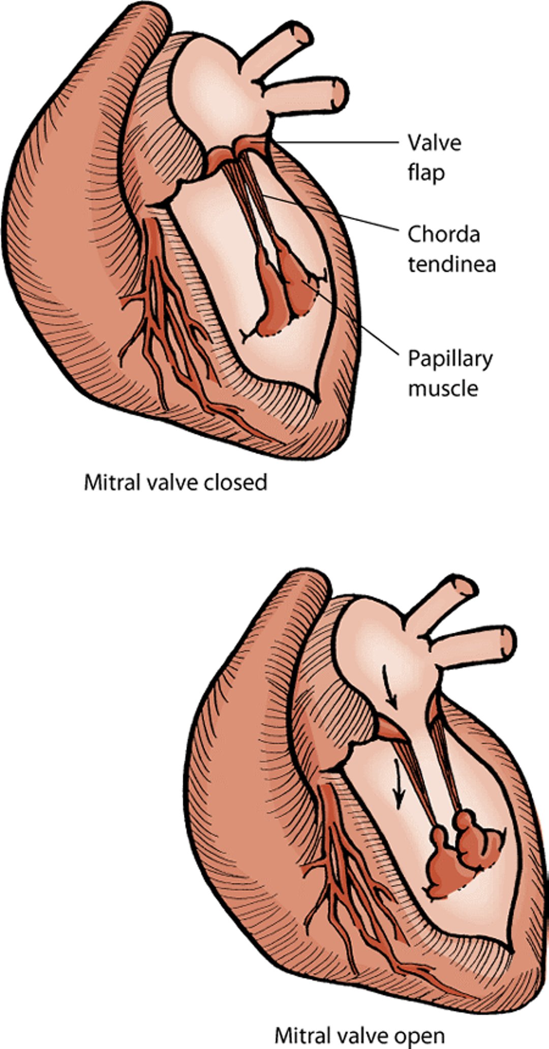
Mitral valve

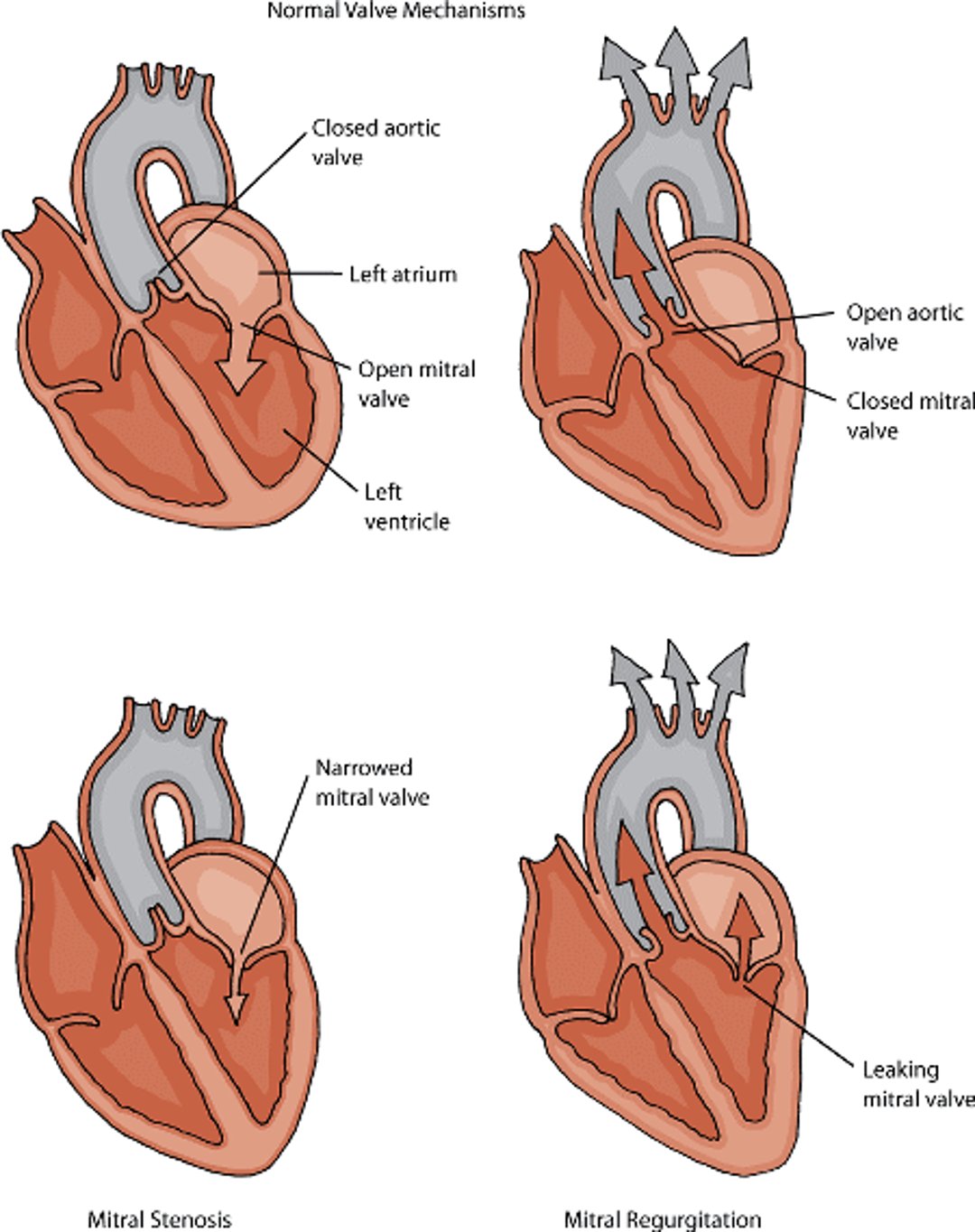
Mitral Stenosis
Mitral valve stenosis is a narrowing of the mitral valve opening caused by abnormalities of the mitral valve. This obstructs blood inflow to the left ventricle. The defect is rare in dogs, but it can occur together with other congenital defects such as subaortic stenosis, mitral valve dysplasia, and pulmonic stenosis (all discussed earlier in this chapter).
Mitral stenosis results in enlargement of the left atrium and an increase in blood pressure within the veins of the lungs. Fluid in the lungs can develop as a consequence. Loss of consciousness due to lack of blood flow to the brain occurs in some dogs with these defects. A low-grade heart murmur can sometimes be detected. If mitral valve dysplasia (see above) is present together with the stenosis, a louder murmur may be heard. X-rays and electrocardiography are useful in showing the effects of stenosis; echocardiography (ultrasonography) can confirm the diagnosis.
Dogs with mitral valve stenosis may be prescribed diuretics (to help eliminate fluid buildup) and put on a low-salt diet. Diuretic use should be carefully monitored because too high a dose can severely reduce blood flow from the heart. Surgery to relieve mitral stenosis may also be an option.
Mitral stenosis and regurgitation

Tricuspid Dysplasia
Tricuspid dysplasia refers to abnormal development or malformation of the tricuspid valve of the heart, allowing regurgitation of blood back into the right atrium. This defect is seen occasionally in dogs at birth. Breeds most likely to have tricuspid dysplasia are Labrador Retrievers and German Shepherds. Rarely, stenosis (narrowing) of the tricuspid valve can be noted. Other defects of the heart may also be noted in affected dogs.
Longterm tricuspid regurgitation leads to volume overload of the right heart, enlarging the right ventricle and atrium. Blood flow to the lungs may be decreased, leading to fatigue and an increased rate of respiration. As the pressure in the right atrium increases, blood pools in the veins returning to the heart, causing an accumulation of fluid in the abdomen.
The more severe the defect, the more obvious the signs will be in affected dogs. Signs of right-sided congestive heart failure, such as accumulation of fluid in the abdomen and limbs, may be seen. A loud heart murmur is very noticeable. Arrhythmias, especially the sudden onset of a very high heart rate, are common and may cause death. Electrocardiography and x-rays may show enlargement of the right ventricle and atrium, while the malformed tricuspid valve and regurgitation can be seen using echocardiography (ultrasonography).
The outlook for dogs with tricuspid dysplasia depends on the severity of the abnormality and degree of regurgitation. Periodic draining of fluid from the abdomen may be needed. Medications such as diuretics, vasodilators, and digoxin may also be prescribed.
Hernias Between the Abdomen and Membrane Surrounding the Heart
An abnormal opening between the abdomen and the membrane surrounding the heart (peritoneopericardial diaphragmatic hernia) occurs as a birth defect in dogs. A diaphragmatic hernia is an abnormal opening in the membrane that separates the abdomen from the chest cavity. The result is a hole through which the abdominal organs can protrude up into the chest. The liver is most commonly herniated, followed by the small intestine, spleen, and stomach. Signs vary, with many patients showing no signs and the defect being discovered only after death. Chest x-rays or specialized contrast x-rays can show the intestine or liver crossing the membrane into the sac surrounding the heart (the pericardium). The diagnosis can also be made using echocardiography (ultrasonography) or contrast tomography (a CT scan). Patients with vomiting, signs of hepatic encephalopathy (a disease of the brain due to a buildup of toxic substances that are normally removed by the liver), trouble breathing, or other adverse conditions resulting from the hernia should have surgery to repair the hernia.
Cor Triatriatum Dexter
Cor triatriatum dexter results from a fibrous membrane dividing the right atrium and has been rarely reported in dogs. The affected atrium is divided into 2 chambers. There may or may not be one or more perforations in the separating membrane, allowing communication between the 2 portions of the atrium. Dogs with severe disease often have signs of right heart failure (such as fluid accumulation in the abdomen). Surgery can be performed to correct this disease.
For More Information
Also see professional content regarding congenital and inherited disorders of the cardiovascular system.


